
Abstract
von Willebrand disease (vWD) is the most common inherited coagulopathy in dogs, particularly in Doberman Pinschers. We developed a pyrosequencing-based assay to estimate the frequency of the c.7437G>A mutation associated with vWD type 1 in the Doberman Pinscher population of Buenos Aires, Argentina. We found a 0.41 frequency for the mutated allele, which varied significantly within families (family 1 = 0.43, family 2 = 0.58, unrelated animals = 0.35). The use of a popular founder male carrier of mutant allele A increased vWD incidence within a family and in the general population. The mode of inheritance was confirmed as autosomal dominant with incomplete penetrance. No differences were found between sexes and coat colors. Pyrosequencing was a good complement to clinical and coagulation tests for vWD type 1 diagnosis and a useful alternative for detecting the c.7437G>A mutation.
von Willebrand factor (vWF) is a protein that acts as a molecular bridge between platelets and subendothelium, containing 3 important functional regions: a collagen-binding region, a platelet-binding region, and a region that facilitates interaction with factor VIII.6,7,16 vWF also acts as a carrier for factor VIII, providing stability in circulation by preventing protease-related degradation. 25
von Willebrand disease (vWD) is the most common type of extrinsic platelet disorder and the most commonly found inherited coagulopathy in dogs. 1 Although vWD is closely associated with Doberman Pinschers, the disease has been identified in 54 breeds.14,15 It was first reported in 1970 in a family of German Shepherd Dogs with clinical signs similar to those in the human disease.3,13
Three types of vWD have been identified in dogs. In vWD type 1, the most widespread type of the disease (accounts for >95% of all cases), there is quantitative deficiency of vWF in circulation. In vWD type 2, which is rare in dogs, there is a qualitative defect of vWF. vWD type 3 is the most severe and least common form, and it is characterized by virtual absence of vWF. 1
The phenotypic diagnosis of vWD is based on clinical signs and laboratory tests. 9 Clinical signs are highly variable among animals. Some dogs do not show evidence of the disease, whereas others may have bleeding from the nose, vagina, urinary bladder, or oral mucous membranes, and less frequently subcutaneous hematomas and hemarthrosis. Disease signs are usually evident even during low-complexity surgeries or after trauma. 11 Because no single laboratory test is sufficiently comprehensive to detect all vWD variants, determination of both vWF concentration and function is important in diagnosis. 9
vWD has been known since the 1970s; however, there are very few epidemiologic reports on this disease. In the Doberman breed, the reported prevalence of vWD type 1 is high: ~50% in the United Kingdom, 73% in the United States, and ~30% in Brazil.2,17 However, to our knowledge, reports on the prevalence of its associated mutations in Argentina and other countries do not exist.
vWD types are determined by different mutations in the vWF gene located on chromosome 27 (NCBI reference sequence NM_001002932.1). In several dog breeds, including Doberman Pinschers, vWD type 1 is associated with a G→A transversion of the last base of exon 43 of the vWF gene (c.7437G>A). The mutation activates a cryptic splice site a few nucleotides upstream of the normal splice site, leading to a frame shift that results in the formation of a truncated protein of 119 amino acids. 24 In dogs, vWD type 1 is considered an autosomal recessive disorder in some breeds, although it is possibly inherited as dominant with incomplete penetrance in others.15,18,22
We estimated the prevalence of c.7437G>A single nucleotide polymorphism, an associated mutation for vWD type 1, in the Doberman Pinscher population of Buenos Aires and surrounding regions (see Supplementary Fig. 3 for an illustration of methods). We developed a pyrosequencing-based assay and further evaluated the mode of inheritance of this disease based on pedigree and clinical analyses.
Our study was reviewed and approved by the Animal Care and Use Committee of the National University of La Plata School of Veterinary Sciences, Argentine (Res.Ref.Exp. 600-007197/11). All experiments were conducted under the guidelines established in The Guide for The Care and Use of Laboratory Animals. 5 Owners signed an informed consent prior to study initiation.
Samples from 89 Doberman Pinscher dogs from the province of Buenos Aires were collected. Dogs were grouped into 2 families and 1 group of unrelated animals: family 1 (n = 34), family 2 (n = 13), and unrelated animals (n = 42). Forty-one of the 89 sampled dogs had available medical records, including surgeries (e.g., ear cropping and tail docking), parturition, and other bleeding episodes.
Blood samples were collected from the cephalic or saphenous vein in a single extraction and stored at −20°C. Tubes for hemogram, platelet count, and DNA extraction contained 6% EDTA. Tubes for kaolin-activated partial thromboplastin time and prothrombin time (PT) determination contained 3.2% sodium citrate. These latter blood samples were immediately centrifuged at 1,000 × g for 15 min, and the citrated plasma was collected, placed in 1.5-mL Eppendorf tubes, and stored at −20°C until analyzed. Additional coagulation parameters evaluated in these dogs included buccal mucosal bleeding time (BMBT), 8 hemogram, PT, and activated partial thromboplastin time. Details of methods used are described in Supplementary Table 3.
Genomic DNA was extracted from blood samples (Wizard Genomic DNA purification kit, Promega, Madison, WI) according to the manufacturer’s recommendations. DNA was genotyped by direct sequencing and pyrosequencing methods as described in Supplementary Table 4. The pyrosequencing assay was designed using the DNA sequence previously reported in GenBank (http://www.ncbi.nlm.nih.gov/genbank/; Supplementary Table 1).
Gene and genotype frequencies and Hardy–Weinberg equilibrium (HWE) were estimated using a population genetics software package. 21 A pairwise chi-squared test was used to compare the gene frequencies obtained within each group. Single factor ANOVA was used to evaluate significant differences in bleeding time between genotypes. Significance was set at p < 0.05.
All dogs were genotyped for the c.7437G>A mutation using the developed pyrosequencing method.19,20 The accuracy of our assay was validated by direct sequencing, and the obtained results showed 100% agreement between the genotyping methods (Fig. 1).
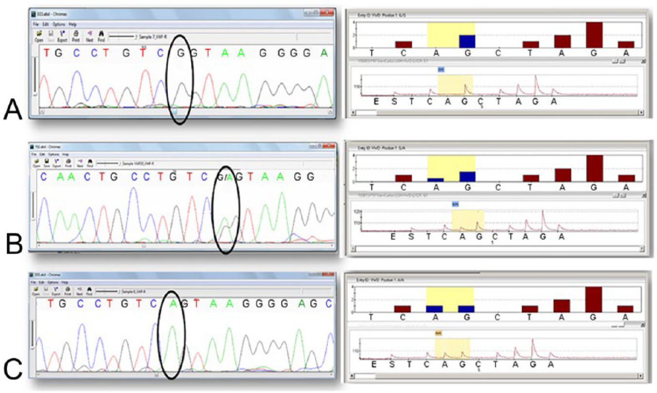
Electropherograms and pyrograms of c.7437G>A mutation obtained by DNA sequencing and pyrosequencing assays, respectively:
Gene and genotype frequencies of all of the study groups were in HWE (total population, p = 0.67; family 1, p = 0.16; family 2, p = 1), with the exception of unrelated animals (p = 0.034; Table 1).
Genotype and c.7437G>A allele frequencies of Doberman Pinscher dogs in the Buenos Aires area for 2 families and unrelated animals.
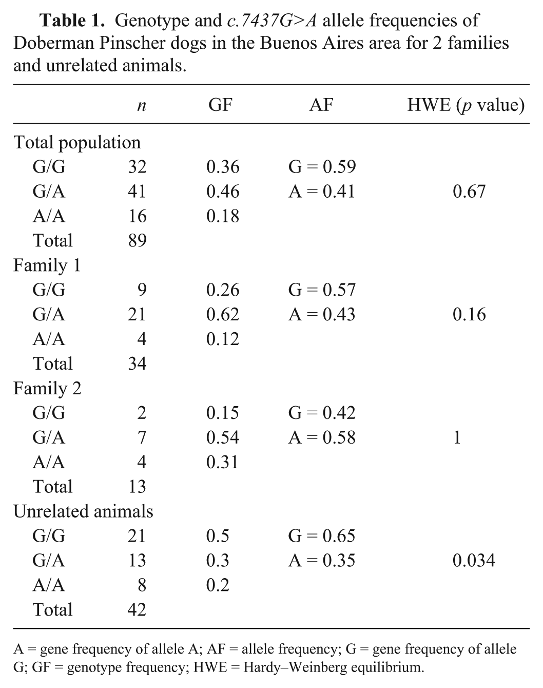
A = gene frequency of allele A; AF = allele frequency; G = gene frequency of allele G; GF = genotype frequency; HWE = Hardy–Weinberg equilibrium.
Analysis of the genetic differentiation of gene frequencies between pairs of groups showed a significant difference between both families and the unrelated animal group (p < 0.01). However, differences between family 1 and family 2 were not significant (p > 0.05). Furthermore, nonsignificant differences in gene frequencies were observed in sex and coat color (brown and black) of the animals (Supplementary Fig. 1).
Data from the 41 medical records of the genotyped animals showed that 3 of 5 homozygous A/A dogs had no evidence of clinical signs; all homozygous G/G animals were normal. In addition, only 6 of 27 heterozygous animals showed clinical signs (Table 2). Hemogram, platelet count, and coagulation test results were within normal ranges in the 41 animals (Supplementary Table 2) and a relationship with genotype (Supplementary Fig. 2) was not present.
Presence or absence of clinical signs according to the c.7437G>A allele frequencies of Doberman Pinscher dogs in the Buenos Aires area.
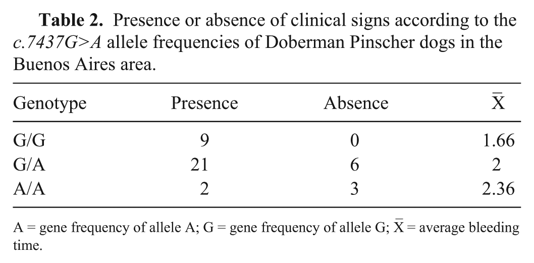
A = gene frequency of allele A; G = gene frequency of allele G;
Determination of differences in bleeding times among the 3 c.7437G>A genotypes by single-factor ANOVA showed that, although G/A and A/A had higher average bleeding times than G/G, these values were not statistically significant (p = 0.403; Table 3).
Single-factor ANOVA: average mucosal bleeding time for Doberman Pinscher dogs with different c.7437G>A alleles.
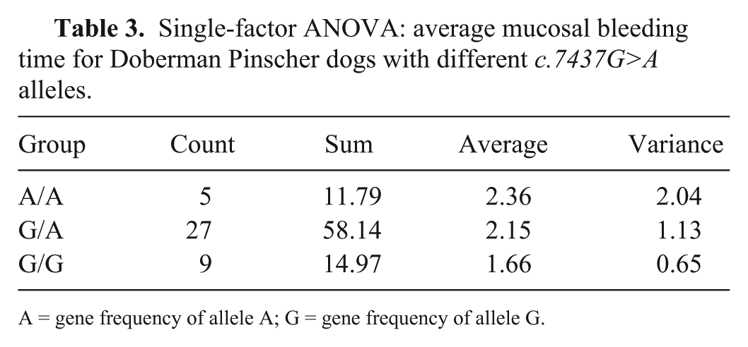
A = gene frequency of allele A; G = gene frequency of allele G.
Based on the parentage data of families 1 and 2, 2 pedigrees were built and jointly analyzed assuming that both families had a common dog ancestor with genotype A/A (Fig. 2). Genotyping data from these pedigrees were consistent with Mendelian inheritance; all individuals with G/A and A/A genotypes had parents carrying the mutant allele. Furthermore, none of the G/G animals showed signs or laboratory findings consistent with a coagulopathy. As expected, the presence of a common ancestor homozygous for the mutant allele explained the higher A gene frequencies observed in these families relative to the unrelated animals.
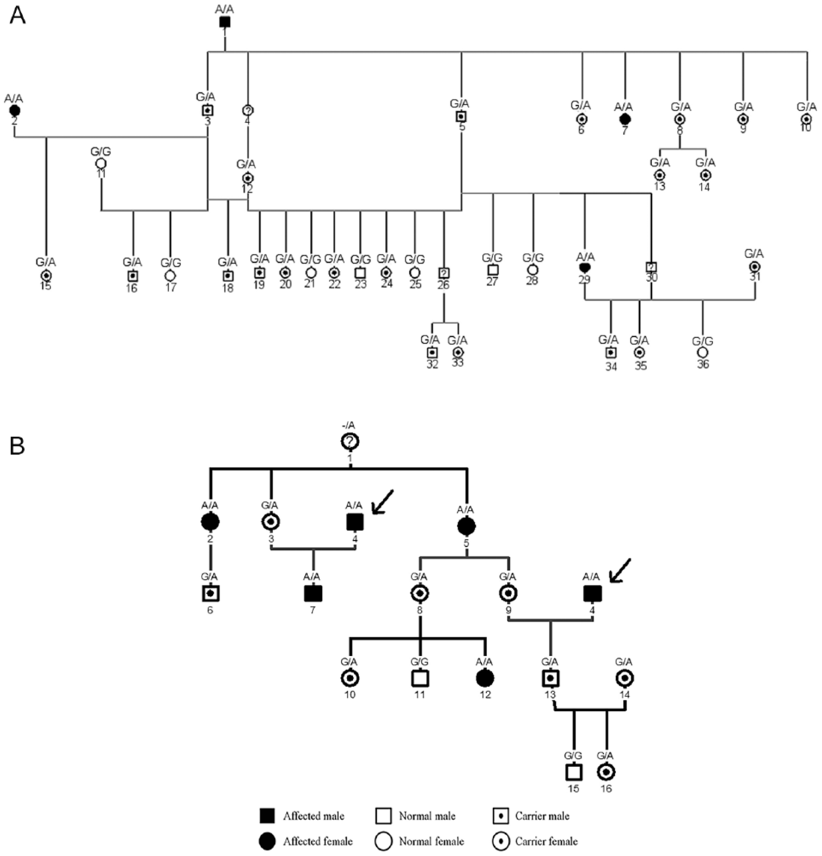
Pedigree analysis of
Laboratory confirmation of vWD is partly based on measurements of vWF antigen. Factor VIII coagulant activity may be reduced or within the normal range in the plasma of affected animals. Furthermore, vWD diagnosis includes the measurement of vWF antigen using ELISAs. The concentration of vWF antigen is consistently low in dogs with vWD. 1 Type 2 vWD in people may be characterized by dysfunctional, rather than deficient, protein. 10 A Laurel 1 rocket immunoelectrophoresis assay is an alternative and less sensitive method than ELISA to measure vWF. 7 Thus, the results of clinical analyses and laboratory methods are usually variable and not specific for the diagnosis of animals with vWD. In order to identify the genetic cause of vWD, the proposed causal mutation of this disease had been mapped. 24 With this information, 2 different tests have been developed based on the detection of the proposed causal mutation using PCR-RFLP (restriction fragment length polymorphism) 24 or real-time PCR. 12 We developed a low-cost and high-throughput specific pyrosequencing method. This assays allowed us to genotype samples from Doberman Pinschers with 100% concordance with the direct sequencing method.
Two inherited modes have been proposed for vWD type 1 in dogs: autosomal recessive, or dominant with incomplete penetrance.4,15,18,22 Based on our clinical records and BMBT, our results showed that only animals with at least one mutant allele had clinical signs, suggesting a mode of inheritance of dominance with incomplete penetrance consistent with the majority of previous reports. This type of inheritance also explains the occurrence of the mutated allele at a higher gene frequency than vWD.
Previous work reported that vWD prevalence was influenced by different factors, such as sex and coat color. Higher prevalence has been reported in vWD-affected females, 17 whereas other authors have reported a higher frequency in vWD-affected males. 23 In contrast, we found no differences in allele frequency between sexes. An association between diluted coat colors and vWD has been reported 18 ; this hypothesis could not be tested because our study included only black and brown animals.
Our results showed a frequency of 0.41 for the mutated allele c.7437G>A in Doberman Pinschers in the Buenos Aires region, lower than the 0.47 observed in the United States (https://goo.gl/P1ePkm).This average value can vary significantly within families. As shown by pedigree analysis, the introduction of a popular male from family 1 into family 2 clearly increased the frequency of the mutant allele in the second family. The increased frequency of the mutated allele in family 2 showed the negative impact of introducing an asymptomatic homozygous A/A dog into the breed, and the importance of genotyping to prevent the spread of the mutated allele.
Our pyrosequencing-based assay for the detection of the c.7437G>A vWD-associated mutation is a better alternative than direct sequencing and real-time PCR, allowing the genotyping of up to 96 samples in 20 min, and the results were easier to interpret. This assay is a useful complement to clinical and functional studies for vWD diagnosis.
Footnotes
Acknowledgements
We thank A Arizmendi, C Baccino, D Arias, D Posik, and A Di Maggio for their assistance.
Declaration of conflicting interests
The authors declared no potential conflicts of interest with respect to the research, authorship, and/or publication of this article.
Funding
Research for this paper was supported by Consejo Nacional de Investigaciones Científicas y Técnicas (CONICET) and Universidad Nacional de La Plata (UNLP).
References
Supplementary Material
Please find the following supplemental material available below.
For Open Access articles published under a Creative Commons License, all supplemental material carries the same license as the article it is associated with.
For non-Open Access articles published, all supplemental material carries a non-exclusive license, and permission requests for re-use of supplemental material or any part of supplemental material shall be sent directly to the copyright owner as specified in the copyright notice associated with the article.