
Abstract
A 22-y-old Quarter Horse gelding was presented to the University of Illinois Veterinary Teaching Hospital for evaluation of increased heart rate and mild colic signs. Rectal examination revealed a large left perirenal mass. Abdominal ultrasonography further confirmed this finding. Thoracic ultrasonography indicated multifocal irregularities on the pleural surface suggestive of consolidation and possibly masses in the lungs. The animal was euthanized. Autopsy findings included a large, firm, expansile, gelatinous retroperitoneal mass that surrounded both kidneys, as well as nodules with similar morphology in the lungs, liver, intestinal mesentery, cecum, and caudal mesenteric artery. Histologically, the masses were composed of neoplastic stellate-to-spindloid cells in abundant mucinous stroma. Neoplastic cells exhibited strong immunoreactivity for vimentin and were negative for pancytokeratin (A1/A3), CD3, CD20, melan A, and synaptophysin. Mucinous stroma was strongly positive with alcian blue and weakly positive with periodic acid–Schiff histochemical staining. These findings are consistent with metastatic myxosarcoma. Myxosarcoma is a rare neoplasm in horses, and metastasis to tissues other than sentinel lymph nodes has not been described previously to our knowledge.
A 559-kg, 22-y-old Quarter Horse gelding was presented to the University of Illinois Veterinary Teaching Hospital Equine Medicine service (Urbana, IL) for evaluation of increased heart rate and mild colic signs. The animal had been treated with trimethoprim–sulfadiazine and metronidazole the previous week. Serial chemistry profiles from the referring veterinarian indicated increasing hypercalcemia over the prior 3 wk (3.95 mmol/L [15.8 mg/dL] wk 1; 4.15 mmol/L [16.6 mg/dL] wk 2; 4.88 mmol/L [19.5 mg/dL] wk 3; reference interval [RI]: 2.63–3.25 [10.5–13 mg/dL]). On physical examination, marked ventral edema including the preputial sheath was noted. Rectal examination suggested a mass in the left dorsal quadrant involving the left kidney; the rectum was noted to deviate to the right and ventrally. Abdominal ultrasonography further confirmed a left-sided perirenal mass. Thoracic ultrasonography indicated multifocal irregularities in the pleural surface suggestive of consolidation and possibly masses in the lungs. Venous blood gas analysis (Stat Profile Critical Care Xpress, NOVA Biomedical, Waltham, MA) indicated metabolic acidosis (pH = 7.38, RI: 7.4–7.45) with notable hypercalcemia (ionized 2.38 mmol/L, RI: 1.17–1.37 mmol/L), hyponatremia (121 mmol/L, RI: 134–138 mmol/L), hypokalemia (3.5 mmol/L, RI: 3.8–4.5 mmol/L), and hypochloremia (96 mmol/L, RI: 102–105). Increased urea (8.2 mmol/L [23 mg/dL], RI: 4.3–6.1 [12–17 mg/dL]) and creatinine (221 µmol/L [2.5 mg/dL], RI: 97–150 µmol/L [1.1–1.7 mg/dL]) concentrations and hyperlactatemia (0.53 mmol/L [4.8 mmol/L], RI: 0–0.13 mmol/L [0–1.21 mmol/L]) was present. Cytology from the abdominal fluid revealed a large number of red blood cells with a scant number of vacuolated macrophages, neutrophils, and lymphocytes (nucleated cell count: 0.78 cells × 109/L [867 cells/µL], erythrocyte count: 466 × 109/L [466,000 cells/µL], total protein: 18 g/L, specific gravity: 1.017).
The high suspicion of neoplasia was discussed with the owners based on the rectal and ultrasonographic findings, as well as the persistent and increasing hypercalcemia. The owners elected euthanasia based on the gelding’s age and poor prognosis. The gelding was euthanized with an overdose of pentobarbital, and the body was submitted for autopsy.
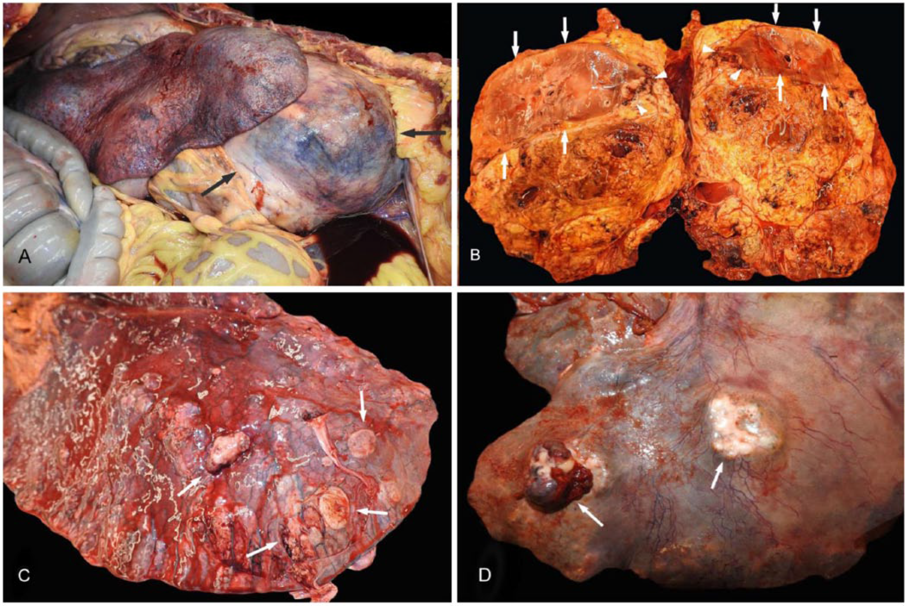
Upon gross examination, the peritoneal cavity contained ~50 L of serosanguineous fluid mixed with a moderate amount of fibrin, which adhered loosely to the serosae. In addition, numerous fibrous adhesions were present throughout the peritoneal cavity, multifocally between viscera and body wall. The retroperitoneal cavity contained a yellow-to-tan-to-black, multinodular mass (Fig. 1A) that was 42 × 27 × 30 cm and weighed 21 kg. On cut section, the mass contained moderate amounts of clear-to-yellow, mucinous material. The mass completely encompassed both kidneys and infiltrated the caudal pole of the left kidney (Fig. 1B). The adrenal glands could not be identified. There were nodules of up to 6 × 4 × 4 cm within the mesentery. A mass within the lumen of the caudal mesenteric artery at the level of the base of the cecum adhered firmly to the tunica intima. A 7 × 4 × 3 cm intramural mass was present within the base of the cecum. The lung contained nodules of up to 7 × 6 × 6 cm (Fig. 1C). Nodules of up to 20 × 10 × 5 cm were present within the liver (Fig. 1D). Additional gross findings included numerous fibrous adhesions within the thoracic cavity, and moderate enlargement of the pituitary gland.
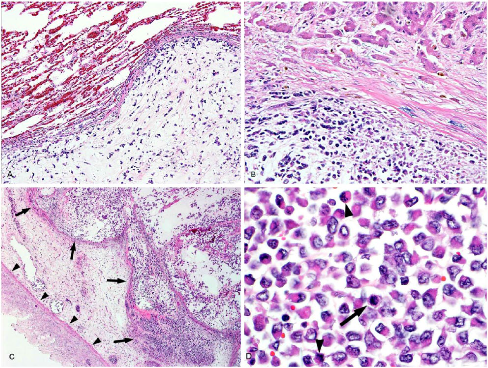
Tissues were fixed in 10% neutral-buffered formalin, routinely processed, and embedded in paraffin; 5-µm thick sections were slide-mounted and stained with hematoxylin and eosin. Histologic examination of the retroperitoneal mass revealed a moderately cellular, poorly demarcated, multilobular, and infiltrative neoplasm (Fig. 2A). Lobules were separated by fibrovascular tissue and composed of stellate-to-spindloid neoplastic cells that often formed bundles and streams in a moderate amount of colorless space mixed with wispy, eosinophilic-to-amphophilic material. Neoplastic cells had variably distinct cell borders, and a moderate amount of eosinophilic, granular cytoplasm. Nuclei were round-to-ovoid and contained coarsely stippled chromatin and 1–2 prominent nucleoli (Fig. 2B). Moderate-to-marked anisocytosis and anisokaryosis were observed. Occasional multinucleate neoplastic cells contained up to 5 nuclei. Approximately 7 mitotic figures were observed within 10 high-power fields. Multifocal, moderate-to-marked hemorrhage and necrosis were observed throughout examined sections. Masses within the mesentery, mesenteric lymph nodes, caudal mesenteric artery, cecum (Fig. 3C), liver (Fig. 3B, 3D), and lung (Fig. 3A) contained neoplastic cells similar to those described for the retroperitoneal mass, and were interpreted to be metastases from the retroperitoneal mass. Additional histologic findings included moderate lymphoplasmacytic enterocolitis, moderate membranous glomerulonephropathy with proteinosis, moderate renal tubular necrosis, interstitial fibrosis and hemorrhage, moderate pars intermedia adenomatous hyperplasia, thyroid adenomas in the right thyroid gland, and marked interstitial fibrosis within the left thyroid gland.
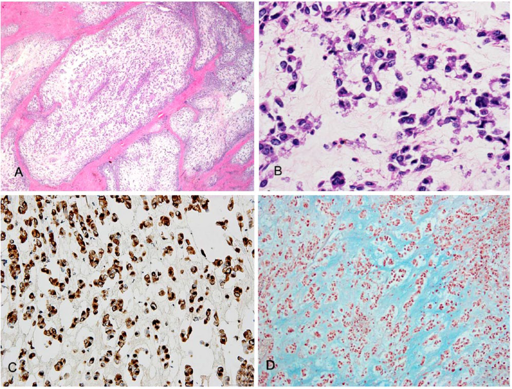
Immunohistochemistry and additional staining was performed to further characterize the neoplasm. Neoplastic cells within the retroperitoneal mass showed strong immunoreactivity to vimentin (clone V9 pre-dilute antibody, mouse monoclonal, IgG1/kappa, catalog PM048, Biocare Medical, Concord, CA; Fig. 2C) and no immunoreactivity to pancytokeratin (A1/A3; pre-dilute cocktail antibody, mouse monoclonal, IgG1, catalog PM011, Biocare Medical), CD3 (concentrated antibody, rabbit polyclonal, dilution: 1:150, catalog CP215, Biocare Medical), CD20 (epitope specific rabbit antibody, dilution 1:1000, catalog RB-9013-P, Thermo Fisher Scientific, Fremont, CA), melan A (clone A103 concentrated antibody, mouse monoclonal, IgG1, dilution: 1:60, catalog ACI 3114, Biocare Medical), or synaptophysin (clone 27G12 concentrated antibody, mouse monoclonal, IgG1, dilution 1:100, catalog CM371, Biocare Medical). Mixed colorless space and wispy eosinophilic stroma between neoplastic cells was strongly positive with alcian blue staining (Fig. 2D), and weakly positive with periodic acid–Schiff (PAS) staining. Neoplastic cells within the lung were strongly immunoreactive to vimentin and negative for pancytokeratin and synaptophysin. The colorless space and wispy eosinophilic stroma between neoplastic cells within the lung stained blue with alcian blue staining. Based on morphologic and staining characteristics, the neoplasm was diagnosed as a primary retroperitoneal myxosarcoma with metastasis to the lung, liver, mesentery, mesenteric lymph nodes, caudal mesenteric artery, and cecum.
Myxosarcomas are malignant neoplasms of fibroblastic origin, and are composed of spindloid-to-stellate mesenchymal cells that produce abundant myxomatous matrix that is rich in glycosaminoglycans. 10 Myxosarcomas exhibit similar histologic characteristics to their benign counterpart, myxomas, but have more apparent cellular and nuclear pleomorphism, including bizarre mitotic figures. 12 In addition, myxosarcomas are capable of metastasis; myxomas do not metastasize. 10
The retroperitoneal space contains the kidneys, adrenal glands, ureters, nervous tissue, blood vessels (aorta, caudal vena cava), lymphatics, and adipose and connective tissue. Neoplasms may arise from any of these tissues. Renal, ureteral, and adrenal tumors are rare in horses.11,16 Other very rare retroperitoneal tumors that have been reported in horses include extra-adrenal sympathetic paraganglioma, 8 hemangiosarcoma, 9 and a single case of primary retroperitoneal tumor. 13 The myxosarcoma described in our case was differentiated from other mesenchymal neoplasms, including primary retroperitoneal tumor, by the production of abundant mucinous matrix.
Many different types of neoplasms produce mucin, including tumors of both mesenchymal and epithelial origin. Use of both alcian blue and PAS stains will differentiate between mesenchymal mucins and epithelial-derived mucins. Mesenchymal mucins are positive with alcian blue staining, but negative to weakly positive with PAS staining. Epithelial-derived mucins are positive for both stains.1,15 Our case exhibits a staining pattern that is consistent with mucin produced by mesenchymal cells. Mesotheliomas can also produce mucinous stroma and have been reported in equids; however, they have a more epithelioid morphology and exhibit positive immunoreactivity for cytokeratin in addition to positive immunoreactivity to vimentin.2,10 Myxosarcomas have mesenchymal morphology and do not exhibit immunoreactivity to cytokeratin.
Myxosarcomas have been described previously in animals. Previous publications document occurrence in the skin, mandible, heart, lungs, and abdominal peritoneum.1,5-7,14,15 Myxomas and myxosarcomas have been reported to affect both foals and adult horses. Myxomas are rare tumors in equid that have been reported to affect the bone and soft tissues of the head, the heart, and bronchi. Myxosarcomas are exceedingly rare in equids and have been reported in the gingiva, mandible, and cecum.3,10,14 Three cases of cecal myxosarcomas have been described previously, all of which metastasized to regional lymph nodes3,4; primary retroperitoneal myxosarcoma with metastasis to thoracic and abdominal viscera has not been reported previously to our knowledge.
Footnotes
Acknowledgements
We thank the diagnostic receiving and histology staff, as well as all anatomic pathology faculty not listed as authors from the Veterinary Diagnostic Laboratory, College of Veterinary Medicine, University of Illinois at Urbana-Champaign, for their assistance with this case.
Declaration of conflicting interests
The authors declared no potential conflicts of interest with respect to the research, authorship, and/or publication of this article.
Funding
The authors received no financial support for the research, authorship, and/or publication of this article.