
Abstract
We investigated possible age-related differences in coagulation profiles in bovine species by means of rotational thromboelastometric (ROTEM) analysis. We evaluated hemostasis by ROTEM in newborn Piemontese calves at birth (T0), 8 d (T8), and 15 d (T15) of age and compared the ROTEM results obtained in 16 newborn calves with 28 adult Piemontese cattle. Hemostasis was evaluated using standard coagulation tests and ROTEM analysis, obtaining in-TEM, ex-TEM, and fib-TEM profiles. Statistically significant differences in the ROTEM profiles of newborn calves were found between T0 and T8 and between T0 and T15 (p < 0.05) but not between T8 and T15. Differences between ROTEM profiles of calves and adults were statistically significant at T0 (p < 0.05) but no differences were found at T15 (p < 0.05). Hence, ROTEM reference intervals for adult cattle can be used to evaluate profiles in Piemontese calves ≥8 d of age.
Introduction
The rate of development of the hemostatic system differs among mammals. At birth, hemostasis is immature in the lamb, intermediate in the foal and the pig, and relatively mature in the rabbit. In human infants, the coagulation profile takes several weeks to reach adult values.1,6 In newborn calves, activated partial thromboplastin time (aPTT) and prothrombin time (PT) values are within the normal adult range in most studies.2,4,6,11 The coagulation system of newborn calves appears to be generally efficient, and the small differences from adults do not affect hemostatic function. For example, the plasma activities of factors VII, VIII:C, IX, X, XI, and fibrinogen concentration are at the lower end of the normal adult range, although the levels of the anticoagulant proteins α2-macroglobulin and antithrombin are markedly lower in calves than in adult cattle.2,4,6,11
Conventional coagulation tests rely on technologies that evaluate single steps of coagulation or single plasma factors. Because hemostasis is a dynamic process that also involves cellular elements, viscoelastic techniques, such as rotational thromboelastometry (ROTEM), may better represent hemostasis in vivo. 12 Such techniques require a whole blood sample for analysis and are able to measure clot formation kinetics (the time needed to make a clot), evaluate the mechanical properties of clot (clot firmness), and determine the rate of clot dissolution (fibrinolysis).8,9 Blood samples are processed by adding specific reagents to generate ROTEM profiles: the in-TEM profile for the intrinsic pathway; the ex-TEM profile for the extrinsic pathway; and the fib-TEM profile correlated to functional fibrinogen levels.
Thromboelastography and ROTEM analysis have been applied to evaluate hemostasis in numerous species; however, few studies in cattle have been reported to date.3,5,8,9,14 The analytical performance of ROTEM and normal reference intervals have been defined in adult cattle and calves in a study population of 48 adult cattle (15 Piemontese females and 33 males of different beef breeds) and 14 Holstein–Friesian calves (6 mo old). 5 The bovine thromboelastogram presented several features distinct from equine and canine tracings, and ROTEM was an accurate assay for the evaluation of hemostasis in cattle. In a later study, the same authors applied ROTEM to investigate the hemostatic effect of low-dose dexamethasone treatment in Friesian calves. 3 Finally, a 2014 study established reference intervals for thromboelastography in dairy cows in different lactation periods (≤30 d postcalving, 31–99 d postcalving, and ≥100 d postcalving) and tested sample stability up to 100 h. 14
Given the increasing application of viscoelastic techniques in cattle, we thought it useful to determine whether there are differences in ROTEM assay results in relation to animal age, breed, or sex, as reported for other biochemical parameters. We therefore evaluated hemostasis by ROTEM in newborn Piemontese calves at birth and 8 and 15 d of age, and compared the ROTEM results between the calves and adult Piemontese cattle.
Materials and methods
The study protocol was approved by the local Ethical and Animal Welfare Committee of the Department of Veterinary Science, University of Turin. Calves were judged healthy on physical examination, complete blood count (CBC), and standard coagulation profile (PT, aPTT, and fibrinogen). Calves were excluded if born premature, by caesarean section or dystocia, had received pharmacological treatment, or were noted to have clinical signs of hypocoagulability. ROTEM analysis was performed in adult Piemontese cattle judged healthy on physical examination, CBC, basic biochemical profile, and standard coagulation profile. Animals were excluded if they had received pharmacological treatments within 1 mo before the start of the study, were noted to have clinical signs of hypocoagulability, were in the final month of pregnancy, or in the lactation period.
Whole blood samples were collected by jugular venipuncture (20-ga needle) once from the adults and from the calves within 24 h of birth (T0) and then at 8 d (T8) and 15 d (T15) of age. Samples that were difficult to obtain (e.g., repeated venipuncture attempts, needle repositioning, or interruption of blood flow into the tube) were discarded, and blood draws were taken from the contralateral jugular vein. Whole blood was divided into 3 test tubes: 2 containing 3.2% trisodium citrate a for analysis of hemostasis, and 1 containing K3-EDTA b for CBC and analysis of a fresh blood smear. c An additional tube was used to obtain serum d for biochemical analysis e in the adult cattle.
Secondary hemostasis was evaluated by a standard coagulation profile (PT, aPTT, and fibrinogen) using plasma. f For ROTEM analysis, g whole blood samples were stored at room temperature in 3.2% trisodium citrate tubes and analyzed 30 min after collection following the PROVETS guideline. 12 Analyses were performed according to the manufacturer’s instructions, using the automated pipette included with the instrument kit, which dispenses 300 μL of blood and 20 μL of activator or inhibitor in a cup for each test run of 30 min. 7 For each sample, in-TEM, ex-TEM, and fib-TEM profiles were obtained to evaluate the intrinsic pathway (with activation by ellagic acid), h the extrinsic pathway (with tissue factor activation), i and fibrinogen function (platelets inactivated with cytochalasin D), l respectively.
The following parameters were recorded for each profile: clotting time (CT; in s); clot formation time (CFT; in s); maximum clot firmness (MCF; in mm); and alpha angle (α; in degrees [°]). The profiles are presented as reaction curves (Fig. 1). CT represents the first phase of fibrin formation, from activation of the test to a clot amplitude of 2 mm; this parameter is mainly affected by the concentration of plasma coagulation factors and coagulation inhibitors (e.g., antithrombin or drugs).8,9 CFT expresses the velocity of clot formation and corresponds to the time necessary for the clot to increase from 2 to 20 mm; it is affected predominantly by platelet count and function and by fibrinogen activity. MCF, the maximum firmness the clot reaches and maximum amplitude reached during the test, is determined by both platelet count and function and fibrin formation in the presence of factor XIII.8,9 The α angle corresponds to the slope of the tangent on the elasticity curve; it describes the kinetics of clot formation and is affected predominantly by platelet count and function and fibrinogen.
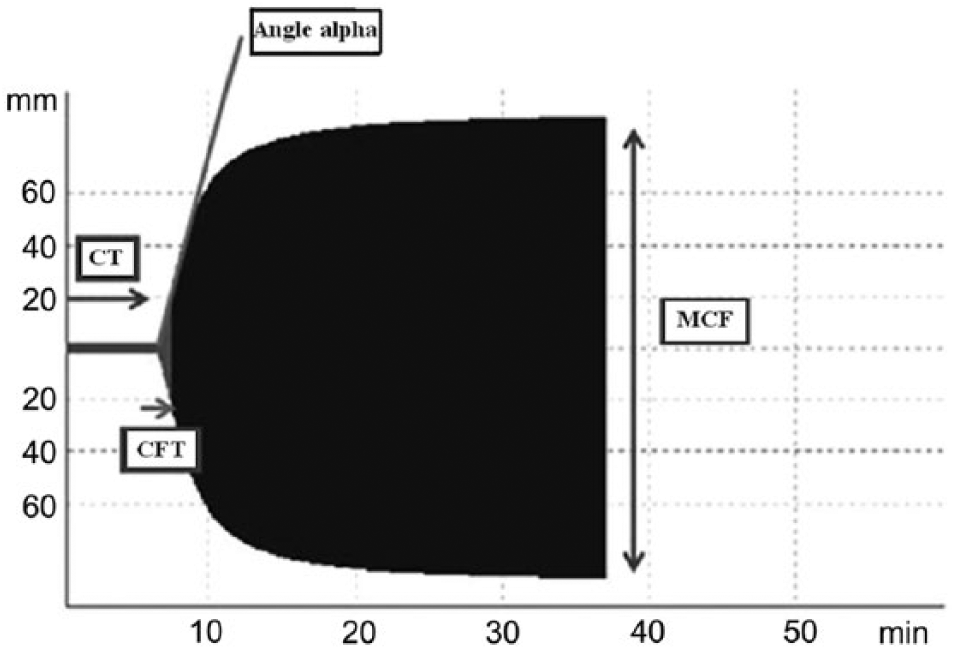
Example of a TEMogram (ex-TEM profile). CFT = clot formation time; CT = clotting time; MCF = maximum clot firmness; TEM = thromboelastometry.
The data were entered into an ad hoc database. n The normal distribution of data was verified by means of a test for normality based on skewness and on kurtosis. To verify the presence of differences among the 3 groups of calves, when the assumption of normality was respected, an analysis of variance (ANOVA) for repeated measures was performed; otherwise, the Friedman nonparametric 2-way ANOVA was used. In the case of comparison between 2 groups, the Mann–Whitney 2-sample statistic was performed. Significance was set at p < 0.05.
Results
Sixteen Piemontese calves born on the farm of the Department of Veterinary Science, University of Turin were enrolled. One calf showed an alteration on the standard coagulation profile (increased PT and aPTT) and was excluded. The final study sample was 15 calves (7 male and 8 female). There were no differences in ROTEM profiles between female and male calves (Table 1). There were statistically significant differences in the 3 profiles of the samples obtained at the 3 times. In the in-TEM profile, MCF was significantly increased at T0 versus T8 (p = 0.01); there were no statistically significant differences between T0 and T15 or between T8 and T15. In the ex-TEM profile, CFT was significantly prolonged at T0 versus T8 (p = 0.001) and T15 (p = 0.005), and the α angle was significantly smaller at T0 than at T8 (p = 0.001) and at T15 (p = 0.004); there were no statistically significant differences between T8 and T15 for any of the parameters. In the fib-TEM profile, only the MCF value was significantly lower at T0 than at T8 (p = 0.001) and at T15 (p = 0.02); there were no statistically significant differences between T8 and T15.
Results of rotational thromboelastometric analysis (ROTEM) analysis in Piemontese calves (n = 15).*
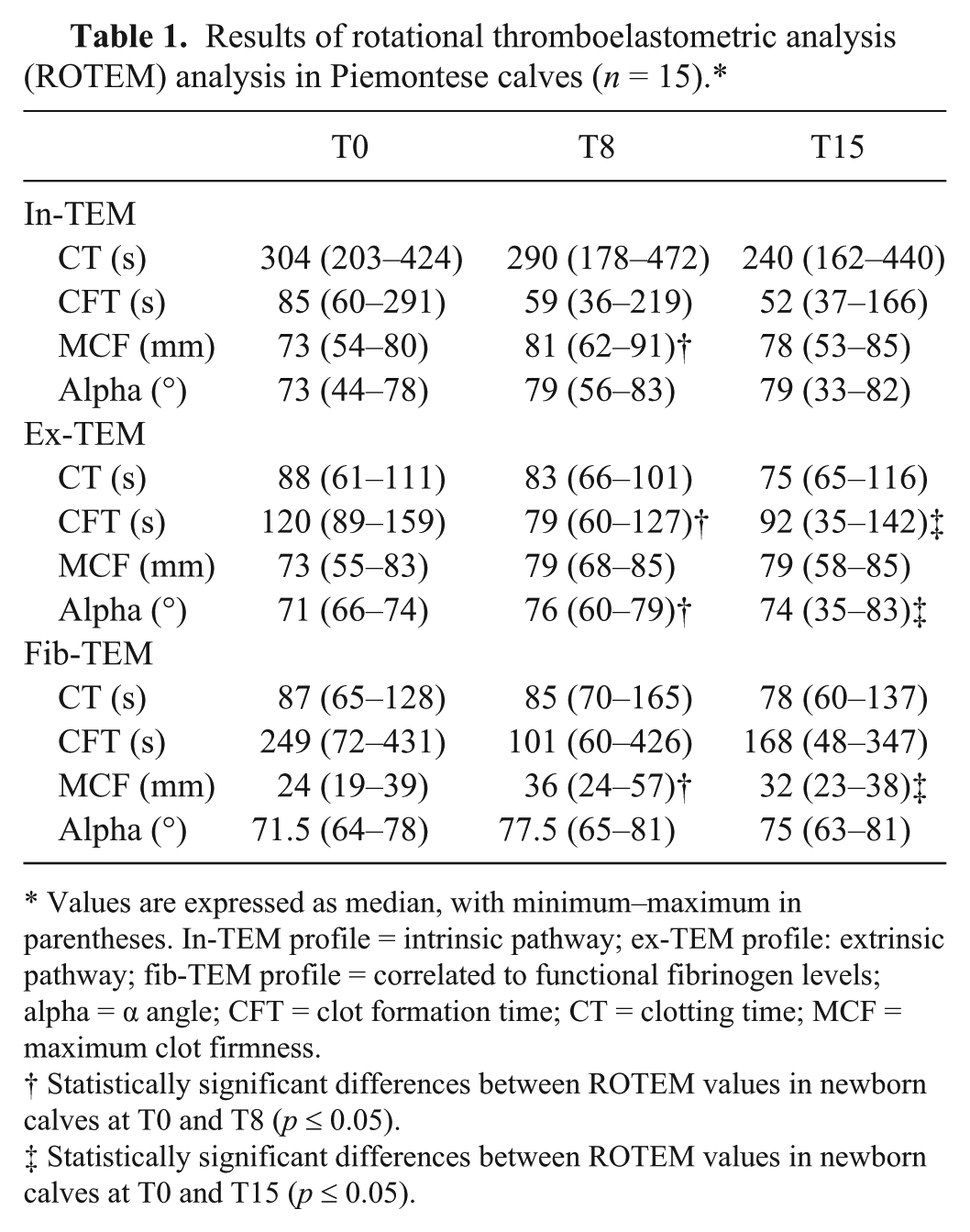
Values are expressed as median, with minimum–maximum in parentheses. In-TEM profile = intrinsic pathway; ex-TEM profile: extrinsic pathway; fib-TEM profile = correlated to functional fibrinogen levels; alpha = α angle; CFT = clot formation time; CT = clotting time; MCF = maximum clot firmness.
Statistically significant differences between ROTEM values in newborn calves at T0 and T8 (p ≤ 0.05).
Statistically significant differences between ROTEM values in newborn calves at T0 and T15 (p ≤ 0.05).
The aPTT was prolonged at T0 versus T8 (p = 0.0001) and T15 (p = 0.0001) but there were no statistically significant differences between T8 and T15; there were no significant differences in PT between any of the 3 times (Table 2). Fibrinogen concentration was significantly lower at T0 than at T8 (p = 0.009).
Standard coagulation profile in Piemontese calves at T0, T8, and T15 (n = 15).*
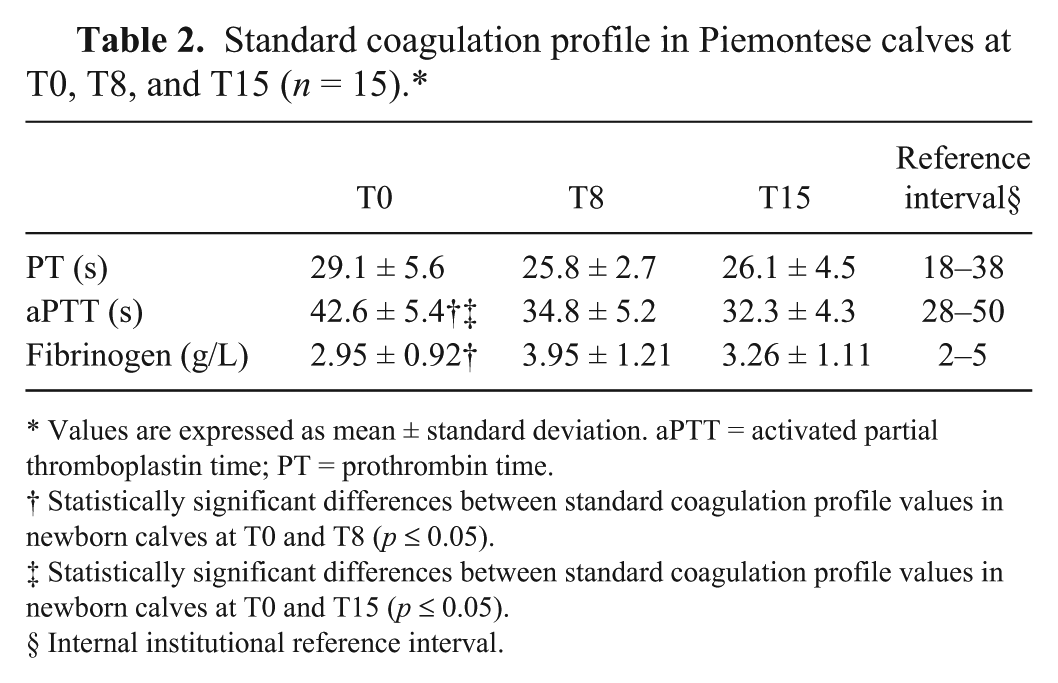
Values are expressed as mean ± standard deviation. aPTT = activated partial thromboplastin time; PT = prothrombin time.
Statistically significant differences between standard coagulation profile values in newborn calves at T0 and T8 (p ≤ 0.05).
Statistically significant differences between standard coagulation profile values in newborn calves at T0 and T15 (p ≤ 0.05).
Internal institutional reference interval.
Statistically significant differences in CBC values were noted (Table 3). Hematocrit (HCT) was significantly higher at T0 than at T8 (p = 0.002) and at T15 (p = 0.002) and at T8 than at T15 (p = 0.005). Hemoglobin (Hb) was significantly higher at T0 than at T8 (p = 0.032) and at T15 (p = 0.004) and at T8 than at T15 (p = 0.026). Platelet count was significantly lower at T0 than at T8 (p = 0.035); there were no statistically significant differences in platelet count between T0 and T15 or between T8 and T15.
Complete blood count (CBC) in Piemontese calves with statistically significant differences between T0, T8, and T15 (n = 15).*
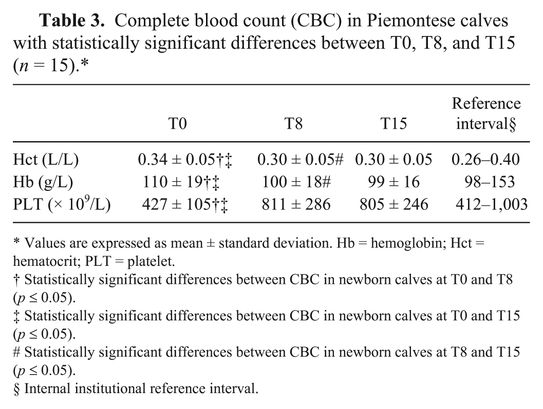
Values are expressed as mean ± standard deviation. Hb = hemoglobin; Hct = hematocrit; PLT = platelet.
Statistically significant differences between CBC in newborn calves at T0 and T8 (p ≤ 0.05).
Statistically significant differences between CBC in newborn calves at T0 and T15 (p ≤ 0.05).
Statistically significant differences between CBC in newborn calves at T8 and T15 (p ≤ 0.05).
Internal institutional reference interval.
The total number of adult Piemontese cattle was 28 animals (15 female and 13 male, median age 19 mo, range 18–78). Statistically significant differences in ROTEM results were observed between adults and calves at T0 (Table 4): MCF on the in-TEM profile was significantly decreased (p = 0.009), CFT on the ex-TEM profile was significantly prolonged (p = 0.029), and the α angle was significantly smaller in the calves than in the adults (p = 0.01); CT and CFT on the fib-TEM profile were significantly prolonged (p = 0.004 and p = 0.003, respectively); and MCF and the α angle were significantly smaller (p = 0.008 and p = 0.034, respectively) in the calves. There were no statistically significant differences in ROTEM results between the adults and the calves at T15 (Table 4).
Rotational thromboelastometric analysis (ROTEM) analysis in newborn calves and adult Piemontese cattle.*
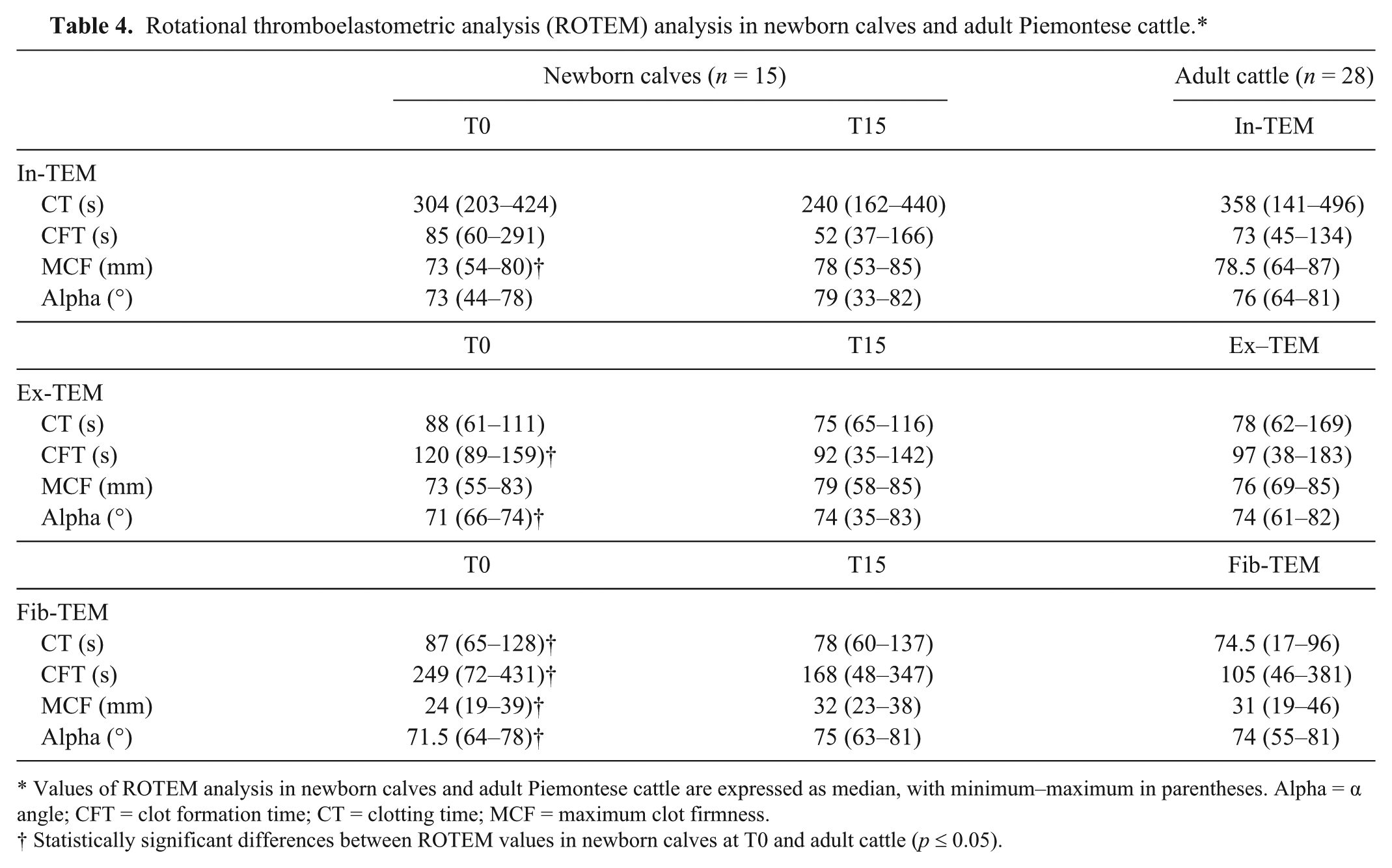
Values of ROTEM analysis in newborn calves and adult Piemontese cattle are expressed as median, with minimum–maximum in parentheses. Alpha = α angle; CFT = clot formation time; CT = clotting time; MCF = maximum clot firmness.
Statistically significant differences between ROTEM values in newborn calves at T0 and adult cattle (p ≤ 0.05).
Discussion
The ROTEM tracings at birth were similar to those at 8 and 15 d of age in newborn Piemontese calves. Significant differences in the in-TEM, ex-TEM, and fib-TEM profiles were observed only at T0 versus T8 and versus T15. MCF in the in-TEM profile was lower at T0 than at T8. The ex-TEM profile showed prolonged CFT and a smaller α angle at T0 versus T8 and T15. Because CFT, MCF, and α angle are predominantly affected by platelet number and function, and by fibrinogen, the results from samples obtained at T0 were probably influenced by the low platelet number and fibrinogen level present at T0.8,9 Furthermore, MCF in the fib-TEM profile was lower at T0 than at T8 and at T15. Because platelet function was inhibited, only the fibrinogen contributed to MCF: the fibrinogen concentration was low at T0 and increased over the next 15 d.8,9
Analysis of CBC results showed higher HCT and Hb levels and a lower platelet count at birth than at 8 and at 15 d of age. This observation is consistent with findings from a previous study.10,15 Erythrocytes are less fragile and larger in fetal calves than in adult cattle. 15 After birth, the mean corpuscular volume of erythrocytes progressively decreases from fetal values of 97 fL to 37 fL during the first 8–12 wk of age, and then declines again until ~2 y of age. 15 Differences in hematological parameters measured at the 3 times could partly account for the differences in the ROTEM results at T0. Indeed, a study using canine whole blood samples showed that ROTEM results were influenced by coagulation factors, platelet and fibrinogen concentrations, as well as by red blood cell mass. 13 Platelet, fibrinogen, erythrocyte, and Hb concentration were significantly correlated with CFT, α angle, and MCF. 13 The relationship between HCT and coagulability, as measured via ROTEM, has been suggested as an artefact of the plasma volume loaded into the tool. 13 Samples with a high HCT provide a lower mass of coagulation factors participating in the ROTEM reaction given a decrease in the liquid component of the blood, thus causing apparent hypocoagulability. 7
Our finding of a low platelet number at birth is shared by others in their study on Holstein dairy calves, which reported that platelet number increased markedly from birth to day 14 and then rose slightly up to day 84 of life. 10
Comparison of the standard coagulation profiles showed statistically significant differences in aPTT and fibrinogen. Compared with subsequent measurements, aPTT at birth was prolonged, but the final value fell within the normal adult interval for our laboratory. This result is in line with published data that report a standard coagulation profile within the adult range for newborn calves.4,6,11 The fibrinogen level was low at birth, increased significantly at 8 d, and then decreased at T15 (differences not statistically significant), as found by others, who reported a below normal adult range for fibrinogen at birth, followed by a marked increase during the first 7 d of life and a decrease at day 30. 6
Comparison of ROTEM results of calves at T0 with the results of adult cattle showed differences similar to comparing results of calves at T0 and T8 or T0 and T15; no differences were found between ROTEM results in calves at T15 and adults. The low platelet count, fibrinogen level, and HCT concentrations at birth might have contributed to the differences in the ROTEM tracings between the calves and the adults. Our findings indicate that reference ROTEM intervals for newborn calves should be used at birth, whereas reference intervals of adult cattle can be used to evaluate a ROTEM analysis in calves ≥8 d of age.
The limitations of our study were that the sample size was too small to derive reference intervals and the population included only Piemontese cattle. Further studies are needed to determine whether there are breed-related differences in ROTEM analysis.
Footnotes
a.
Vacutainer 3.2% buffered sodium citrate, BD, Franklin Lakes, NJ.
b.
Venosafe K3-EDTA, Terumo, Tokyo, Japan.
c.
ADVIA 120 hematology, Siemens Healthcare Diagnostics, Tarrytown, NY.
d.
Venosafe with gel and clot activator, Terumo, Tokyo, Japan.
e.
ILAB 300 plus, Clinical Chemistry System, Instrumentation Laboratories, Milan, Italy.
f.
Coagulometer StART, Diagnostica Stago, New York, NY.
g.
ROTEM, Tem International, Munich, Germany.
h.
In-TEM (partial thromboplastin phospholipid made of rabbit brain (chloroform extract), ellagic acid, buffer, preservatives in small glass vials), Tem International, Munich, Germany.
i.
Ex-TEM (recombinant tissue factor and phospholipids, heparin inhibitor, preservatives, and buffer in small glass vials), Tem International, Munich, Germany.
l.
Fib-TEM (Cytochalasin D/DMSO solution 0.2 mol/l CaCl2 in HEPES buffer pH 7.4, preservative in glass vials), Tem International, Munich, Germany.
m.
Manual ROTEM, 2002, 2004 ROTEM gamma; PENTAPHARM, Munich, Germany.
n.
Stata statistical software: release 11, StataCorp, College Station, TX.
Declaration of conflicting interests
The authors declared no potential conflicts of interest with respect to the research, authorship, and/or publication of this article.
Funding
The authors declared that they received no financial support for their research and/or authorship of this article.