
Abstract
Salmonella enterica subsp. enterica serovar Abortusequi is a pathogen restricted to horses. Our investigation targeted 4 draft horses (9–10 months old) kept on a Japanese farm that had suffered an outbreak of S. Abortusequi abortion. The 4 horses were suspected to be carriers of the bacterium owing to their high agglutination titers (≥1:2,560) in tube agglutination testing. The owners’ on-farm observations confirmed that the horses had no apparent abnormalities, and S. Abortusequi was not isolated from their blood, rectal swabs, or sternal bone marrow fluid at antemortem investigation. However, at autopsy, all horses displayed the following: suppurative aneurysm of the cranial mesenteric artery with heavy infection with Strongylus vulgaris larvae; heavy intestinal parasitic infection with Gasterophilus intestinalis, Parascaris equorum, Anoplocephala perfoliata, and S. vulgaris; and enlargement of the systemic lymph nodes. In each case, large numbers of S. Abortusequi were isolated from the anterior mesenteric artery thrombus. The thrombus isolates harbored a single virulence plasmid, and the pulsed-field gel electrophoresis profiles of the isolates were identical not only to each other but also to those of Japanese enzootic strains of S. Abortusequi. These results reveal that parasitic aneurysms of the cranial mesenteric artery should be considered an important possible site of carriage of S. Abortusequi in horses. The results also suggest high clonality of the isolated serovar in the horse population in Japan.
Keywords
Salmonella enterica subsp. enterica serovar Abortusequi is a pathogen restricted to horses. 19 The organism causes mainly abortion but can also cause suppurative arthritides, multiple abscesses, orchitis, and septicemia, with the disease syndrome being referred to as equine paratyphoid in Japan. 1 The disease was first described in 1893 in the United States. 12 Although the disease had virtually disappeared by the 1950s in the United States, 12 it has been reported sporadically in Italy, 6 Croatia, 15 and Argentina5,7 since the 1970s. The disease has been more frequently confirmed in Japan. 1
A carrier state for S. Abortusequi has been described in horses,2,13,16 and carrier animals have been implicated as initial sources of infection in outbreaks in nonendemic areas.2,11,15 Generally speaking, detection of the organism in feces is the gold standard for detecting carriers of Salmonella spp. 10 However, negative results for fecal detection have been reported on farms where an outbreak of S. Abortusequi infection occurred. 17 It is therefore difficult to specify the source of outbreaks.5,15 In addition, the epidemiology and ecology of S. Abortusequi are not yet well understood, even though this pathogen was first studied in the 1890s. 12 In this report, we present some interesting results regarding the site of carriage of S. Abortusequi in horses, with the results being obtained from an investigation of the pathology and etiology of an outbreak of S. Abortusequi infection in a nonendemic area of Japan.
The investigation targeted 4 draft horses (9–10 months old) kept on a farm in Iwate Prefecture, Japan, where an outbreak of S. Abortusequi abortion had occurred in October 2008. Three of the horses were colts, whereas the fourth was a filly. This was the first case in this prefecture since 1989. The horses were suspected to be carriers of S. Abortusequi because of their high agglutination titers (≥1:2,560) against S. Abortusequi in tube agglutination testing, performed on 6 November 2008 using a previously described methodology. 3 On 28 November 2008, the horses were transferred from the farm to the Epizootic Research Station at the Japan Racing Association’s Equine Research Institute in Tochigi Prefecture for further investigation. The horses were not given any antimicrobial agents during the outbreak. No S. Abortusequi organisms were isolated from any clinical specimens, although blood, rectal swabs, and sternal bone marrow fluid were submitted for bacteriologic investigation. When these horses arrived at the institute, their agglutination titers were still high at ≥1:1,280 despite the fact that their owners had noticed no abnormalities on farm. These horses were therefore considered to be carriers of S. Abortusequi.
One horse was euthanized for autopsy on 4 December 2008 and the remaining 3 on 9 December. Euthanasia was performed in accordance with the Guidelines for Animal Rights and Medical Ethics (Japan Racing Association Euthanasia Guideline, November 1999). Complete postmortem examinations were performed, and the organs were inspected grossly. At autopsy, all horses displayed the following: a suppurative aneurysm and a large thrombus in the cranial mesenteric artery with a heavy infection of Strongylus vulgaris larvae (L4; Fig. 1); heavy intestinal parasitic infections with Gasterophilus intestinalis, Parascaris equorum, Anoplocephala perfoliata, and S. vulgaris; and systemic enlargement of lymph nodes. Mild suppurative bronchopneumonia in both cranial lung lobes was observed in 3 of the 4 horses. Bacteriologic investigations were performed of the principal organs (liver, spleen, heart, kidney, and lung), lymph nodes (mesenteric lymph nodes, inguinal lymph nodes, cecal nodes, colonic lymph nodes), intestinal contents (ileum and colon), Peyer patches, uterine mucosal swab (in the mare), and the thrombi in the cranial mesenteric artery. Isolates of S. Abortusequi–like organisms were obtained from the thrombus in the cranial mesenteric artery (4 horses), a colonic lymph node (3 horses), a cecal lymph node (2 horses), the liver (2 horses), lung (1 horse), and Peyer patches (1 horse). Particularly large numbers of S. Abortusequi–like organisms were isolated from the thrombi. No other Salmonella sp. organisms were isolated from any of the specimens. The isolates were characterized by a commercial identification kit, a and the profile code of the kit for all the isolates from the thrombus in each horse was 430450247. This code was not applicable in the database of the kit. The isolates were serotyped in accordance with the Kauffmann–White scheme by using Salmonella typing cranial sets for O and Vi antigen typing b and for H antigen typing. c The isolates were confirmed by serotyping as S. Abortusequi (O 4,12:–:e,n,x).
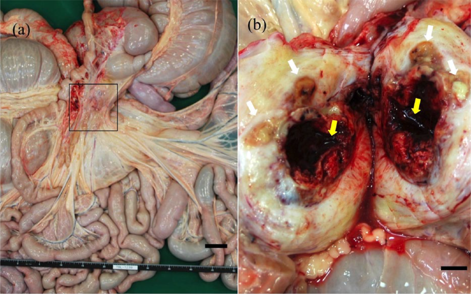
External appearance (
Histopathology revealed large numbers of inflammatory cells only in the thrombi. Some of these cells, which were phagocytizing S. Abortusequi, were observed in the thrombi by immunohistochemical staining using anti–O4 Salmonella-specific rabbit serum d (Fig. 2).
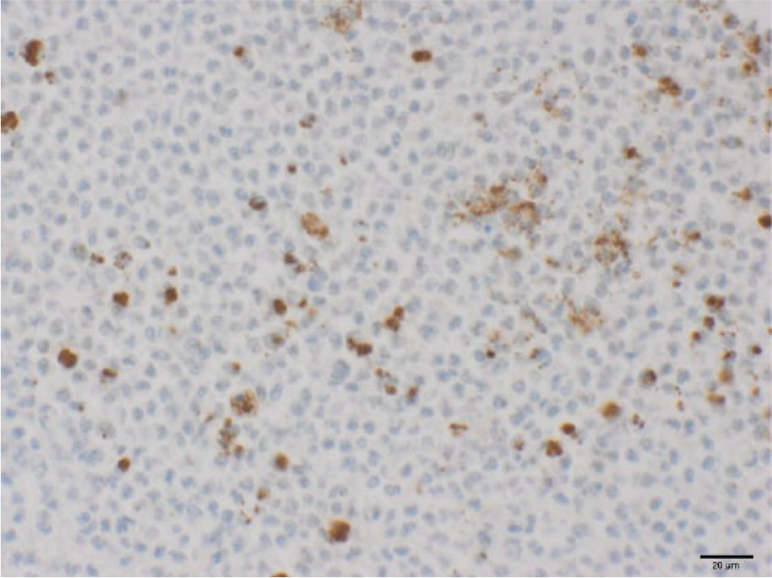
Immunohistochemical appearance of thrombus of aneurysm of the cranial mesenteric artery. Many inflammatory cells are present in the thrombus, and many were found to be phagocytizing Salmonella enterica subsp. enterica serovar Abortusequi organisms. Immunohistochemical staining by using anti–O4 Salmonella-specific rabbit serum for O-serotyping. Bar = 20 µm.
Plasmid DNA of the isolates was extracted as previously described. 14 Electrophoresis was performed with 0.8% agarose gel. e The gel was stained with ethidium bromide solution for 15 min, de-stained with distilled water for 60 min, and photographed under an ultraviolet transilluminator (Fig. 3). A single plasmid was observed in the 4 isolates obtained from the thrombus in the cranial mesenteric artery of each horse.
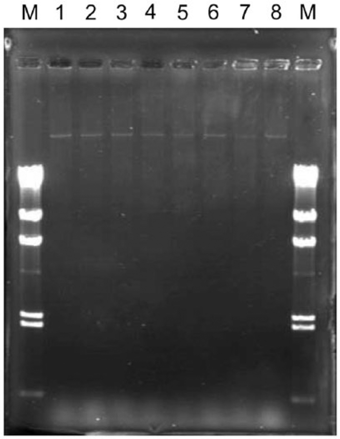
Plasmid profile of the reference strains and the isolates obtained from the horses. Lane M: lambda ladders; lane 1: R91 (Mongolia, 1988); lane 2: CSA-1 (Croatia, 1994); lane 3: S.abor-17 (Japan, 1987); lane 4: Sal-243 (Japan, 2007); lanes 5–8: isolates obtained from the thrombus in the parasitic aneurysm of each horse in our study.
Pulsed-field gel electrophoresis (PFGE) analysis was performed. The DNA for PFGE analysis was prepared by using a commercial kit f in accordance with the manufacturer’s instructions. The restriction enzyme used was XbaI. g PFGE was performed with a commercial system h under the following conditions: 6 V/cm for 20 h at 14°C with an initial switch time of 5.3 s and a final switch time of 49.9 s at an angle of 120° using a 1.2% gel. i After completion of electrophoresis, the PFGE gel was stained with ethidium bromide and photographed as described above. Four reference strains were used in the PFGE analysis: strain 91R was isolated in 1988 in Mongolia, 1 strain CSA-1 in 1994 in Croatia, 15 strain S.abor-17 in 1987 in Kushiro, Hokkaido Prefecture (~300 km from the farm where S. Abortusequi infection is enzootic), and strain Sal-243 in 2007 in Hidaka, which is also in Hokkaido Prefecture (~300 km from the area of Japan where S. Abortusequi infection is not endemic). The PFGE profiles of the isolates and the reference strains are shown in Figure 4. Identical profiles were obtained from not only all of the isolates obtained in the current study but also from all of the Japanese enzootic strains. In contrast, the PFGE profiles of the Mongolian and Croatian isolates differed from each other and from those of all of the Japanese isolates.
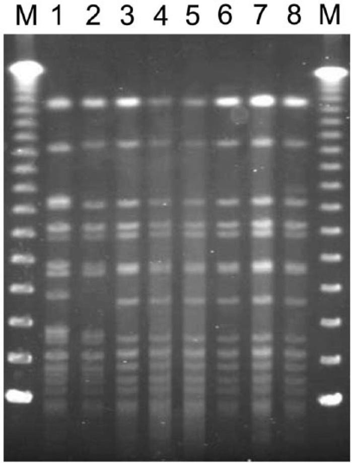
Pulsed-field gel electrophoresis profiles of Salmonella enterica subsp. enterica serovar Abortusequi isolates. Lane M: Lambda ladders; lane 1: 91R (Mongolia, 1988); lane 2: CSA-1 (Croatia, 1994); lane 3: S.abor-17 (Japan, 1987); lane 4: Sal-243 (Japan, 2007); lanes 5–8: isolates obtained from the thrombus in the parasitic aneurysm of each horse in the study.
Host-adapted or restricted serovars (e.g., Typhi, Abortusovis, Typhisuis, Dublin, Choleraesuis, Gallinarum, and Pullorum) are more likely than non–host-restricted serovars to produce a carrier state in host animals. 19 The gallbladder, mammary tissue, and supramammary lymph nodes have been reported as sites of carriage of S. Typhi, 9 splenic macrophages have been reported to carry S. Dublin, 18 and the reproductive tract to carry S. Pullorum. 20 Although an early Japanese study found that sternal bone marrow fluid was adequate as a specimen for isolating S. Abortusequi organisms from carrier horses on antemortem investigation, 4 we did not isolate any organisms from this fluid. Instead, we isolated a number of organisms from a thrombus in a suppurative aneurysm of the cranial mesenteric artery that was heavily infected with S. vulgaris larvae. The cranial mesenteric artery of the horse is a site favored by S. vulgaris during its larval migration; parasitism by the larvae causes endothelial damage, with subsequent thrombus formation. 8 Presumably, thrombus formation and vascular wall roughening by S. vulgaris would have helped to establish the carrier state by trapping the bacteria in the lesion at the aneurysm. In addition, all the horses were heavily infected with other intestinal parasites. The stress of these infections may have helped to establish the carrier state. According to their owners’ on-farm observations, the horses had no obvious abnormalities, and they were autopsied 7 weeks after the last case of abortion in the outbreak. Our results suggest that parasitic aneurysm of the mesenteric artery is an important site of carriage of S. Abortusequi in horses with asymptomatic paratyphoid.
Identical PFGE profiles were observed in all of the Japanese strains, both the enzootic isolates and the isolates obtained from the horses. A previous study 1 found, by PFGE and fluorescent amplified-fragment length polymorphism fingerprinting, that there was a close relationship among S. Abortusequi isolates of equine origin in Japan and that carrier horses introduced from the enzootic area were a cause of outbreaks in nonendemic areas. In the current study, the horses were probably infected directly or indirectly by contaminated materials from aborted fetuses, placentas, and lochia of infected horses on the farm. However, the route by which the organism entered the farm could not be determined because epidemiologic information such as the recent movement histories of horses on the farm could not be obtained. However, the results of our molecular analysis revealed that the S. Abortusequi isolates that caused the outbreak probably belonged to the clone maintained in the horse population of Japan.
In conclusion, parasitic aneurysm of the cranial mesenteric artery caused by S. vulgaris larvae may play an important role as a site of carriage of S. Abortusequi in the horse. Not only the control of S. Abortusequi organisms but also the control of parasitic infection, for example, by administering adequate anthelmintics and optimizing health, might help to decrease the carrier state of S. Abortusequi infection in endemic areas. Although the route of transmission to the farm could not be determined here, our results suggest that the isolates were part of a clone maintained in Japan’s horse population.
Footnotes
Authors’ contributions
H Niwa contributed to acquisition, analysis, and interpretation of data, and drafted the manuscript. S Hobo contributed to conception and design of the study; drafted the manuscript; and critically revised the manuscript. Y Kinoshita and M Muranaka contributed to acquisition and analysis of data and drafted the manuscript. A Ochi and T Ueno contributed to analysis of data and drafted the manuscript. K Oku and K Hariu contributed to acquisition of data and drafted the manuscript. Y Katayama contributed to conception and design of the study and critically revised the manuscript. All authors gave final approval, and agreed to be accountable for all aspects of the work in ensuring that questions relating to the accuracy or integrity of any part of the work are appropriately investigated and resolved.
a.
API 20 E, Sysmex bioMérieux, Tokyo, Japan.
b.
Salmonella antisera “SEIKEN” set 1, Denka Seiken Co. Ltd., Tokyo, Japan.
c.
Salmonella antisera “SEIKEN” set 2, Denka Seiken Co. Ltd., Tokyo, Japan.
d.
Denka Seiken Co. Ltd., Tokyo, Japan.
e.
Agarose H14 “TAKARA”, Takara Bio Inc., Shiga, Japan.
f.
CHEF Genomic DNA plug kit, Bio-Rad Japan, Tokyo, Japan.
g.
Takara Bio Inc., Shiga, Japan.
h.
CHEF Mapper XA Chiller System, Bio-Rad Japan, Tokyo, Japan.
i.
SeaKem Gold agarose, Lonza Rockland Inc., Rockland, ME.
Declaration of conflicting interests
The author(s) declared that they have no conflict of interests with respect to the research, authorship, and/or the publication of this article.
Funding
The author(s) received no financial support for the research, authorship, or publication of this article.