
Abstract
Klebsiella pneumoniae is an important emerging pathogen in humans, particularly the invasive hypermucoviscosity (HMV) phenotype. In addition, the organism is an important public health concern because of nosocomial infections and antimicrobial resistance. Nonhuman primates in captivity are susceptible to Klebsiella, particularly when a stress factor is involved. Infections vary depending on the species but can cause significant morbidity and mortality in these animals. The objective of this study was to describe a case of bronchopneumonia and bacteremia caused by Klebsiella pneumoniae in a free-ranging golden-headed lion tamarin (Leontopithecus chrysomelas) caught and maintained in quarantine during a translocation program for conservation purposes. An adult male, that had showed emaciation and apathy, was clinically examined and, despite being provided supportive therapy, died 2 days after onset of clinical signs. At postmortem examination, generalized bilateral pneumonia and pericarditis were observed. Tissue samples were fixed in 10% formalin for histology, and pulmonary tissues and cardiac blood were collected for microbiologic diagnostic procedures. Bacteria that were shown to be HMV K. pneumoniae subsp. pneumoniae strains were isolated from the pulmonary fluids and cardiac blood in pure cultures. Severe bronchopneumonia was the main pathological finding. The consequences of the confirmed presence of the HMV phenotype of K. pneumoniae subsp. pneumoniae in this wildlife species for human, animal, and ecosystem health should be determined. These results demonstrate the importance of quarantine and potential pathogen screening during wildlife translocation procedures.
Epidemiological studies of potential pathogens in wild animals, either in situ or ex situ, are important for the implementation of programs for the prevention, control, and monitoring of diseases and for developing public and animal health policies. In addition, the knowledge of pathogens that impact human health and wildlife conservation has become increasingly important within the One Health concept.10,11
The genus Klebsiella, which consists of Gram-negative, rod-shaped bacteria, that are nonmotile and usually encapsulated, belongs to the family Enterobacteriaceae.20,25 Klebsiella pneumoniae is associated with significant morbidity and mortality in nonhuman primates (NHPs) housed in captivity. Nonpathogenic strains are widely distributed in nature as free-living forms in the soil and water or as commensals in the intestinal tract of humans and animals, constituting a normal fecal and oral microbiome in many NHPs.19,25 However, situations that trigger stress, such as transport, quarantine, malnutrition, and overcrowding, seem to predispose animals to the disease. 25
The diseases caused by K. pneumoniae infection vary depending on the host species, but pneumonia and septicemia are both associated with outbreaks in humans and NHPs.13,24 Meningitis, peritonitis, and cystitis have been described in captive Old and New World monkeys. 25 Klebsiella pneumoniae is an important emerging nosocomial human pathogen (Gozalo A, Montoya E. Klebsiella pneumoniae infection in a New World nonhuman primate. Laboratory Primate Newsletter 1991;30:13–15. Available from: http://www.brown.edu/Research/Primate/lpn30-2.html). 20 Consequently, it is also a relevant public health issue, particularly because of antimicrobial resistance. 20
In addition, a distinctive human clinical syndrome of invasive K. pneumoniae, characterized by primary liver abscesses and frequently associated with bacteremia and complications, such as meningitis, endophthalmitis, lung abscess, or fasciitis, has been described. These invasive strains of K. pneumoniae are highly associated with the hypermucoviscosity (HMV) phenotype.3,16 Old World NHPs in captivity are also affected by invasive HMV K. pneumoniae, which is a significant threat to the health of these animals. 16
Golden-headed lion tamarins (Leontopithecus chrysomelas; GHLTs) are an endangered species, and the endemic population in the state of Bahia (Brazil) is in sharp decline. 17 It is likely that in the year 2000, several GHLT groups were improperly released in an urban Atlantic forest remnant in Rio de Janeiro state and are currently considered an exotic invasive species in this location. Because of the risk of hybridization with local and endangered Leontopithecus species (Leontopithecus rosalia, golden-lion tamarin), environmental authorities have proposed the translocation of this invasive GHLT population to their original region, Bahia state.
Pathogens that may affect wildlife are a significant issue for translocation programs (http://www.oie.int/fileadmin/Home/eng/Internationa_Standard_Setting/docs/pdf/WGWildlife/A_Training_Manual_Wildlife.pdf). 18 Such movement can spread pathogens to new areas through the interchange of wildlife, not only impacting the local wildlife but also, in cases of zoonosis, potentially presenting a risk to the human population living in the area.10,11,18 Furthermore, stress generated by translocation may result in the establishment of disease due to conditional pathogens that lived in equilibrium with the host in the wild state. 28 The present case describes the clinical course and main postmortem macro- and microscopic lesions of a GHLT affected by bronchopneumonia caused by K. pneumoniae subsp. pneumoniae during a translocation program.
One adult L. chrysomelas (GHLT 43) male was captured along with his group of 5 individuals at Serra da Tiririca State Park (22°56′04.28″S, 43°02′22.66″W; Niterói, Rio de Janeiro, Brazil) during a translocation program of the invasive GHLT population. The animals were subjected to a clinical examination and housed in quarantine for 30 days to conduct a comprehensive sanitary evaluation, including complete hemogram, serum biochemistry, and survey of selected pathogens and infections. The animal was negative for all pathogens and infections screened, including Klebsiella spp. However, a rectal swab from another animal from the same group was positive for K. pneumoniae.
Because of technical and operational limitations, the group was not translocated, and 6 months after leaving the quarantine and transfer to an enclosure in the Rio de Janeiro Primatology Center, the animal (GHLT 43) became very thin, apathetic, and depressive, with bristling of the fur on the face and head. Clinical examination revealed a respiratory infection, and broad-spectrum antibiotic therapy with benzathine benzylpenicillin, procaine benzylpenicillin, benzylpenicillin potassium, dihydrostreptomycin, and streptomycin, a was administered together with supportive therapy, including anti-toxic fluids b and heating of the environment. The animal died 2 days after clinical onset and was submitted for postmortem examination.
The major findings at postmortem examination were emaciation and bronchopneumonia (Fig. 1A, 1B). Samples from the majority of organs and tissues were collected, fixed in 10% buffered formalin, and processed according to routine histologic procedures. 22 Cut sections of 4–5 µm were stained using hematoxylin and eosin. The microscopic examination revealed a bacterial process leading to a severe multi-focal to coalescing fibrinosuppurative bronchopneumonia with the presence of a large number of foamy macrophages and intracytoplasmic and free coccobacilli (Fig. 1C, 1D). Other major histological findings included moderate fibrinosuppurative pericarditis, splenic lymphoid depletion associated with moderate histiocytosis, and mild diffuse hepatic degeneration.
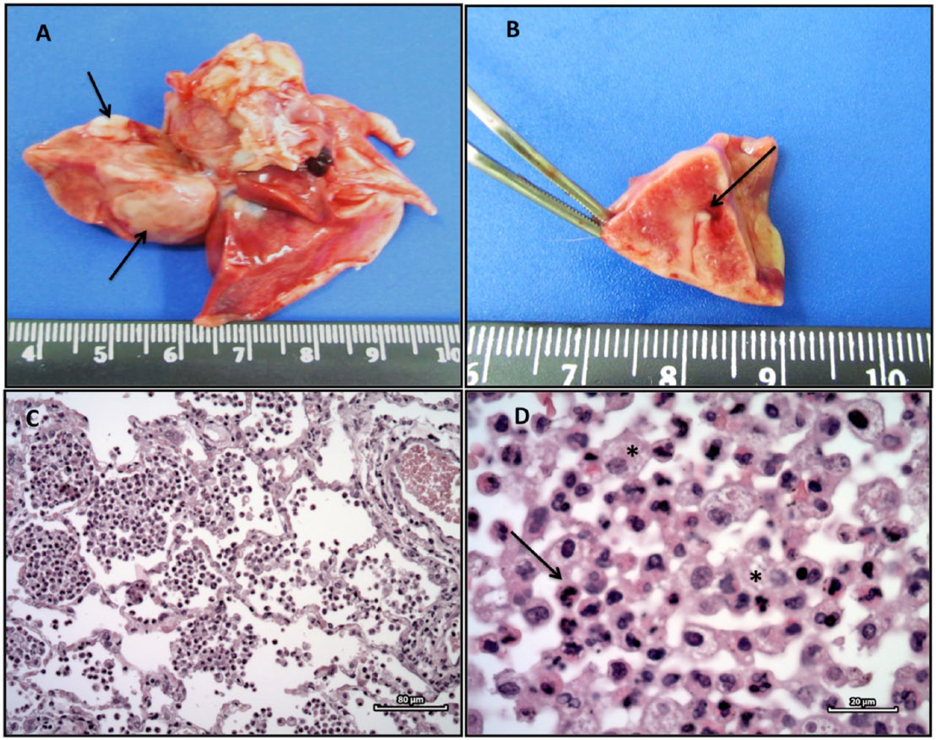
Pneumonia and bacteremia in a golden-headed lion tamarin (Leontopithecus chrysomelas) caused by Klebsiella pneumonia subsp. pneumonia.
Pulmonary fluids and heart blood were aseptically sampled, cultured on brain heart infusion c and MacConkey agar plates, d and incubated aerobically at 37°C. After 24 hr, a pure culture of a bacterium with excessively mucoid colony appearance was detected from both samples.
The identification of bacteria was performed using a commercial identification kit, e and antimicrobial susceptibility testing was performed according to international standards 8 using the disk diffusion method against 12 different antibiotics f belonging to 6 different classes as follows: beta-lactams (ampicillin, amoxicillin, cephalexin, cefoxitin, and ceftiofur); fluoroquinolones (enrofloxacin and ciprofloxacin); aminoglycosides (streptomycin and gentamicin); tetracycline; chloramphenicol; and sulfonamide–trimethoprim. Breakpoints that were not defined in the veterinary documentation 7 were taken from other standard guidelines. 6
Isolates from pulmonary fluids and cardiac blood were identified as K. pneumoniae subsp. pneumoniae. To confirm the hypermucoviscosity phenotype, a string test 4 was performed, with both strains showing a positive reaction. For the susceptibility to the antimicrobial agents tested, both strains were resistant only to aminopenicillins (ampicillin, amoxicillin), which was not unexpected because Klebsiella spp. are intrinsically resistant to these antibiotics. 2
Although not isolated from the fecal sample of the GHLT of this case report, K. pneumoniae subsp. pneumoniae was recovered from the rectal swab of one individual of the same group as soon as it arrived at quarantine in good health. Thus, the intestinal microbiota of apparently healthy animals deserves attention during capture, quarantine, and translocation processes because infections caused by Klebsiella spp. tend to occur primarily in individuals with depressed immune systems. 12 Newly captured free-ranging animals and those kept in captivity have high rates of stress and consequently increased cortisol, which can cause decreased immunity leading to the onset of disease. 14 However, Klebsiella spp. are part of the normal enteric microbiota, and the presence of this genus does not normally impair the translocation process.5,21
From an epidemiological perspective, it is important to determine the origin of an infection, particularly in endemic and epidemic nosocomial outbreaks of Klebsiella spp. infections to improve the management of these outbreaks. 20 More than one-third of the GHLTs sampled in our study showed Klebsiella spp. in their intestinal microbiota, suggesting that the animals were in contact with this bacterium in the wild before reaching captivity (Iovine RO, et al. Klebsiella spp. is part of the natural intestinal microbiota and a potential disease pathogen in wild golden-headed lion tamarins (L. chrysomelas). European Wildlife Disease Association Conference, Edinburgh, Scotland, August 2014:25–29). Immunosuppression due to stress during the period of adaptation most likely favored the occurrence of this severe case of bronchopneumonia and bacteremia.
Little is known about the composition of the intestinal microbiota of GHLTs, particularly of the free-ranging individuals, but it is likely that the intestinal microbiota is a reflection of several factors, such as corporal mass, climate, and diet, among others. 15 For free-ranging GHLTs, small vertebrates and insects, such as cockroaches, are a part of their diet in nature. 18 Cockroaches are an important reservoir for infectious pathogens, and studies indicate the transmission of K. pneumoniae by these insects in the human environment. 26
Because this exotic GHLT population inhabits an urban state park surrounded by an extensive human and domestic animal population, attention must be given to the potential transmission of HMV K. pneumoniae among species. Nevertheless, our finding is consistent with previously described studies, indicating that HMV K. pneumoniae is present both in captive and wild animals and that NHPs may be affected, resulting in pulmonary lesions and acute death.25,28
Generally, the gross lesions reported in cases associated with HMV K. pneumoniae infection are pneumonia and septicemia. Sepsis, meningitis, peritonitis, cystitis, mesenteric lymphadenopathy, and hepatic abscesses are also described. 13 Microscopic injuries vary with the extent, severity, and stage of the disease. Briefly, NHPs infected with HMV K. pneumoniae usually exhibit diffuse fibrinopurulent bronchopneumonia, suppurative bronchitis, and pleuritis, with the lung presenting multifocal necrosis and exudation with alveolar congestion, hemorrhage, and edema. 25 In the present case, the microscopic findings are consistent with those described above.13,25
The present case demonstrates the importance of monitoring the health conditions of animals undergoing reintroduction and/or translocation programs to ensure both human and animal health. Particular attention should focus on wildlife habitats in the same area as humans. 13 Moreover, it is also important to monitor the health of animals after release, particularly because the philosophy of zero risk is impossible to achieve. Any translocation is a source of stress, 25 and Klebsiella infection tends to occur in this situation, causing changes in the parasite–host balance and favoring the emergence of disease (Animal movements and disease risk, 5th ed., 2003, http://www.veterinariosvs.org/redvvs/recursosredvvs/docus/Disease_Risk_manual.pdf).
Connections between vertebrate species through the human–animal–ecosystem interface may have consequences for human, animal, and ecosystem health. 11 The primary risk factors for the emergence and spread of diseases include the transport of live animals over long distances, live animal markets, habitat destruction, and the consumption of bush meat, among other factors. 27 Therefore, translocated species may introduce parasites to their new environment, and some of these novel parasites may cause disease in immunologically naive hosts.9,23
In addition, NHPs deserve special attention because of their close relatedness to humans and potential disease exchange. 1 Thus, it is important to identify epidemiologic factors associated with infection in these tamarins to minimize the risk of pathogen transmission in both ex situ and in situ in both animal and human populations. The study of potential pathogens, particularly those related to human–wildlife–ecosystem interface, such as HMV K. pneumoniae, provides relevant data for understanding the dynamic interactions important for environmental health.
Footnotes
Acknowledgements
We would like to thank the Instituto Pri-Matas team and Centro de Primatologia do Rio de Janeiro (CPRJ)/Instituto Estadual do Ambiente (INEA) for their support.
a.
Pentabiótico Veterinário Pequeno Porte, Fort Dodge, Brazil.
b.
Mercepton, atitóxico; Bravet, Rio de Janeiro, Brazil.
c.
BHI, Difco Laboratories Inc., Detroit, MI.
d.
MacConkey plates, Difco Laboratories Inc., Detroit, MI.
e.
API-20E galleries, bioMérieux, Marcy l’Etoile, France.
f.
Cefar Diagnóstica Ltd, São Paulo, Brazil.
Declaration of conflicting interests
The author(s) declared no potential conflicts of interest with respect to the research, authorship, and/or publication of this article.
Funding
The author(s) disclosed receipt of the following financial support for the research, authorship, and/or publication of this article: We received financial support from Fundação de Amparo à Pesquisa do Estado de São Paulo–FAPESP (process no. 2011/08149-8). Renata de Oliveira Iovine had financial support from Coordenação de Aperfeiçoamento de Pessoal de Nível Superior–CAPES. The Golden-Headed Lion Tamarin Translocation Program received funds from Fundação Grupo Boticário de Proteção à Natureza, Lion Tamarin of Brazil Fund, Primate Action Fund, Margot Marsh Foundation, The Mohamed bin Zayed Species Conservation Fund, RBO Energia S.A. (Câmara de Compensação Ambiental/Secretaria do Meio Ambiente Rio de Janeiro), and Tropical Forest Conservation Act/Fundo Brasileiro para Biodiversidade (TFCA/FUNBIO).