
Abstract
The current report describes granulomatous pneumonia due to Spirocerca lupi in 2 free-ranging maned wolves (Chrysocyon brachyurus). Both wolves had multiple, white, 1–1.5 cm in diameter, soft, encapsulated granulomas in the caudal lung lobes, which contained centrally placed parasites on cut sections. Microscopically, there was granulomatous inflammation with numerous intralesional sections of spirurid nematodes. Representative complete adult specimens of nematodes derived from these lesions were submitted for parasitological exam and identified as the spirurid S. lupi. To the authors’ knowledge, there have been no published reports of S. lupi in maned wolves.
Spirocerca lupi (order Spirurida, superfamily Spiruroidea, family Spirocercidae) is a cosmopolitan nematode commonly found in tropical and subtropical regions where it is regarded as a typical parasite among carnivores.2,16,21 Adult parasites usually reside in the caudal esophagus, where females release embryonated eggs that are later shed in feces. The life cycle is complex and, following ingestion of eggs by suitable coprophagous beetles, infective third-stage larvae (L3) develop; transmission to the carnivore definitive host involves foraging on arthropod intermediate or various vertebrate paratenic hosts.8,21 In the carnivore, L3 released into the gastric lumen penetrate the gastric mucosa, migrate through the gastric and gastroepiploic arteries to the celiac artery, and abdominal and thoracic aorta, and later reach the caudal esophagus, where larvae mature to the L4 stage and to adult worms.
Dogs are frequently hosts of S. lupi, with the esophagus and aorta being the most commonly affected organs, where infections may present as granulomatous esophagitis, vasculitis, aneurysm, thrombosis, and rupture of the aorta.9,13 Additionally, lungs, trachea, heart, diaphragm, vertebrae, stomach, intestine, kidney, pancreas, subcutaneous tissues, and spinal cord can be rarely affected.1,2,8,10,12,15–18,20 Regardless of the affected tissues, chronically infected organs can develop malignant neoplasms, usually of mesenchymal origin.9,19
To the authors’ knowledge, there have been no published reports of S. lupi in maned wolves (Chrysocyon brachyurus). The current report describes the anatomic pathology findings of granulomatous pneumonia due to S. lupi in 2 maned wolves from the central-western region of Brazil.
Two free-ranging adult, male maned wolves, each with a history of trauma (hit by a car) were submitted for necropsy. The first submitted maned wolf (wolf 1) was found dead at the margin of a highway in Brasilia, Federal District. The second submitted maned wolf (wolf 2) arrived at the teaching veterinary hospital of University of Brasilia with paralysis of pelvic limbs and paresis, with reduced reflexes of the thoracic limbs. Radiographic exam revealed complete vertebral fracture in L1. This animal was euthanized due to the poor prognosis.
The lungs had multiple, white, 1–1.5 cm in diameter, soft, encapsulated nodules with few 5–6 cm in length and 0.1 cm in diameter adult nematodes in the caudal lung lobes (Fig. 1). In addition, 1 large, red nematode, compatible with Dioctophyma renale, was found in the right kidney of both wolves. Samples from the pulmonary nodules and from multiple other organs (liver, kidney, urinary bladder, heart, brain, stomach, and adrenal glands) were collected and fixed in 10% buffered formalin for histology processing (i.e., paraffin-embedded and stained with hematoxylin and eosin). The lung nematodes were collected from both wolves and preserved in 70% alcohol and deposited for identification in the U.S. National Parasite Collection (Agricultural Research Service, U.S. Department of Agriculture, Beltsville, Maryland; voucher specimens are retained under USNPC no. 107256.00).
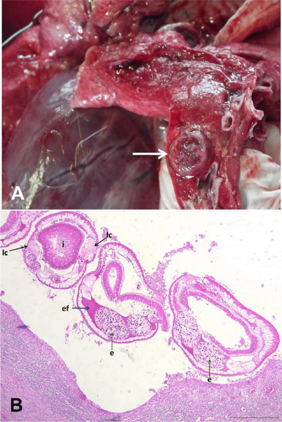
Maned wolf (Chrysocyon brachyurus), lung.
Microscopically, the center of the granulomas was composed of amorphous necrotic areas with many cross-sections of adult nematodes. The parasites were 950 μm in maximum diameter, had an eosinophilic cuticle, prominent lateral chords, coelomyarian musculature, a prominent pseudocoelom containing eosinophilic homogeneous material, a well-developed large intestine with high columnar epithelium and brush microvillar border, and a uterus filled with small, thick-shelled, ovoid eggs. These findings are consistent with a spirurid nematode. There was moderate to severe focally extensive infiltration of lymphocytes, epithelioid macrophages, plasma cells, neutrophils, and few eosinophils around central areas of necrosis. There were multifocal areas with moderate edema, congestion, and fibrin accumulation within alveoli. Parasitological identification revealed 2 male and 1 gravid female adult spirurids with body length, mouth, esophagus, and spicule lengths compatible with S. lupi.
In dogs, the median age of animals infected by S. lupi and presenting clinical signs of spirocercosis is 4–5 years old, but younger animals (≤1 year old) may be affected.5,16 Both wolves from the current report were adults.
The cause of death was trauma, and the lung parasites were incidental findings in both cases. Dogs infected with S. lupi can develop spondylitis of thoracic vertebrae and present similar clinical signs as described in wolf 2. 16 However, there were no gross or radiographic changes to suggest spondylitis in either wolves, and histology of vertebrae was not performed. Clinical outcomes of S. lupi infection may vary greatly depending on the stage of parasitism, erratic migrations, and secondary complications. 21 In dogs, the main clinical signs include regurgitation, vomiting, and dysphagia due to the esophagic granulomas. 21 Aberrant migrations, as noted in the present study, may result in different clinical presentations, such as respiratory, neurological, and locomotor signs. 10 Sudden death associated with hemothorax has been described when there is rupture of the aorta or other major blood vessels.10,13 Respiratory signs are observed in some cases with mediastinitis, pleuritis, or pyothorax. These lesions are caused by parasite migration to the esophagus through the aortic wall. 14 Aspiration pneumonia is seen in animals with regurgitation due to esophageal obstruction by S. lupi granulomas.16,21 Aberrant migration to the vertebral canal affecting spinal cord in dogs has only been rarely reported.8,20 In dogs, S. lupi granulomatous lesions can frequently develop into sarcomas.9,19 No neoplastic lesions were observed in either wolf from the current report.
Pulmonary granulomas in these cases might be a result of an erratic migration of S. lupi larvae or adults. The stimulus for either directional or aberrant migration is still not well understood. 21
The granulomatous parasitic pneumonia presented by these wolves must be differentiated from other lung parasitic pneumonias in canids, such as those resulting from infection by Angiostrongylus vasorum, Filaroides martis, Filaroides hirthi, Crenosoma vulpis, and Eucoleus aerophilus. Differentiation is based on location and severity of lesions and parasite morphology. The morphological, microscopic, and parasitological aspects were unequivocal in identification of S. lupi as the etiology of the granulomatous pneumonia in both maned wolves. Histologically, it was possible to identify nematodes with typical morphological features of spirurids. 11 Definitive identification of S. lupi was done through parasitological identification of adult worms. Other lung disorders should also be considered in the differential diagnosis, such as primary or metastatic neoplasms.
There is no description of S. lupi infection in maned wolves among parasitological studies of wild carnivores from Brazil and Bolivia or elsewhere, to the authors’ knowledge.3,4,6,7,22 Further epidemiologic and pathologic studies are necessary to determine the pathogenesis and prevalence of S. lupi infection in maned wolves in the central-western region of Brazil.
Footnotes
Declaration of conflicting interests
The author(s) declared that they have no conflicts of interest with respect to their authorship or the publication of this article.
Funding
The author(s) received no financial support for the research, authorship, and/or publication of this article.