
Abstract
A 1-cm-diameter nodule was identified in the left inguinal mammary gland of a 9-year-old male maned wolf (Chrysocyon brachyurus). The mass was surgically excised and examined histologically. Microscopically, the neoplasm consisted of papillary proliferations of epithelial cells on well-defined fibrovascular stalks. A myoepithelial layer was located between the single layer of epithelial cells and the fibrovascular stalk. This histologic appearance was compatible with a diagnosis of simple ductal mammary papilloma. Immunohistochemical staining was positive for p63, cytokeratins AE1/AE3, and estrogen receptors. The clinical and histologic observations in the present case indicate that male maned wolves may develop mammary tumors that are similar to those observed in domestic dogs and humans.
Mammary tumors are the most frequent neoplasms in female dogs and account for about 50% of all histologically diagnosed tumors in dogs. The incidence of mammary neoplasms is increased in older and intact animals. 2,17 In male dogs, mammary neoplasms are rare and generally are observed in animals of advanced age. 19,21 Information regarding the frequency of mammary tumors in free-ranging carnivores is limited. Mammary tumors have been reported previously in a red fox (Vulpes vulpes), 11 black-footed ferrets (Mustela nigripes), 3,12 and a European pine marten (Martes martes). 25
The present study describes the histological and immunohistochemical characteristics of a simple ductal mammary papilloma in a male maned wolf (Chrysocyon brachyurus). The maned wolf is a member of the family Canidae and lives in the South American grasslands and scrub forests of Brazil, northern Argentina, Paraguay, eastern Bolivia, and southeastern Peru. The maned wolf is historically one of the most typical carnivorous species found in central and southern Brazil but is currently listed on the official Brazilian list of species threatened with extinction. 14
A 1.0-cm-diameter nodule was identified in the left inguinal mammary gland of a 9-year-old male maned wolf born at the Zoo-Botanic Foundation of Belo Horizonte, Minas Gerais, Brazil. The mass was surgically excised, and samples were collected for histopathology and immunohistochemistry. The tissues were fixed in 10% neutral buffered formalin solution, trimmed, processed routinely, and embedded in paraffin. The paraffin blocks were sectioned at 3 μm and stained with hematoxylin and eosin for histologic evaluation.
Microscopically, the nodule was composed of an orderly proliferation of epithelial cells characterized by papillary arrangements on well-defined fibrovascular stalks. A layer of myoepithelial cells was observed between the layer of epithelial cells and the fibrovascular stalk. The microscopic appearance of the lesion was compatible with a simple ductal mammary papilloma (Fig. 1). The histologic diagnosis of simple ductal papilloma subsequently was confirmed by 2 additional pathologists following diagnostic criteria for mammary tumors. 15
Immunohistochemistry was performed on replicate tissue sections using monoclonal antibodies against estrogen receptor (1:150 dilution; NCL-LH2 a ), p63 (1:100 dilution; 4A4 b ), and cytokeratins (CK) AE1/AE3 (1:100 dilution; CK AE1/AE3 a ). The reactivity of the antibodies in canine mammary cells was reported previously. 7,19 The sites of primary antibody binding were identified by the streptavidin–biotin–peroxidase complex method c using diaminobenzidine c as the chromogen.
Negative controls consisted of replacing the primary antibody with phosphate buffered saline. The adjacent normal mammary tissue was used as an internal control in each test. Normal mammary gland samples obtained from domestic female dogs were used as positive controls for p63 and CK AE1/AE3 immunoreactivity. One complex adenoma with proven immunoreactivity for estrogen receptors from another female dog was used as a positive control for this immunostaining procedure. The number of cells immunoreactive for p63 and CK AE1/AE3 was scored semiquantitatively as follows: 0 = no stained cells; + = <10% positive cells; ++ = 10–50% positive cells; and +++ = >50% positive cells. The number of estrogen receptor–positive cells was determined as the percentage of stained cells: – = no staining or less than 5% of nuclei stained; + = 5–19% of nuclei stained; ++ = 20–59% of nuclei stained; and +++ = ≥60% of nuclei stained.
In the normal canine mammary gland (positive control), the alveolar and ductal epithelial cells were positive for CK AE1/AE3 and estrogen receptor, and negative for p63. The nuclei of myoepithelial cells uniformly distributed around the ductal and alveolar structures stained positively for p63 (+++) and CK AE1/AE3 (+).
In the simple ductal papilloma from the maned wolf of this report, estrogen receptor immunoreactivity was detected in the nuclei of a small number of epithelial cells (+). Most luminal epithelial and myoepithelial cells had cytoplasmic staining for CK AE1/AE3 (+++;Fig. 2). Immunostaining for p63 (+++) was restricted to the nuclei of myoepithelial cells located between the luminal epithelium and basement membrane. These cells were also positive for CK AE1/AE3 (+; Figs. 2, 3).
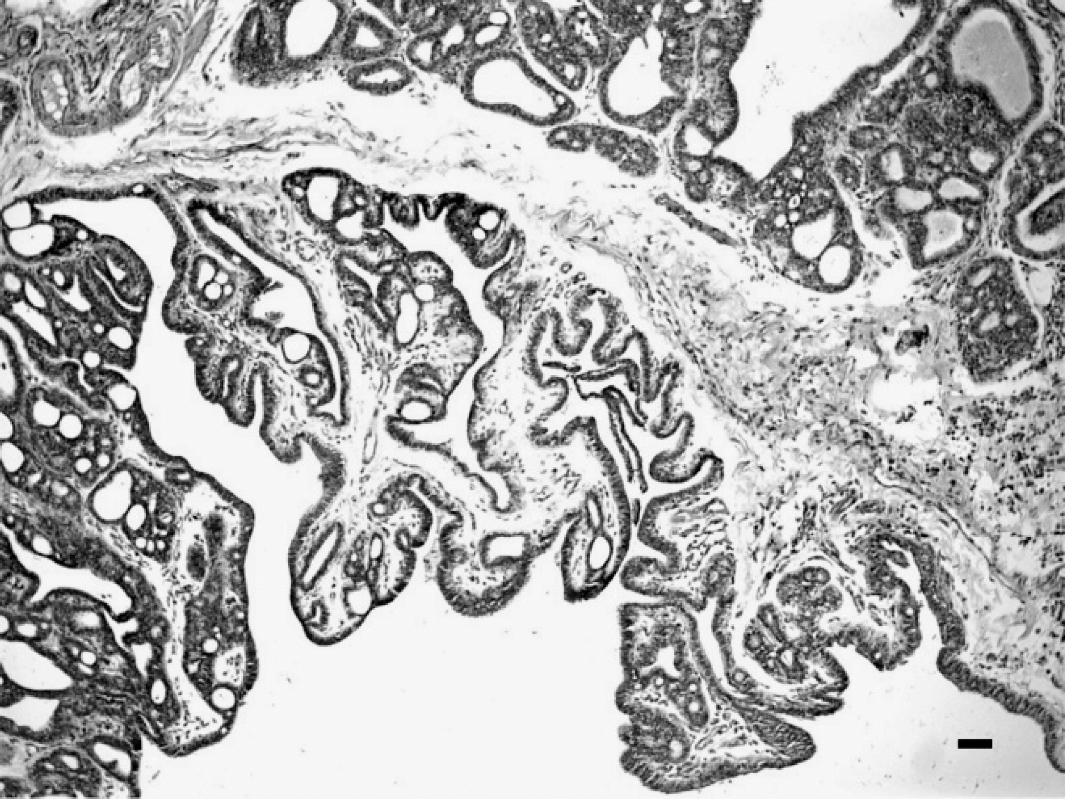
Simple ductal mammary papilloma from a male maned wolf showing papillary proliferation of epithelial cells on well-defined fibrovascular stalks. Hematoxylin and eosin. Bar = 100 μm.
Despite the high incidence of mammary tumors in female dogs, these neoplasms occasionally have been reported in male dogs. 17,23 The life span of the manedwolfis13 years; therefore, the present tumor was diagnosed in an older animal as has been reported for mammary neoplasms in male domestic dogs. 23 Because wild canids are not examined routinely, it is possible that the frequency of mammary tumors might be higher than indicated in reports of peer-reviewed literature. The lack of detection or absence of registration of neoplasms may contribute to the scarcity of information about the occurrence of mammary tumors in wild canids.
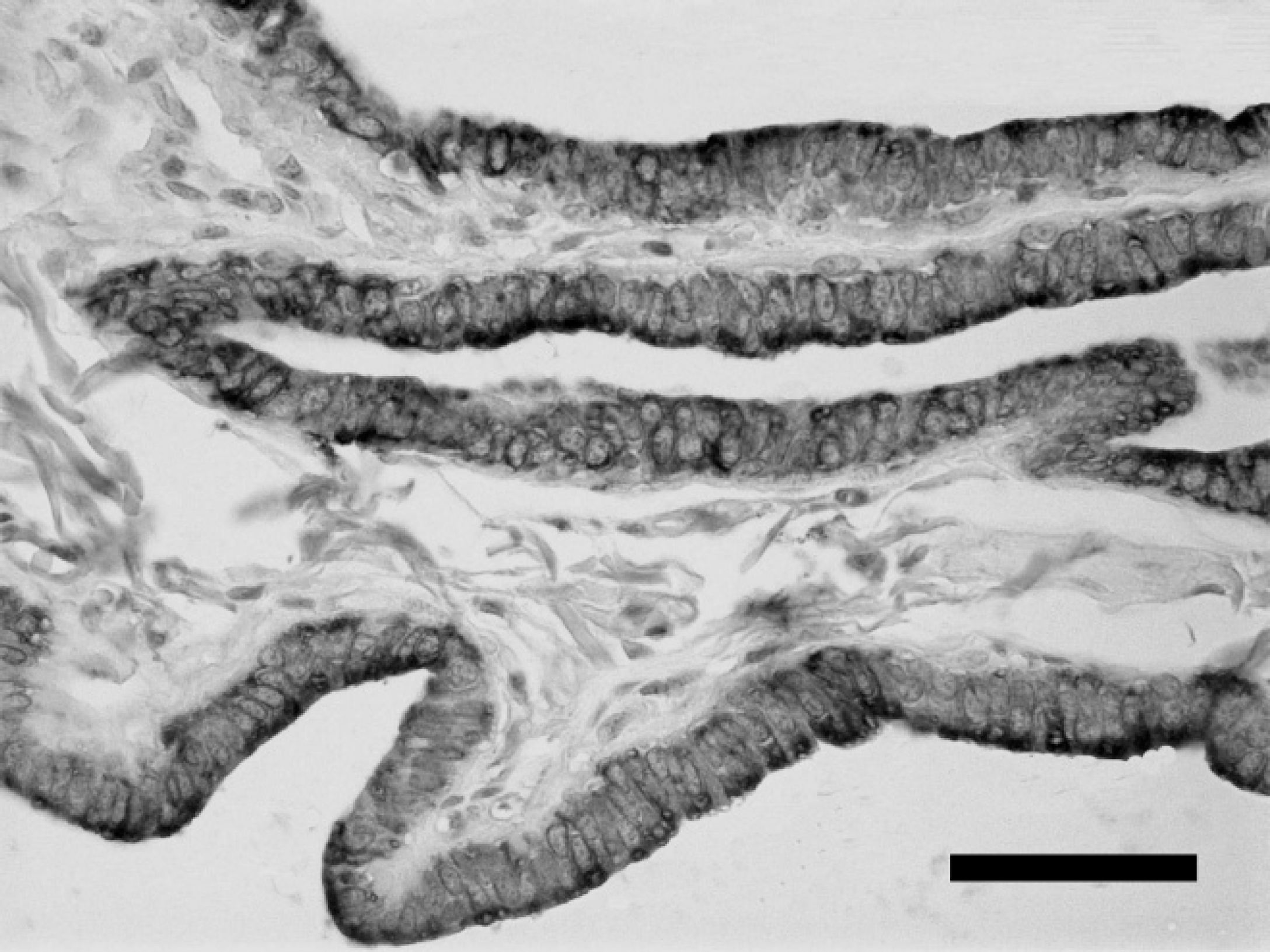
Immunohistochemical reactivity for cytokeratins AE1/AE3 in the epithelial cells of a mammary simple ductal papilloma from a male maned wolf. Bar = 50 μm.
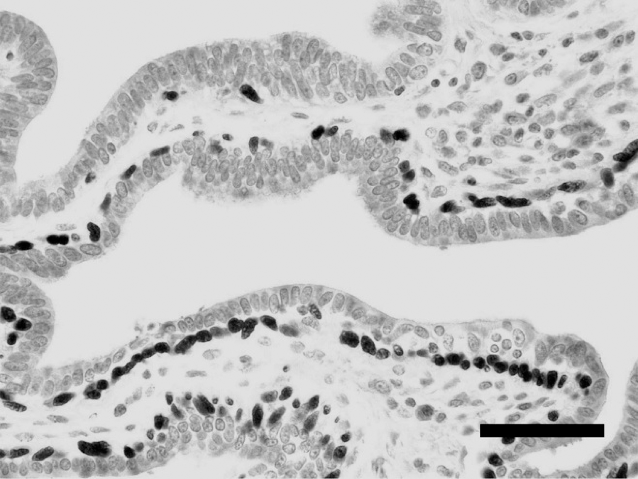
Immunohistochemical reactivity for p63 in the myoepithelial cells of a mammary simple ductal papilloma in a male maned wolf. Bar = 50 μm.
The simple ductal papilloma in the male maned wolf of this report is a benign mammary tumor that usually is diagnosed in female dogs. 15 In humans, all histologic types of mammary neoplasia observed in women also have been described or reported in men. However, mammary tumors in men are predominantly ductal in origin because the male breast normally does not contain lobules. Furthermore, papillary growth is common in male breast carcinomas. 22
Immunohistochemical staining revealed immunoreactivity of neoplastic epithelial luminal cells for CK AE1/AE3, confirming the presence of epithelial cells in the neoplasm. The CK AE1/AE3 antibody identifies the presence of cytokeratins and has been used as a marker of epithelial cells in mammary tumors and other neoplasms. 9
In domestic dogs, the occurrence of complex tumors with luminal epithelial cells and myoepithelial cells is common. 15 The p63 antibody is a specific and sensitive marker to identify myoepithelial cells. 1,7 The absence of p63 immunoreactivity in the interstitial myoepithelial cells confirms that the mammary neoplasm in the maned wolf in the current study is a simple as opposed to a complex neoplasm. Several myoepithelial markers such as calponin, smooth muscle actin, S-100, and cluster of differentiation (CD) 10 can also be used as myoepithelial cell markers. 4,26 The advantages of p63 include its nuclear staining and the lack of cross-reactivity with stromal myofibroblasts. 1 Additionally, the observation of p63 reactivity in myoepithelial cells at the epithelial-stromal junction demonstrated the integrity of the myoepithelium and confirmed the simple papillary nature of this ductal papilloma. The microscopic distinction between papillary adenoma and papillary carcinoma is critical to proper diagnosis. Consensus exists that ductal papillomas retain a continuous myoepithelial layer, whereas papillary carcinomas have focal clusters of or absence of myoepithelial cells. 20
Estrogen receptor immunoreactivity suggests the participation of steroid-derived hormones in the pathogenesis of mammary tumors in wild animals, as has been observed for similar neoplasms of humans and domestic dogs. Estrogen promotes neoplastic growth by stimulating epithelial proliferation through the estrogen receptor. 10 In female dogs, both malignant and benign neoplasms may have estrogen receptors. 5,13 In humans, studies have shown that more than 80% of breast carcinomas in men are estrogen-receptor positive, 8,22 and conditions that increase the exposure to estrogen such as iatrogenic exogenous administration of estrogen, obesity, and testicular diseases increase the risk of developing breast cancer. 6,24 The presence of testicular tumors also may pose a risk for the development of mammary neoplasms in dogs, 16 but this was not the case in the maned wolf in the current report. However, the epidemiologic, histologic, and immunohistochemical findings in this maned wolf suggest that wild canids also may develop mammary neoplasms that are histologically and biologically similar to their counterparts in domestic dogs and humans.
Acknowledgements. The authors thank the National Council of Scientific and Technological Development (CNPq; Brazil), the Brazilian Federal Agency for the Support and Evaluation of Graduate Education (CAPES), and the Research Support Foundation of the State of Minas Gerais (FAPEMIG; Brazil) for financial support.
Footnotes
a.
Novocastra Laboratories Ltd., Newcastle upon Tyne, UK.
b.
NeoMarkers Inc., Fremont, CA.
c.
Dako North America Inc., Carpinteria, CA.