
Abstract
Approximately 8,000 isolates of Streptococcus agalactiae, Streptococcus dysgalactiae, Streptococcus uberis, Staphylococcus aureus, and Escherichia coli, isolated by 25 veterinary laboratories across North America between 2002 and 2010, were tested for in vitro susceptibility to beta-lactam, macrolide, and lincosamide drugs. The minimal inhibitory concentrations (MICs) of the beta-lactam drugs remained low against most of the Gram-positive strains tested, and no substantial changes in the MIC distributions were seen over time. Of the beta-lactam antimicrobial agents tested, only ceftiofur showed good in vitro activity against E. coli. The MICs of the macrolides and lincosamides also remained low against Gram-positive mastitis pathogens. While the MIC values given by 50% of isolates (MIC50) for erythromycin and pirlimycin and the streptococci were all low (≤0.5 µg/ml), the MIC values given by 90% of isolates (MIC90) were higher and more variable, but with no apparent increase over time. Staphylococcus aureus showed little change in erythromycin susceptibility over time, but there may be a small, numerical increase in pirlimycin MIC50 and MIC90 values. Overall, the results suggest that mastitis pathogens in the United States and Canada have not shown any substantial changes in the in vitro susceptibility to beta-lactam, macrolide, and lincosamide drugs tested over the 9 years of the study.
Keywords
Introduction
The U.S. Food and Drug Administration (FDA) has published guidelines for dairy cattle veterinarians on the judicious use of antimicrobial drugs (U.S. FDA/CVM: 2002, Judicious use of antimicrobials for dairy cattle veterinarians. Available at: http://www.fda.gov/downloads/AnimalVeterinary/SafetyHealth/AntimicrobialResistance/JudiciousUseofAntimicrobials/UCM095571.pdf) as has the Canadian Veterinary Medical Association. 5 Responsible stewardship involves activities that aim to minimize the emergence and spread of antimicrobial resistance and preserve the efficacy of current veterinary drugs. Surveillance for antimicrobial susceptibility among pathogens is an important component of stewardship.3,18 Data regarding loss of in vitro susceptibility in pathogens can be used by veterinarians, livestock producers, pharmaceutical companies, and others working to support the health and welfare of animals. It has been noted that “Access to timely and accurate antimicrobial resistance data may also help to prevent inappropriate responses to anecdotal reports of resistance, which can unfairly bias the perceived efficacy of an agent, complicate antibiotic strategies and confuse laboratory testing efforts.” 11
There are few publications of national surveys of the antimicrobial susceptibilities of pathogens isolated from dairy cattle on farms in the United States or Canada.2,3 To address this lack of data, the animal health business unit of Pfizer Inc. (currently operating as Zoetis™) established a Mastitis Pathogen Antimicrobial Susceptibility Surveillance Program in 2002 to monitor the susceptibility of the major mastitis pathogens to antimicrobial drugs used for treatment of mastitis in North America. The objective of the program was to monitor bovine mastitis pathogens that had been isolated from sick animals and report on minimal inhibitory concentration (MIC) distributions over time. The microorganisms tested in the program, Streptococcus agalactiae, S. dysgalactiae, S. uberis, Staphylococcus aureus, and Escherichia coli, have all been reported as major mastitis pathogens in the United States and Canada (USDA, APHIS: 2008, Prevalence of contagious mastitis pathogens on US dairy operations, 2007. #N533.1008. Available at: http://www.aphis.usda.gov/animal_health/nahms/dairy/downloads/dairy07/Dairy07_is_ContMastitis.pdf; CBMRN: 2010, Annual Report 2009–10. Available at: http://www.medvet.umontreal.ca/rcrmb/dynamiques/PDF_AN/About_Us/0910CBMRNAnnualReport.pdf).1,13 In the current study, results of in vitro susceptibility of bovine mastitis pathogens to beta-lactam, macrolide, and lincosamide veterinary antibacterial agents used to treat mastitis are presented.
Materials and methods
Isolates
Between 2002 and 2010, 25 veterinary laboratories submitted isolates of S. agalactiae, S. dysgalactiae, S. uberis, S. aureus, and E. coli to Zoetis (Kalamazoo, Michigan). Not all participating laboratories submitted isolates each year. The laboratories are located in the United States (21 laboratories) and Canada (4 laboratories; Table 1).
Isolate origin and number of isolates per year by region for a 9-year (2002–2010) survey of antimicrobial susceptibility of bacteria that cause bovine mastitis in the United States and Canada.*
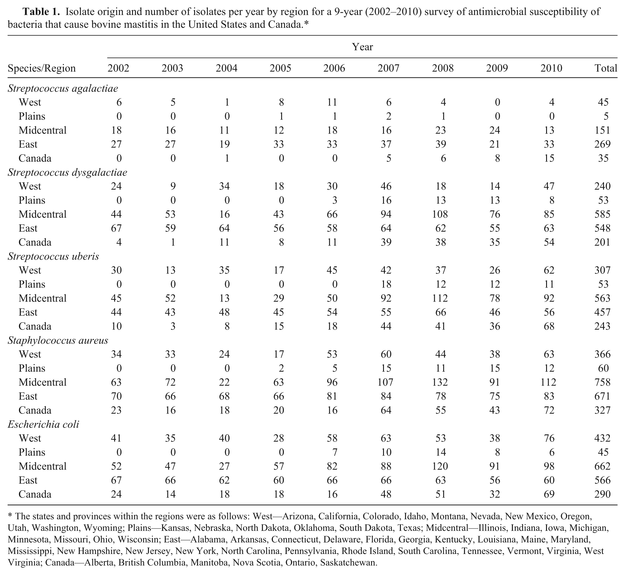
The states and provinces within the regions were as follows: West—Arizona, California, Colorado, Idaho, Montana, Nevada, New Mexico, Oregon, Utah, Washington, Wyoming; Plains—Kansas, Nebraska, North Dakota, Oklahoma, South Dakota, Texas; Midcentral—Illinois, Indiana, Iowa, Michigan, Minnesota, Missouri, Ohio, Wisconsin; East—Alabama, Arkansas, Connecticut, Delaware, Florida, Georgia, Kentucky, Louisiana, Maine, Maryland, Mississippi, New Hampshire, New Jersey, New York, North Carolina, Pennsylvania, Rhode Island, South Carolina, Tennessee, Vermont, Virginia, West Virginia; Canada—Alberta, British Columbia, Manitoba, Nova Scotia, Ontario, Saskatchewan.
The isolates had been recovered from either clinical or subclinical cases of bovine mastitis. The isolates were selected by the laboratories, which had been requested not to use susceptibility as a criterion for submission. Each laboratory was limited to the number of isolates (typically less than 25 isolates of each species) that could be submitted each year, so that there was not an overrepresentation from any geographic region. Laboratories were discouraged from submitting multiple isolates of the same bacterial species from an individual herd to prevent testing of epidemiologically related strains. The majority of the isolates came from animals within the same state or province of the submitting laboratory; however, isolates were also submitted from additional states or provinces. Isolates were identified by the submitting laboratory before shipment to Zoetis. Confirmation or further characterization of isolates, when necessary, was conducted using standard biochemical tests and commercially available identification systems.a,b
Minimal inhibitory concentration determinations
Minimal inhibitory concentrations for all isolates were determined using a broth microdilution system, c which conforms to the standards of the Clinical and Laboratory Standards Institute (CLSI) for testing veterinary pathogens. 6 Antimicrobial agents were selected either as representatives of their antimicrobial class for the purposes of susceptibility testing or based on interest and availability in commercial, approved intramammary preparations. The custom 96-well microtiter panels included doubling dilutions of ampicillin, ceftiofur, cephalothin, erythromycin, oxacillin, the combination drug penicillin–novobiocin, and pirlimycin. Concentration ranges for each antimicrobial drug were chosen to encompass appropriate CLSI quality control ranges and clinical breakpoints wherever possible. 6
All MIC testing was done at 1 of 2 laboratories (Zoetis, Kalamazoo, Michigan or Microbial Research Inc., Fort Collins, Colorado). Direct colony suspensions were used when testing all organisms, and suspensions were prepared to yield a final concentration of bacteria of approximately 5 × 10⁵ colony-forming units/ml. The growth medium for all streptococcal isolates was cation-adjusted Mueller–Hinton broth supplemented with 2.5–5% lysed horse blood. All other isolates were tested with unsupplemented Mueller–Hinton broth. Both laboratories adhered strictly to CLSI standardized methods, including performance of quality control tests. 6
Results
Between 2002 and 2010, 505 isolates of S. agalactiae, 1,627 isolates of S. dysgalactiae, 1,623 isolates of S. uberis, 2,182 isolates of S. aureus, and 1,995 isolates of E. coli were tested for in vitro susceptibility to ampicillin, ceftiofur, cephalothin, erythromycin, oxacillin, penicillin–novobiocin, and pirlimycin, using CLSI-approved methods. The results are presented as MIC frequency distributions, along with the summarized MIC values for 50% of isolates (MIC50) and the MIC values for 90% of isolates (MIC90), in Tables 2–8.
The distribution frequencies of ampicillin minimal inhibitory concentrations (MICs) against bovine mastitis pathogens from the United States and Canada (2002–2010).*
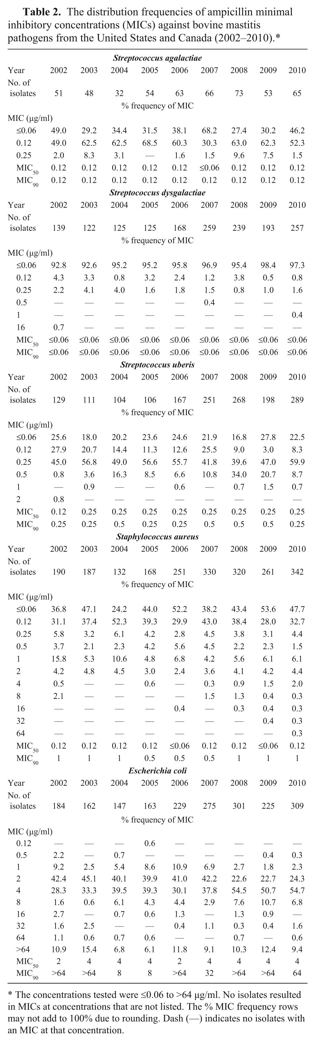
The concentrations tested were ≤0.06 to >64 µg/ml. No isolates resulted in MICs at concentrations that are not listed. The % MIC frequency rows may not add to 100% due to rounding. Dash (—) indicates no isolates with an MIC at that concentration.
The distribution frequencies of ceftiofur minimal inhibitory concentrations (MICs) against bovine mastitis pathogens from the United States and Canada (2002–2010).*

The concentrations tested were ≤0.06 to >64 µg/ml. No isolates resulted in MICs at concentrations that are not listed. The % MIC frequency rows may not add to 100% due to rounding. Dash (—) indicates no isolates with an MIC at that concentration. The CLSI-VAST–approved upper limit for susceptibility was 2 µg/ml, and the lower limit for resistance was 8 µg/ml for ceftiofur against S. agalactiae, S. dysgalactiae, S. uberis, S. aureus, and E. coli. 6
The distribution frequencies of cephalothin minimal inhibitory concentrations (MICs) against bovine mastitis pathogens from the United States and Canada (2002–2010).*
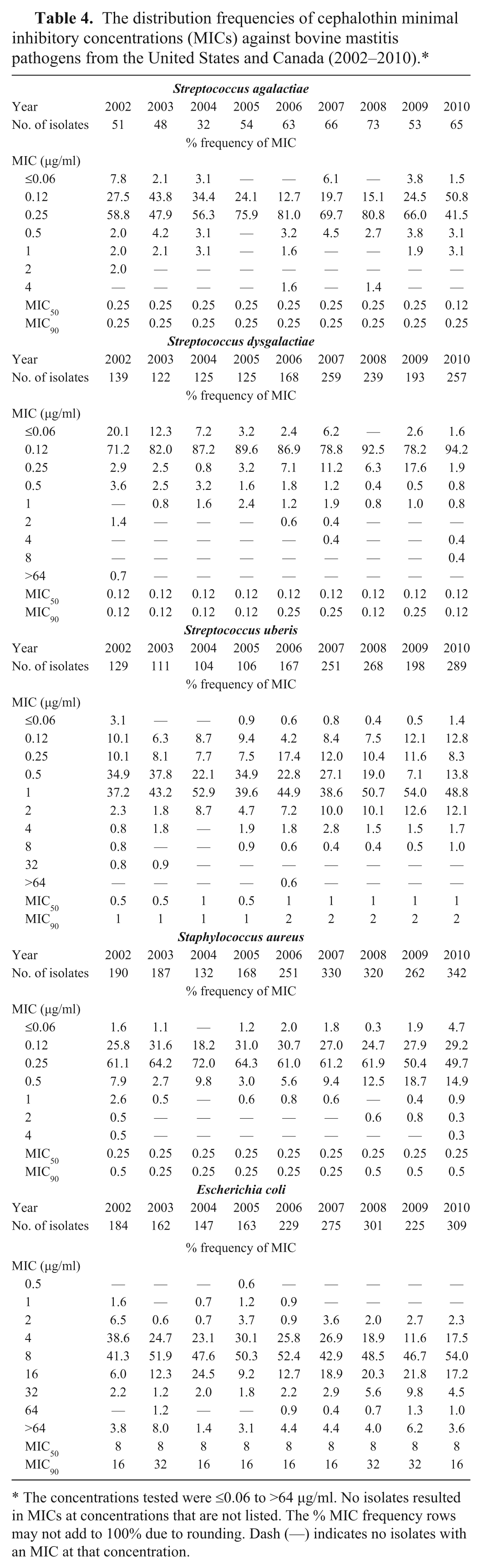
The concentrations tested were ≤0.06 to >64 µg/ml. No isolates resulted in MICs at concentrations that are not listed. The % MIC frequency rows may not add to 100% due to rounding. Dash (—) indicates no isolates with an MIC at that concentration.
The distribution frequencies of erythromycin minimal inhibitory concentrations (MICs) against bovine mastitis pathogens from the United States and Canada (2002–2010).*
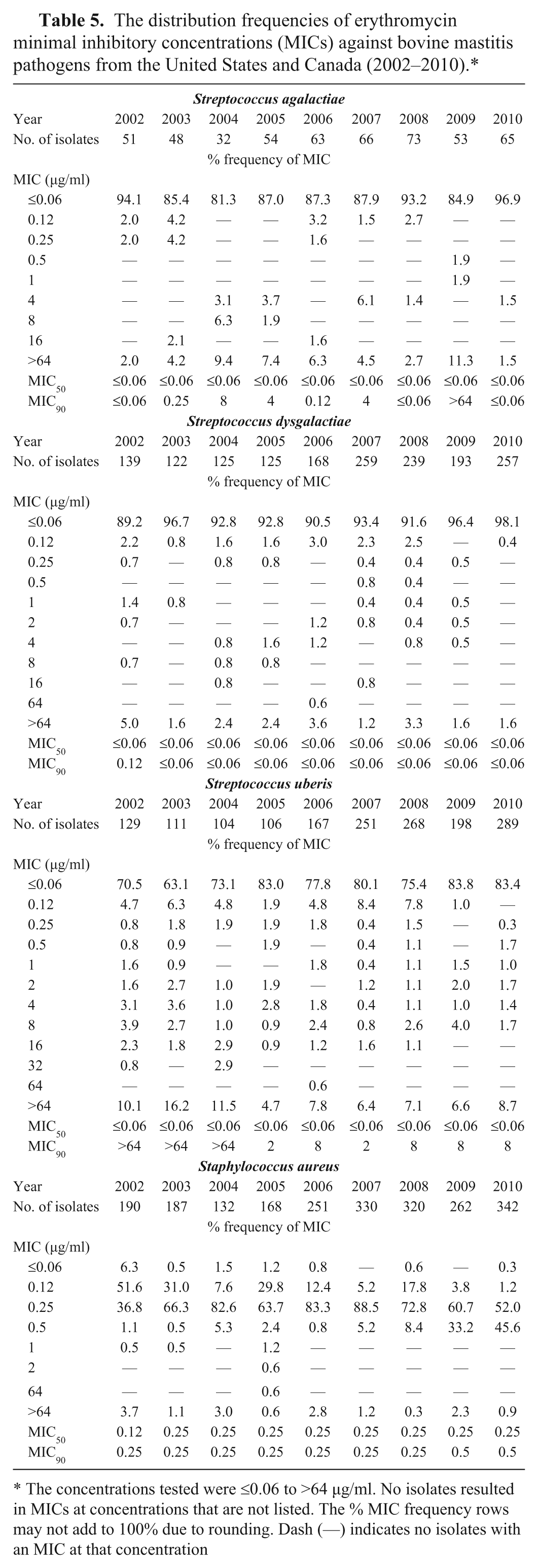
The concentrations tested were ≤0.06 to >64 µg/ml. No isolates resulted in MICs at concentrations that are not listed. The % MIC frequency rows may not add to 100% due to rounding. Dash (—) indicates no isolates with an MIC at that concentration
The distribution frequencies of oxacillin minimal inhibitory concentrations (MICs) against bovine mastitis pathogens from the United States and Canada (2002–2010).*
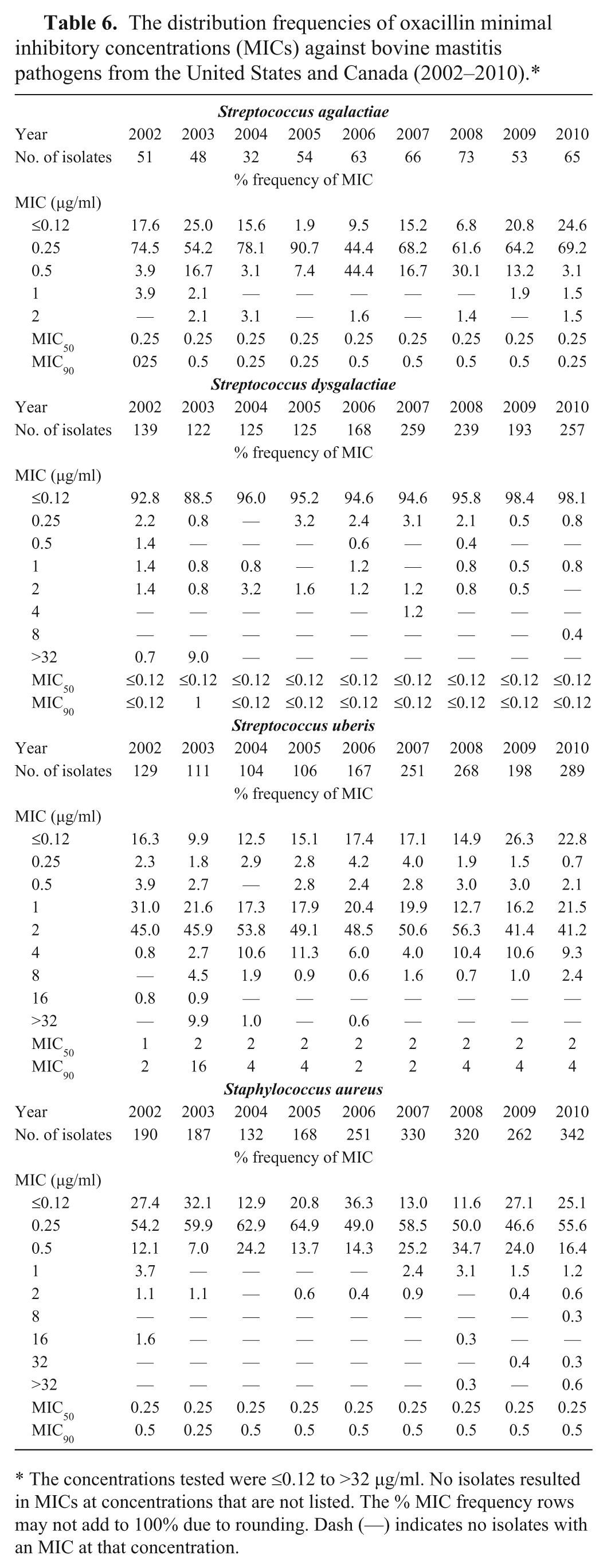
The concentrations tested were ≤0.12 to >32 µg/ml. No isolates resulted in MICs at concentrations that are not listed. The % MIC frequency rows may not add to 100% due to rounding. Dash (—) indicates no isolates with an MIC at that concentration.
The distribution frequencies of penicillin–novobiocin minimal inhibitory concentrations (MICs) against bovine mastitis pathogens from the United States and Canada (2002–2010).*
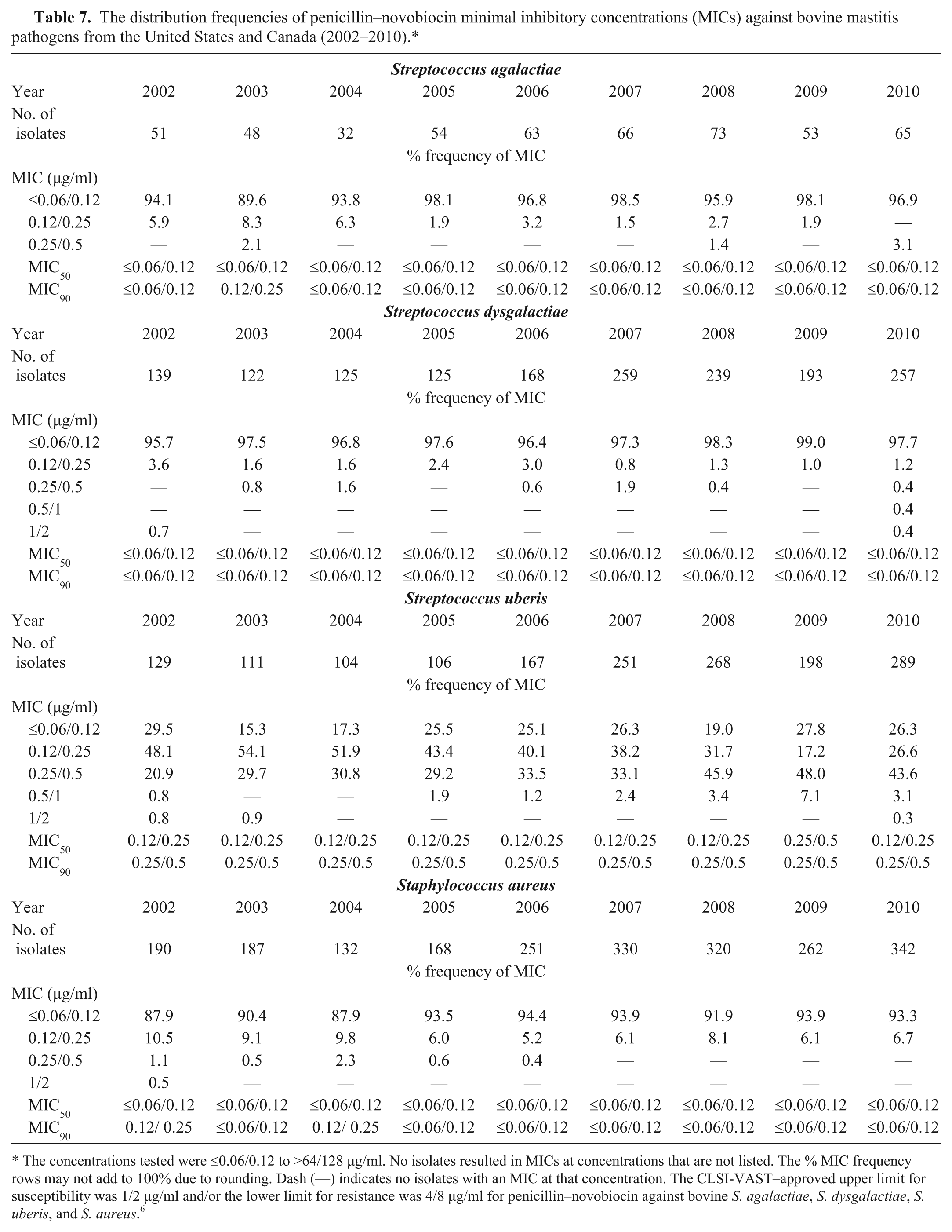
The concentrations tested were ≤0.06/0.12 to >64/128 µg/ml. No isolates resulted in MICs at concentrations that are not listed. The % MIC frequency rows may not add to 100% due to rounding. Dash (—) indicates no isolates with an MIC at that concentration. The CLSI-VAST–approved upper limit for susceptibility was 1/2 µg/ml and/or the lower limit for resistance was 4/8 µg/ml for penicillin–novobiocin against bovine S. agalactiae, S. dysgalactiae, S. uberis, and S. aureus. 6
The distribution frequencies of pirlimycin minimal inhibitory concentrations (MICs) against bovine mastitis pathogens from the United States and Canada (2002–2010).*
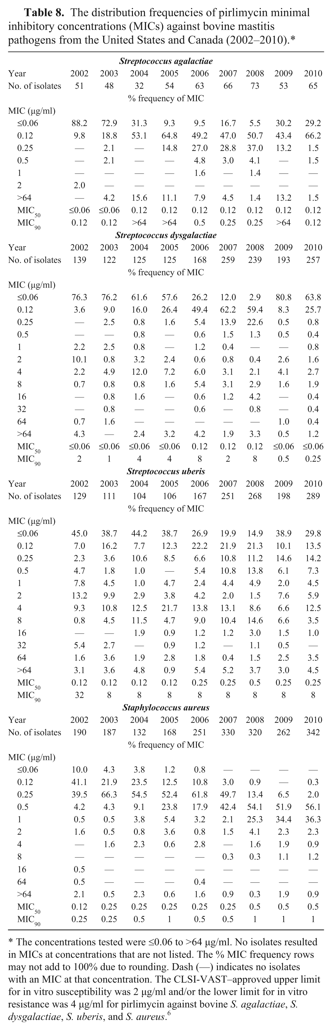
The concentrations tested were ≤0.06 to >64 µg/ml. No isolates resulted in MICs at concentrations that are not listed. The % MIC frequency rows may not add to 100% due to rounding. Dash (—) indicates no isolates with an MIC at that concentration. The CLSI-VAST–approved upper limit for in vitro susceptibility was 2 µg/ml and/or the lower limit for in vitro resistance was 4 µg/ml for pirlimycin against bovine S. agalactiae, S. dysgalactiae, S. uberis, and S. aureus. 6
Ampicillin
The ampicillin MICs against the staphylococci and streptococci isolates were low, and MIC50 and MIC90 values for these pathogens were similar, within a single doubling dilution, over the 9 years of the survey (Table 2). The MIC values of ampicillin against E. coli were high, with MIC90 values of ≥8 µg/ml; however, in each year, at least 50% of the E. coli isolates had ampicillin MICs of ≤4 µg/ml.
Ceftiofur
Streptococci and S. aureus isolated from cows with mastitis demonstrated very high levels of susceptibility to ceftiofur between 2002 and 2010 (Table 3). There was no more than a single dilution difference in the ceftiofur MIC50 or MIC90 values in the streptococci or S. aureus tested over the 9 years, and the MIC frequency distributions showed little variation from year to year. Escherichia coli also showed no change in susceptibility to ceftiofur over the same period. A few E. coli isolates each year showed elevated MICs, but the MIC50 and MIC90 values remained at 0.25 µg/ml and 0.5 µg/ml, respectively, over the 9-year study.
Cephalothin
There were no apparent differences, year to year, in the cephalothin MIC50 and MIC90 values greater than a single doubling dilution among the 3 streptococcal species and S. aureus between 2002 and 2010 (Table 4). The MIC50 value for the E. coli isolates was 8 µg/ml each year of testing, while the MIC90 value was 16 or 32 µg/ml.
Erythromycin
Table 5 provides data on the frequency distribution of erythromycin MICs and the number of isolates tested each year from 2002 to 2010. While there was variability in the distribution of MICs, overall, the data did not provide evidence that there was any obvious increase or decrease in erythromycin susceptibility among the bovine streptococci over the 9 years of the survey. There was no change beyond a single doubling dilution in the erythromycin MIC50 or MIC90 values for S. aureus (Table 5), and the distribution of MICs were similar across the 9 years of the survey.
Oxacillin
Over the 9 years, oxacillin MIC50 and MIC90 values against the streptococci remained unchanged, except in 2003 when increases in the MIC90 of S. dysgalactiae and S. uberis were seen (Table 6). In that year, approximately 10% of these 2 pathogen groups had MICs of >32 µg/ml. This appeared not to be attributable to any particular region or contributing laboratory and remains unexplained. With the exception of a single doubling dilution difference in 2003, the oxacillin MIC50 and MIC90 values for S. aureus were 0.25 µg/ml and 0.5 µg/ml, respectively.
Penicillin–novobiocin
All of the S. aureus and streptococci tested between 2002 and 2010 were susceptible (MIC ≤1/2 µg/ml) to penicillin–novobiocin. There was very little change in the MIC50 or MIC90 values of these Gram-positive species between 2002 and 2010 (Table 7). Escherichia coli showed high MICs, with a penicillin–novobiocin MIC50 value of 16/32 µg/ml, and MIC90 value of 32/64 µg/ml in each year (data not shown).
Pirlimycin
As with erythromycin, there was variability in the distribution of pirlimycin MICs among the streptococci (Table 8). There appeared to be no clear temporal trend in the proportion of streptococcal isolates that were susceptible (MIC ≤2 µg/ml) to pirlimycin. More than 90% of S. aureus isolates remained susceptible (MIC ≤2 µg/ml) to pirlimycin; however, the MIC distribution, MIC50 values, and MIC90 values of S. aureus appears to show a shift toward higher concentrations from 2002 to 2010 (Table 8).
Discussion
There is a need for surveillance information about temporal changes in susceptibility of bovine pathogens to those antimicrobial drugs that are commonly used to treat infections, including mastitis.2,3 Reviews of surveillance studies that looked at antimicrobial susceptibility among mastitis pathogens in the United States 2 and elsewhere 15 noted that these surveys were not recent and that more current data are needed. These reviews2,15 also stated that a longer time frame to study changes in susceptibility is desirable, as are data from a wider geographical area. There are few such programs that monitor susceptibility over a whole country and for multiple, consecutive years. 3 Germany has initiated a surveillance program in which bovine mastitis pathogens (including S. aureus, Streptococcus spp., andE. coli) are monitored for susceptibility to a variety of drug classes, including penicillins, cephalosporins erythromycin, and lincosamides (Bundesamt für Verbraucherschutz und Lebensmittelsicherheit [BVL]: 2008, GERMAP 2008 Antibiotika-Resistenz und -Verbrauch [Antibiotic resistance and consumption]. In German. Available at: http://www.bvl.bund.de/SharedDocs/Downloads/08_PresseInfothek/Germap_2008.pdf?__blob=publicationFile).21,22 The French RESAPATH program monitors mastitis pathogens from across France each year using disk diffusion testing and susceptibility and resistance criteria established by the Antimicrobial Committee of the French Society for Microbiology (Comité de Antibiogramme de la Société Française de Microbiologie; RESAPATH: 2009, . Available at: http://www.resapath.anses.fr/SITE_RESAPATH_WEB\uploadfiles\files\Documents\2008RESAPATHAnnualReportGB[id_doc=167].pdf; RESAPATH: 2010, 2009 annual report. Available at: http://www.resapath.anses.fr/SITE_RESAPATH_WEB\uploadfiles\files\Documents\2009RESAPATHAnnualReportGB[id_doc=167].pdf; RESAPATH: 2011, 2010 http://www.resapath.anses.fr/SITE_RESAPATH_WEB\uploadfiles\files\Documents\2010RESAPATHAnnualReportGB[id_doc=167].pdf). Staphylococci and streptococci isolated from cows with mastitis are monitored for antimicrobial susceptibility in Finland. In that program, erythromycin and clindamycin, but not pirlimycin, are included in the testing panel for S. aureus, S. uberis, and S. dysgalactiae (FINRES-Vet 2005–2006 Finnish veterinary antimicrobial resistance monitoring and consumption of antimicrobial agents. Evira publications 22/2007. Available at: http://www.evira.fi/portal/en/about+evira/publications/?a=view&productId=178). The Swedish veterinary program, SVARM, periodically surveys the antimicrobial susceptibility to cephalothin, penicillin, and oxacillin in S. aureus from cows with subclinical mastitis (SVARM: 2009, SVARM 2008. Available at: http://soapimg.icecube.snowfall.se/strama/Svarm%202008.pdf), and data have been reported for the years 2001, 2002, 2005, and 2008 (SVARM: 2010, SVARM 2009. Available at: http://www.sva.se/upload/Redesign2011/Pdf/Om_SVA/publikationer/Trycksaker/1/Svarm%202009.pdf). However, there have been no similar data published for mastitis pathogens in North America. The data reported herein provide comprehensive multinational information about the in vitro susceptibility of mastitis pathogens, isolated from dairy cattle across the United States and Canada, to antimicrobial drugs that are commonly used to treat mastitis.
Overall, there did not appear to be any substantial changes in the distribution of MICs to the antimicrobial drugs tested in this 9-year study of mastitis pathogens. Previously reported high levels of in vitro susceptibility of mastitis pathogens to penicillins8,9,14,16 appear, from the data in the current survey, to have been sustained over time. While S. aureus isolates with oxacillin MICs of 8 µg/ml were seen occasionally (<0.5% of the total number of S. aureus tested), the data do not suggest that there has been any substantial increase in the occurrence in U.S. or Canadian dairy cattle of methicillin-resistant S. aureus between 2002 and 2010. The high levels of susceptibility to ceftiofur seen among the Gram-positive cocci and E. coli in the current survey are similar to those reported among mastitis pathogens in other North American and European studies (RESAPATH: 2009; RESAPATH: 2010; RESAPATH: 2011).8,9,14,16,21,23 These previous studies demonstrated low cephalothin MICs among U.S. and Canadian streptococci and S. aureus with MICs similar to those reported in the current study. Patterns of susceptibility among the Gram-positive cocci to erythromycin and pirlimycin did not change substantially between 2002 and 2010, and the levels of susceptibility among S. aureus 14 and the environmental streptococci12,19 were comparable to those reported in other similar, but more regional, studies in the United States. While these results are reassuring that substantial changes in susceptibility among mastitis pathogens are not occurring, continued vigilance and surveillance is needed to be assured that this remains the case in the future.
There has been considerable discussion as to the most useful presentation of data obtained from susceptibility programs.7,20 In vitro data are often summarized using such values as MIC50, MIC90, median, mode, or range of MIC values. However, these statistics may not reflect the sometimes subtle shifts in MICs over time that may provide a more sensitive indicator of changes in susceptibility. Other summary values for describing MIC susceptibility data utilize clinical breakpoints or epidemiological cutoff values, but specific veterinary interpretive criteria have not been established for all drugs approved for use in treatment of mastitis. While the CLSI Subcommittee on Veterinary Antimicrobial Susceptibility Testing (CLSI-VAST) has approved veterinary-specific interpretive criteria for ceftiofur, penicillin–novobiocin, and pirlimycin against the Gram-positive mastitis pathogens tested in the surveillance program reported herein, only ceftiofur has interpretive criteria against E. coli. 6 In addition, no specific CLSI interpretive criteria exist for cephalothin, oxacillin, ampicillin, or erythromycin, against bovine mastitis pathogens. Alternatively, for surveillance purposes, the epidemiological cutoff point, where available, may provide a measure of any shifts in MIC and the potential for reduction or loss of in vitro susceptibility in the pathogen. The use of these different methods for MIC interpretation has been described4,20; however, even when a clinical breakpoint or epidemiological cutoff has been established, categorization of isolates as susceptible, intermediate, or resistant may provide an incomplete picture of what is occurring within a population of pathogens, especially over time.
An additional challenge is the potential for interpretive criteria, be they clinical breakpoints or epidemiologic cutoff values, to be changed by standards committees, such as CLSI, and the resulting complication of interpreting historical data in light of these revised criteria. Such difficulties can be minimized by publication of the full MIC distribution for each specific “bug-drug” combination. 20 While such publication does result in large data tables, this is the approach that has been taken in the current article as it enables a number of summary values to be examined. It is also the approach that has been taken with a number of national surveillance programs that examine the antimicrobial susceptibility of zoonotic and commensal enteric bacteria, such as in Canada (CIPARS: 2010, Final report 2007. Available at: http://www.phac-aspc.gc.ca/cipars-picra/pdf/cipars-picra-2007-eng.pdf; CIPARS: 2011, Final report 2008. Available at: http://www.phac-aspc.gc.ca/cipars-picra/2008/pdf/cipars-picra-2008-eng.pdf), the United States (NARMS: 2010, 2007 executive report. Available at: http://www.fda.gov/AnimalVeterinary/SafetyHealth/AntimicrobialResistance/NationalAntimicrobialResistanceMonitoringSystem/ucm209340.htm), and Denmark (DANMAP: 2011, 2010–Use of antimicrobial agents and occurrence of antimicrobial resistance in bacteria from food animals, foods and humans in Denmark. Available at: http://www.danmap.org/Downloads/~/media/Projektsites/Danmap/DANMAPreports/Danmap_2010.ashx).
Antimicrobial resistance surveillance programs are subject to a number of limitations, including sampling bias.3,10,18 In a study of published antimicrobial resistance surveillance studies, 17 the authors concluded that sampling bias and the failure to address multiple occurrences are common, and that case definition and laboratory practices and procedures may also influence the validity of the results. It is important that the isolates tested are representative of the population of interest. For the current mastitis surveillance program, isolates were collected from across the United States and Canada, especially from those states and provinces that contain large proportions of the national dairy herd populations (Table 1). The U.S. veterinary diagnostic mastitis laboratories that submitted isolates for this surveillance program are located in states in which more than 90% of the U.S. dairy cattle are found. The Canadian isolates came from laboratories in provinces with a substantial number of dairy operations. However, many, possibly most, bovine mastitis pathogens are not isolated and cultured, raising the possibility that the isolates from these state and provincial laboratories may not represent the whole population of pathogens. In addition, information was not available on any recent antimicrobial drug therapy that the animals may have received and so the isolates may have included bacteria from animals that had not been treated or had been treated and failed treatment.
In conclusion, the present report provides an extensive survey, in terms of geography and time, of the susceptibilities of North American bovine mastitis pathogens to beta-lactam, macrolide, and lincosamide drugs. Monitoring in vitro susceptibilities of clinical isolates can provide an early warning for the emergence of resistance. 21 The data indicate that there were no substantial numerical changes in the in vitro susceptibility of bovine mastitis pathogens from dairy cattle in Canada and the United States to these drugs during the years 2002–2010. The data support the conclusion of previous reports 15 that scientific evidence derived from local and regional studies in the United States and elsewhere does not support widespread, emerging resistance to common antimicrobial drugs among mastitis pathogens, even to drugs that have been used in dairies for many years.
Footnotes
Acknowledgements
The authors would like to thank the following veterinary diagnostic laboratories for their generous assistance by providing the bacterial isolates for this study: Arizona Dairy Herd Information Association, Cornell University (Quality Milk Production Services), High Plains Veterinary Outlet, Indiana Animal Diagnostic Laboratory (Purdue University), Iowa State University, Louisiana State University (Hill Farm Research Station), Michigan State University, Ohio Department of Agriculture, Pennsylvania State University, Texas A&M (College Station), University of Tennessee, Udder Health Systems Inc., University of California, Davis (VMTRC-Tulare), University of Guelph, University of Illinois, University of Minnesota (Laboratory for Udder Health), University of Montréal, Prairie Diagnostic Services Inc., University of Wisconsin (Madison), University of Wisconsin (Barron), Vermont Agency of Agriculture, Food & Markets Laboratory, and Washington State University. The authors would also like to thank Microbial Research Inc., as well as colleagues who provided thoughtful comments and review of this article.
a.
API Microbial Identification kits, bioMérieux Inc., Durham, NC.
b.
Biolog Microbial Identification System, Hayward, CA.
c.
Sensititre Division, Trek Diagnostic Systems Inc., Thermo Scientific, Cleveland, OH.
Declaration of conflicting interests
The author(s) declared the following potential conflicts of interest with respect to the research, authorship, and/or publication of this article: Cynthia J. Lindeman, Lacie Johansen, and Lisa M. Mullins are employed by Zoetis Inc. (formerly a business unit of Pfizer Inc.). Ellen Portis is a former employee of Zoetis Inc. Gillian A. Stoltman (Waterwood Consulting LLC), a former employee of the animal health business unit of Pfizer Inc., is currently providing consultation services for Zoetis Inc.
Funding
The author(s) disclosed receipt of the following financial support for the research, authorship, and or publications of this article: This study was funded by the animal health business of Pfizer Inc. (currently operating as Zoetis Inc.).