
Abstract
Lymphoma and/or leukemia was diagnosed in 26 camelids (20 alpacas and 6 llamas) out of 110 camelid neoplasia archived January 1995 through January 2012 at the Colorado State Veterinary Diagnostic Laboratories (CSU-VDL). Some of the tumors presented a diagnostic challenge because they could not be distinguished on the basis of gross or microscopic morphology. Immunohistochemistry using a T-cell marker (cluster of differentiation [CD]3), a B-cell marker (paired box protein [PAX]-5), a leukocyte integrin beta-2 marker (CD18), and a neuroendocrine marker (synaptophysin) was employed to help differentiate between lymphoma and other malignant round cell tumors. Alpaca lymphomas presented as either juvenile disseminated lymphoma in crias ≤2 years of age (n = 8) or adult multicentric lymphoma and/or leukemia (n = 12). Lymphomas in alpacas were of T-cell origin (n = 13), non–B-cell, non–T-cell origin (n = 4), B-cell origin (n = 2), or myelogenous leukemia (n = 1). Abdominal organs, predominantly the liver, were commonly involved in both the crias and adult alpacas. Lymphomas in llamas presented as either adult multicentric lymphoma of B-cell origin in animals younger than 7 years of age (n = 2), or T-cell lymphoma (n = 2), and non–B-cell, non–T-cell lymphoma (n = 1) in animals 7 years of age or older. The thorax was commonly involved in llamas, with infiltration of neoplastic cells into hilar and mediastinal lymph nodes. A rare type of lymphoma diagnosed in the llamas was cutaneous, epitheliotrophic T-cell lymphoma (n = 1).
Keywords
Lymphoma has been reported to be the most common neoplasia in camelids from different parts of the world.1,8,13 Studies revealed that there was a significant difference in the age of alpacas (range: 0.2–2 years, median: 0.8 years) and llamas (range: 0.3–15 years, median: 5.5 years) diagnosed with lymphoma.9,13 Immunohistochemical characterization of lymphomas has been undertaken in small numbers of camelids.1,7,13 Analysis of greater numbers of round cell tumors is still needed to better investigate any relationship between immunophenotype, age, sex, organ distribution, and clinical or pathological diagnosis. The purpose of the current study is to report the pathologic and immunophenotypic characteristics of lymphoma and/or leukemia in alpacas and llamas archived January 1995 through January 2012 at Colorado State University Veterinary Diagnostic Laboratory (CSU-VDL; Fort Collins, Colorado).
Immunohistochemical staining was performed on an automated staining platform a using an alkaline phosphatase red kit b as chromogen. Primary antibodies and methods of antigen retrieval are listed in Table 1. Species distribution, age, and sex of affected animals; organ involvement; and immunohistochemical characteristics of the archived tumors are summarized in Table 2.
Sources of immunohistochemistry primary antibodies and antigen retrieval methods used in malignant round cell tumor diagnosis.
Species, age, and sex of affected animals and summary of pathologic and immunophenotypic characteristics of malignant round cell tumors (lymphoma) diagnosed in alpacas and llamas submitted to Colorado State University Veterinary Diagnostic Laboratory (Fort Collins, Colorado) from 1995 to 2012.*
CM = castrated male; F = female; m = month(s); y = year(s); U = unknown age; LNs = lymph nodes; ND = not determined; ++++ = strongly positive; +++ = moderately positive; ++ = slightly positive; + = a few scattered cells are positive; +/– = suspicious; – = negative.
Biopsy or necropsy tissue from only 1 organ was submitted for histologic examination, but multicentricity was implied in the history.
Malignant round cell tumors are restricted in the present study to typical lymphoma, leukemic lymphoma, myeloid leukemia, and lymphoblastic non–B-cell, non–T-cell lymphoma (n = 26, 20 alpacas and 6 llamas), all of which were the most common neoplasias (out of a total of 110) diagnosed at CSU-VDL between 1995 and 2012. Lymphoma and leukemia had an overall prevalence of 22.2% and 23.6%, respectively, of camelid neoplasia if proliferative lesions are to be excluded. Alpaca lymphomas (77%) are overrepresented in the current study, which is consistent with previous reports.9,13,15 Statistical analyses revealed a clear predisposition of female camelids (total number of 76 females and 41 males from both species) to develop neoplastic/proliferative lesions (one-sided P = 0.0006) while female predisposition to develop lymphomas was not that highly significant (one-sided P = 0.0577).
Juvenile lymphomas in alpacas 2 years of age or younger (range: 0.1–2 years, median: 0.9 year) represented 40% of the alpaca lymphomas. The disseminated nature of juvenile lymphoma of T-cell origin and the wide variety of tissues involved suggests that the syndrome has many parallels with juvenile or calf lymphoma.3,4 Affected alpacas had bilateral peripheral lymphadenopathy, often marked abdominal organomegaly, and occasional leukemic profile (neoplastic cells in vascular spaces) with bone marrow involvement in 25% of the cases (the percentage could have been higher if affected animals had been submitted in whole for necropsy rather than sending just 1 organ for histologic evaluation). True thymic involvement, however, was not detected in any of the CSU-VDL cases, but the base of the heart was involved in 1 case.
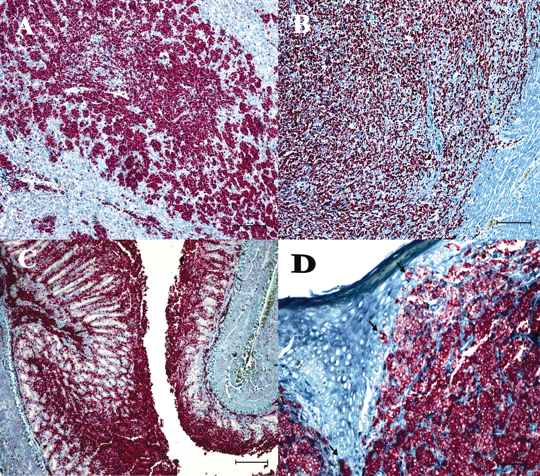
Cells from a lymphoma in the lung of an 11-month-old alpaca stained neither for T-cell nor B-cell markers (Fig. 1), which suggests the possibility of non–B-cell, non–T-cell origin (lymphoblasts), given that camelids in prior studies developed these lymphoblastic tumors very early in neonatal life or even perinatally.4,7,9,10 In human beings, non–B, non–T cells with lymphoblast morphology are classified into 4 categories: 1) cluster of differentiation (CD)7+ stem cell lymphoma, 2) blastic natural killer lymphoma (B-NKL), 3) myeloid/NK precursor cell leukemia (M/NKL), and 4) CD4+CD56+ hematodermic malignancy. 6 The adult alpaca multicentric lymphomas described herein (Fig. 2) were very similar to the disseminated forms described in the literature.1,5,7,13 Acute myeloid leukemia with multilineage dysplasia, previously diagnosed at CSU-DVL, was reported to occur in an 18-year-old female alpaca (not included in the current study because the tissue blocks could not be retrieved) and is somewhat similar to the case reported herein. 12
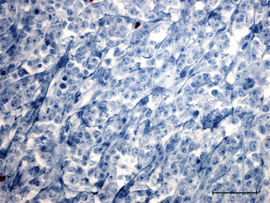
Eleven-month-old alpaca; lung; non–B-cell, non–T-cell lymphoma. Neoplastic cells show negative immunoreactivity against cluster of differentiation (CD)3, which only stains rare mature lymphocytes interspersed among neoplastic cells. Bar = 100 µm.
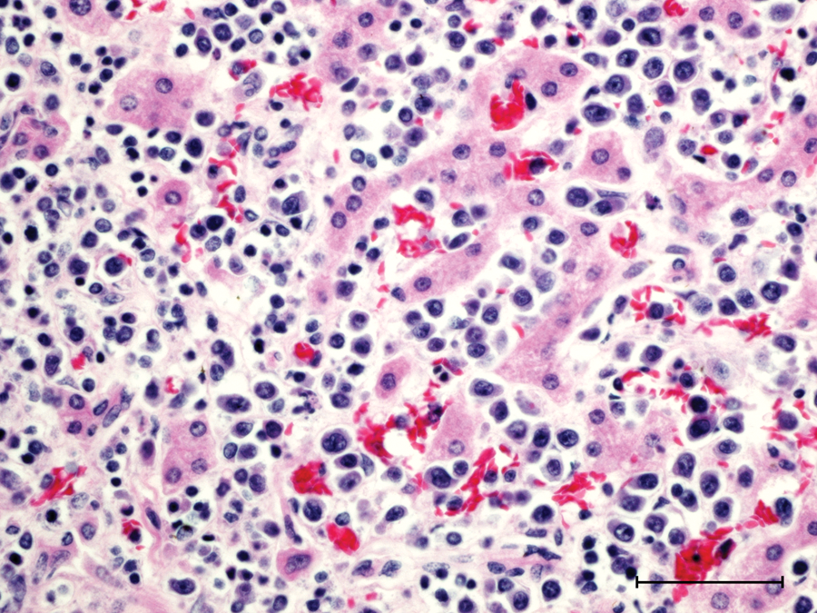
Three-year-old alpaca; liver; leukemic T-cell, B-cell rich lymphoma. Immature lymphocytes diffusely fill hepatic sinusoids and replace and dissect hepatic plates. Bar = 100 µm.
Lymphomas in llamas in the current study occurred at an older age than alpacas, with only adult animals (range: 3–21 years, median 7 years) affected. The multicentric adult lymphoma in the llamas showed more predilection for the thorax, with rare involvement of respiratory organs, particularly trachea, a finding corroborated by other studies.2,7,14 The lymphomas were B-cell lymphomas (40%), T-cell lymphomas (40%), and non–B-cell, non–T-cell lymphomas (20%).
In both species, liver was the most commonly involved parenchymatous organ (Figs. 2, 3A), followed by the spleen, peripheral and visceral lymph nodes, kidneys, and lungs. Involvement of myocardium (Fig. 3B), pancreas, and gastric chamber 1 (C1; Fig. 3C) was evident in 1 alpaca, and spinal cord and skeletal muscles of the hind limb was identified in only 1 llama.
Immunohistochemistry.
Epitheliotrophic cutaneous lymphoma of T-cell origin in the eyelid of an aged llama was observed in the current study. The tumor is similar to “mycosis fungoides-like” disease in human beings and dogs, with formation of typical “Pautrier abscesses” (Fig. 3D). 3 Subcutaneous lymphomas reported to occur in alpacas in a previous study were not typed and did not show epidermal tropism. 11 Prior studies confirmed the lack of retroviral etiology via electron microscopic studies.1,9
The current study summarizes the pathologic and immunophenotypic characteristics of 26 camelid lymphomas. The clinical disease and postmortem findings in 20 alpacas and 6 llamas can be broadly categorized into 2 syndromes in each species: T-cell juvenile disseminate (40%) and adult multicentric lymphomas in alpacas (60%) and adult multicentric (83%) and epitheliotrophic (17%) lymphomas in llamas. Further classification of camelid lymphoma and/or leukemia, especially the lymphoblast non–B-cell, non–T-cell lymphomas, merits further studies using human markers, namely, CD7, B-NKL, M/NKL, and CD4+CD56+, to better characterize these tumors.
Footnotes
Acknowledgements
The author would like to thank Todd Bass and Bruce Cummings for their technical help and invaluable expertise in immunohistochemistry and Alexandra Harvey for assistance with archived tissue retrieval.
a.
Benchmark Immunostainer, Ventana Medical Systems Inc., Tucson, AZ.
b.
UltraView Alkaline Phosphatase Red Kit, Ventana Medical Systems Inc., Tucson, AZ.
c.
Leukocyte Antigen Biology Laboratory, Davis, CA.
d.
Protease I, Ventana Medical Systems Inc., Tucson, AZ.
e.
Dako North America Inc., Carpinteria, CA.
f.
Target Retrieval Solution (pH 6.0), Dako North America Inc., Carpinteria, CA.
g.
BioCare Medical, Concord, CA.
h.
Ventana Medical Systems Inc., Tucson, AZ.
Declaration of conflicting interests
The author(s) declared no potential conflicts of interest with respect to the research, authorship, and/or publication of this article.
Funding
The author(s) declared that they received no financial support for their research and/or authorship of this article.