
Abstract
A hunted free-ranging female red deer (Cervus elaphus) from a region near the Nahuel Huapi National Park, Northern Patagonia, Argentina, had a focally extensive peribronchial lymphoid proliferative lesion in the lung characterized by formation of multiple follicles, with prominent germinal centers lacking mantle zone cells and antigen-related polarity. On examination of immunohistochemically stained tissues, a predominance of B cells (cluster of differentiation [CD]20 positive) with only a few scattered T cells (CD3 positive) were present. The histologic and immunohistochemical characteristics are consistent with follicular lymphoma, which is frequently seen in human beings and less frequently in domestic animals.
Follicular lymphomas are slowly progressive tumors of germinal center B cells that are histologically recognized by characteristic follicular architecture, lack of antigen-related polarity, and fading mantle cell cuffs. 4 Such neoplasms have been described in human beings, and, among animals, are most frequently seen in cats and cattle and less commonly in dogs. 4 While in human beings this type of neoplasm represents up to 25% of all lymphomas diagnosed, it is thought that less than 1% of lymphomas diagnosed in animals are of the follicular type. 4 Furthermore, follicular lymphomas have not been reported in the lung of any animal species. The present study describes the diagnosis of what appears to be a primary lung follicular lymphoma in a free-ranging red deer (Cervus elaphus).
An approximately 2-year-old, 100 kg, female red deer was shot by a hunter in the Nahuel Huapi National Park, Northern Patagonia, Argentina. No obvious clinical signs or external gross abnormalities were reported by the hunter. The body was transported to a meat smoking facility where veterinary inspection was performed. During this process, one of the authors of the current article (E. Chang Reissig) had access to the carcass and abdominal and thoracic viscera, which were examined as part of a health surveillance project of wild ungulates performed in the Nahuel Huapi National Park. No significant gross abnormalities were observed during the gross examination of the visceral organs, lymph nodes, or the carcass. Samples of liver, heart, spleen, kidneys, lymph nodes (prescapular, popliteal, bronchial, mediastinal, submandibular, retropharyngeal, and mesenteric), forestomachs, abomasum, small and large intestine, and both lungs (all lobes) were collected and fixed by immersion in 10% buffered formalin (pH 7.2) for several weeks before being embedded in paraffin wax, sectioned at 4 µm, and stained with hematoxylin and eosin. Immunohistochemistry for cluster of differentiation (CD)3, CD18, CD20, CD79, and pancytokeratin was performed using an avidin–biotin conjugate (ABC) immunostaining method following the instructions of the manufacturer a and standard operating procedures of the Veterinary Medical Teaching Hospital at the University of California, Davis. Citrate buffer steam was used for antigen retrieval, followed by serum block, secondary antibody against each corresponding species, and detection steps. Primary antibodies were rat monoclonal against CD3, b mouse monoclonal against CD18, b CD79, a and pancytokeratin, c and rabbit polyclonal against CD20. d Lymph node and skin from a healthy deer were used as positive and negative controls for the leukocyte and epithelial markers, respectively.
Histologically, a single mass located eccentrically around a bronchiole was observed in the lung (Fig. 1). At the submicroscopic level, this mass was approximately 0.5 mm in diameter, roughly circular, well-circumscribed, and encapsulated, and was compressing the adjacent parenchyma and the lumen of an adjacent bronchiole. Histologically, the mass was composed of multiple prominent germinal centers with a more or less uniform population of predominantly small to intermediate size cleaved lymphocytes. The lymphocytes had densely stained nuclei and usually no nucleoli; scant, pale cytoplasm (Fig. 2); and resembled the centrocytes of low-grade follicular lymphoma as seen in dogs. 4 The nuclei of these cells were approximately 8–10 µm in diameter. The small centrocytes were mixed with a much smaller number of large round cells of open nuclei with peripheralized chromatin and 1–3 nucleoli (Fig. 2). The round cells were interpreted to be centroblasts, which are considered to be the main cell type in high-grade follicular lymphoma. 4 The nuclei of the centroblasts were approximately 14–18 µm in diameter. There was mild to moderate anisocytosis and anisokaryosis, and the mitotic rate was less than 1 per high power field (Fig. 2). The follicular cells were closely aggregated, with the smaller centrocytes being uniform in size, but of irregular shape. The follicles were closely facetted and separated from each other by thin fibrous trabeculae that contained blood vessels. Identical cell types were present in every follicle. Most of the follicular cells showed strong positive cytoplasmic staining for CD20 (Fig. 3), and only a few cells showed weak to moderate cytoplasmic staining for CD79 (Fig. 4). Randomly scattered throughout the neoplasm were a few round cells that showed moderate cytoplasmic positive staining for CD3 (Fig. 5), while CD18 and pancytokeratin (Fig. 6) were negative. No antigen-related polarity or mantle cells were observed around the follicles. No readily visible necrosis, apoptosis, inflammatory infiltrates, stainable body macrophages, or hemorrhage were apparent. The parenchyma surrounding the mass was atelectatic, but apart from this, no other histological abnormalities were observed in any of the sections of lung examined.
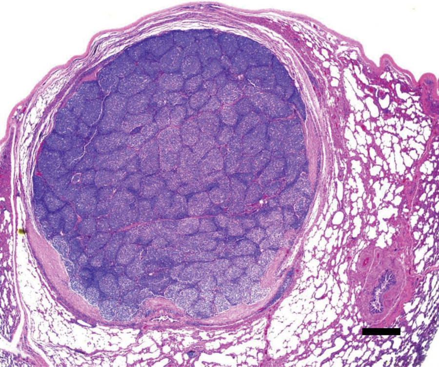
Red deer (Cervus elaphus). Follicular lymphoma located eccentrically around a bronchiole and compressing the surrounding pulmonary parenchyma. Observe the absence of mantle cells cuff and of stainable body macrophages. Hematoxylin and eosin. Bar = 100 µm.
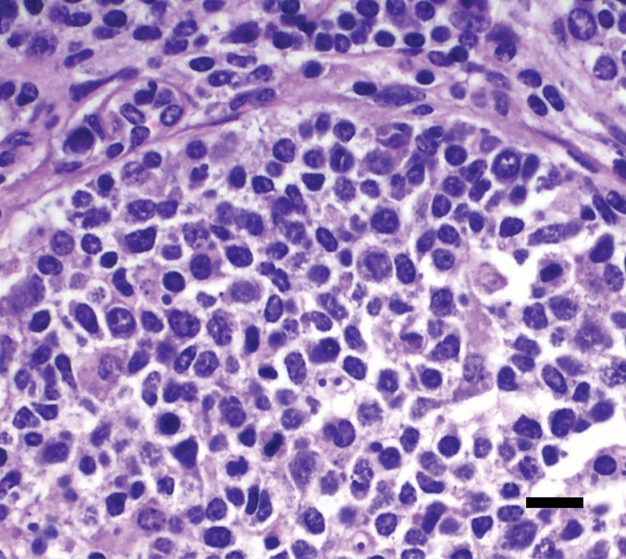
Red deer (Cervus elaphus). High magnification of the follicular lymphoma depicted in Figure 1. Observe the monomorphic population of mostly small lymphocytes and the absence of stainable body macrophages. Hematoxylin and eosin. Bar = 20 µm.
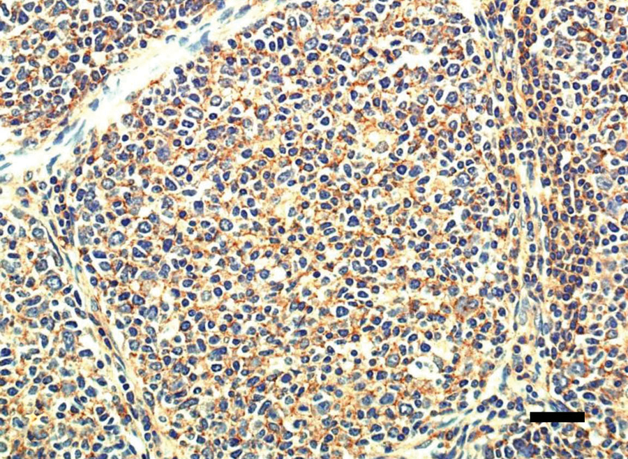
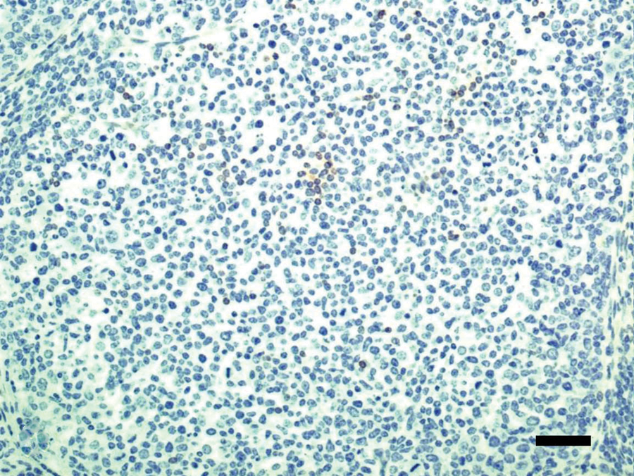
Red deer (Cervus elaphus). Cluster of differentiation (CD)20 staining. Most cells are positively stained. Bar = 50 µm.
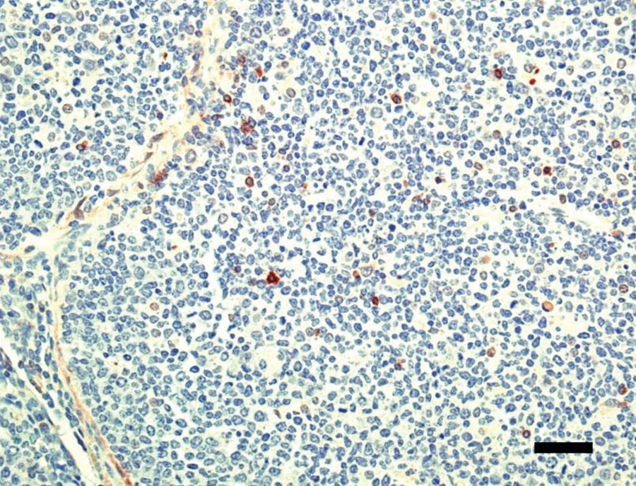
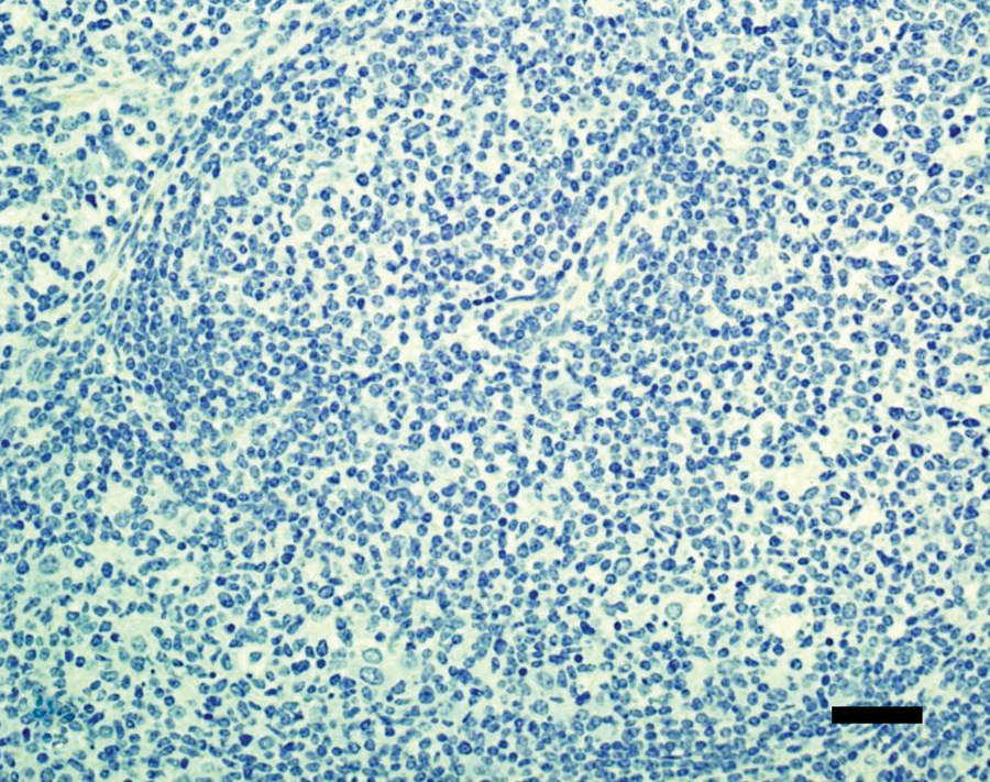
Red deer (Cervus elaphus). Cluster of differentiation (CD)79 staining. Only a reduced number of cells is positively stained. Bar = 50 µm.
Red deer (Cervus elaphus). Cluster of differentiation (CD)3 staining. Only a reduced number of cells is positively stained. Bar = 50 µm.
Red deer (Cervus elaphus). Pancytokeratin staining. No staining is observed. Bar = 50 µm.
No metastasis or other neoplastic changes were observed in other organs examined. Few, approximately 100 µm × 50 µm protozoal cysts with morphology consistent with Sarcocystis spp. were present within cardiomyocytes, but such parasites were not associated with significant tissue damage or inflammation.
Based on the histological morphology of packets of lymphoid cells compressing the pulmonary parenchyma and the adjacent bronchiole, plus the bimorphic nature of the cell population, the absence of mantle cell cuffs and apoptotic cells, the lack of antigen-related polarity in the germinal centers, and the positivity of the cells to CD20, the neoplasm was diagnosed as follicular lymphoma. 4 The cell morphology described in the current case is compatible with cases of human follicular lymphoma where the cells inactivate the apoptotic gene, with the consequence that there are no dying cells and hence no macrophages are seen, such as one would expect to observe in benign follicles with progressive B-cell selection and frequent cell death. 4 By definition, follicular lymphomas are neoplasms of follicular center B cells (centrocytes and centroblasts) that have a follicular pattern and in which the centrocytes fail to undergo apoptosis, due to chromosomal rearrangement that prevents the turning of the anti-apoptotic gene. 4
Multiple samples from all lobes of lungs, lymph nodes, liver, spleen, and most other visceral organs of the red deer were collected and examined, and no lesions were observed apart from the neoplasia described herein. It is therefore likely that this neoplasia was unicentric. Strong positivity for CD20 indicated that this was a B-cell lymphoma. Only a few cells were positive for CD79. The results indicate that, in deer, CD20 seems to be a better marker for B cells than CD79, or alternatively, that CD79 is not strongly expressed in lymphocytes of this species. No information about the specificity of the antibodies used is available for deer tissues. However, deer tissues were used as positive and negative controls with the expected results and the authors believe that this provides reasonable proof of the specificity of these antibodies for the target cells.
Follicular lymphomas are predominately of lymph node origin.4,5 However, extranodal lymphoid tissue including spleen, oropharynx, bone marrow, liver, and less commonly nonlymphoid organs have been involved.4,5 The current case is particularly unique because of the pulmonary location and presence of the largest tumor formation around a bronchiole, which suggests that the origin of the neoplasia was in the cells of the bronchus-associated lymphoid tissue.
The main differential diagnosis for follicular lymphoma is benign follicular hyperplasia. 1 In the present case, follicular hyperplasia was ruled out because the follicles were more or less uniform in size (follicular hyperplasia usually has more variation in size) and there was no mantle cell cuff around each germinal center. In addition, there was no antigen-related polarity present that would indicate a benign process, and all follicles had the same cellular composition.2,3 Also, as noted above, the moderate mitotic activity, which is characteristic of follicular hyperplasia, was absent. Finally, there was absence of the starry-sky pattern of stainable body macrophages, normally seen within benign germinal centers. 3
In the current case, no clinical signs were reported by the hunters in the brief period during which this animal was observed before it was shot. Although this cannot be considered a clinical examination, the fact that this animal was in very good nutritional condition suggests that no major clinical alterations occurred, as loss of condition tends to be one of the first indications of chronic health problems in wild animals. 1 Follicular lymphomas are indolent lesions that may reach quite large size, and it is thought that, in animals, the lesions might transform into more aggressive diffuse large B-cell lymphosarcoma with time, an outcome usually seen in human beings.2,4 It is possible that this might have been the case with the deer in the current study had it not been killed at this stage of the neoplasm development.
Footnotes
Acknowledgements
The authors thank Weiss Smoking Plant, Bariloche, Argentina, for allowing access to the deer carcass, and the National Park Administration (APN) for a research permit (no. 721).
a.
Dako North America, Carpinteria, CA.
b.
Dr. P. Moore, Department of Pathology, Microbiology and Immunology, School of Veterinary Sciences, University of California, Davis, CA.
c.
Biocare Medical, Concord, CA.
d.
Thermo Scientific, Fremont, CA.
Declaration of conflicting interests
The author(s) declared no potential conflicts of interest with respect to the research, authorship, and/or publication of this article.
Funding
The author(s) disclosed receipt of the following financial support for the research, authorship, and/or publication of this article: This work was financially supported by the Wildlife Health Fund, Wildlife Conservation Society, United States; the Rufford Small Grant for Nature Conservation, Rufford Foundation (RSG 3802-07 and 5738-1), United Kingdom; and the California Animal Health & Food Safety Laboratory, University of California, San Bernardino, Davis, CA. Elizabeth Chang Reissig was supported by a Doctoral Fellowship of the National Council of Scientific and Technical Research, Argentina.