
Abstract
Ischial callosities have received little attention in veterinary medicine even though they are distinguishing anatomic organs. The organs are characterized by a pair of hairless pads of thickened epidermis, located bilaterally in the gluteal region, which overlay the tuberosities of the ischia of all Old World monkeys, gibbons, and siamangs. The current report describes a case of reactive amyloidosis associated with ischial callosititis in a rhesus macaque (Macaca mulatta). Amyloid A (AA) protein was found in the liver, spleen, small intestine, mesenteric lymph nodes, and ischial callosities by histology, Congo red stain, and immunohistochemistry. Confocal microscopy showed that many cluster of differentiation (CD)68-positive macrophages within the ischial callosities contained intracellular AA protein, which suggests that CD68-positive macrophages have an important role in the pathogenesis of reactive amyloidosis in nonhuman primates. The normal histology of ischial callosities of rhesus macaques is also documented in this report.
Keywords
Amyloidosis is a group of disorders caused by extracellular deposition of misfolded proteins that lead to impair organ function. 4 More than 25 different proteins have been isolated from amyloid deposits.4,19 Reactive, or secondary, amyloidosis is one of the most common amyloid diseases in human beings and animals, in which misfolded circulating serum amyloid A (SAA) deposits are found in 1 or more organs.2,11 Serum amyloid A is one of the acute phase reactants produced mainly by hepatocytes, and regulated by interleukin (IL)-1, IL-6, and tumor necrosis factor (TNF). 3 Reactive amyloidosis has been reported in human patients with rheumatoid arthritis and other chronic inflammatory diseases. 10
Ischial callosities (ICs) are a pair of hairless pads of thickened epidermis that are located bilaterally in the gluteal region and overlay the tuberosities of the ischia of all Old World monkeys, gibbons, and siamangs. 17 Ischial callosities provide comfort and stability while sitting on thin branches during feeding, sleeping, grooming, resting, and locomoting in the peripheral branch zone. 16 In macaques, ICs are modified skin composed of strikingly thickened and cornified stratum corneum with a subcutaneously dense fibro-fatty plate that fuses into the underlying periosteum by connective tissue. 9 However, ICs have not received much attention in veterinary medicine. The present study describes a case of chronic ischial callosititis and associated reactive amyloidosis in a rhesus macaque (Macaca mulatta). The normal morphology of ICs from age-matched rhesus macaques was also documented for the purpose of comparative pathology.
A 15-month-old, male, India-origin rhesus macaque presented with diarrhea and dehydration. Upon physical examination, the animal was found to have suboptimal body condition with a body condition score of 2 out of 5. Two months after presentation, the diarrhea had resolved temporarily, and bilateral purulent ulcerations of ICs were noted on physical examination. Over the course of 4 subsequent months, the animal was treated with multiple courses of antibiotics, guided by culture and sensitivity. Drugs administered included cephalexin, enrofloxacin, clindamycin, chloramphenicol, and gentamicin. The diarrhea relapsed several times, and the lesions of ICs never resolved. A mixed population of Staphylococcus haemolyticus, Proteus mirabilis, Escherichia coli, and Morganella morganii were consistently isolated from the ulcers of the ICs. Euthanasia was elected due to nonresponsiveness to antibiotic treatment and deterioration of physical condition.
Upon necropsy, the animal was in poor body condition with bilateral, diffuse, severe necrohemorrhagic and ulcerative ischial callosititis peripherally surrounded by a large amount of dark crust (Fig. 1). The colon had smooth, pale mucosa and contained partially digested food material. The mesenteric lymph nodes were markedly enlarged. The spleen and liver were mildly enlarged. All other organs were grossly unremarkable.

Ischial callosities, India-origin rhesus macaque (Macaca mulatta); bilateral ulcerative ischial callosititis.
Histologically, the ICs were diffusely, severely ulcerated, and covered by a thick mat of fibrin, cellular debris, numerous degenerated neutrophils, and admixed hemorrhage and scattered colonies of mixed bacteria (Fig. 2A). Moderate osteonecrosis of the tuberosities of the ischia was also noted at this site. Within less affected areas, the stratum corneum was disorganized by undulated and fragmented keratin, cellular debris, and many intracorneal pustules. The epidermis was markedly thickened by prominent acanthosis, prominent rete ridge formation, and moderate parakeratotic hyperkeratosis. Subcutaneously, the fibro-fatty zone was markedly decreased to completely lost, replaced by fibroblasts and collagen, admixed with numerous nondegenerate neutrophils, many regularly spaced new vessels, and small numbers of lymphocytes, plasma cells, and macrophages (granulation tissue). Multifocally, there were variably sized aggregates of variable amounts of amorphous, smudgy to finely fibrillar eosinophilic, acellular substance. Similar deposits were also noted in the space of Disse in the liver, the white pulp of the spleen, the lamina propria of the small intestine, and the lymphoid follicles of the mesenteric lymph nodes. The spleen and mesenteric lymph nodes had diffuse, moderate lymphoid depletion.
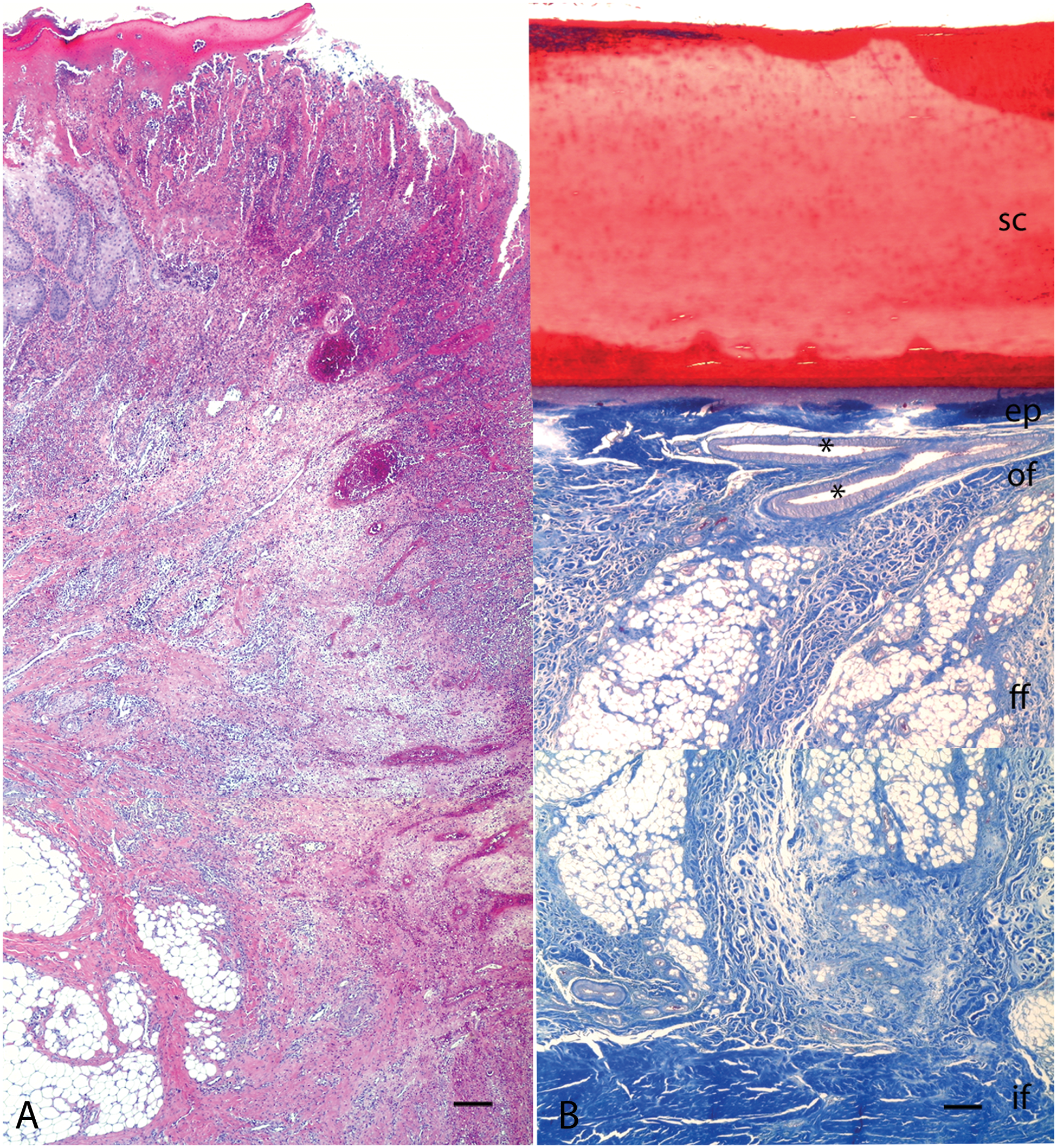
Ischial callosities of India-origin rhesus macaque (Macaca mulatta).
Congo red stains demonstrated that the eosinophilic deposits in the liver, spleen, small intestine, mesenteric lymph nodes, and ICs were congophilic and exhibited apple-green birefringence with polarized light. Immunohistochemical staining was performed on 4-µm thick, formalin-fixed, and paraffin-embedded tissue sections using a standard streptavidin–biotin method and diaminobenzidine as the detection chromogen with a hematoxylin counterstain. The acellular, hyaline substance in the ICs and liver was strongly positive for AA (Fig. 3). To co-localize macrophages and amyloid, multicolor confocal microscopy was performed using anti-cluster of differentiation (CD)68 antibody a specific for monocytes-macrophages and an anti-AA antibody. a Confocal microscopy was performed using a confocal microscope equipped with three lasers. b Individual optical slices represent 0.2 mm, and 32–62 optical slices were collected at 512 × 512 pixel resolution. Softwarec,d was used to assign colors to the channels collected: HNPP/Fast Red, e a substrate that fluoresces red when exposed to a 568-nm wavelength laser; Alexa 488, f which fluoresces green; and Alexa 633, f which fluoresces blue. Many CD68-positive cells were also positive for intracytoplasmic AA in the IC lesions (Fig. 4). Rare Kupffer cells in the liver were intracytoplasmic AA positive.
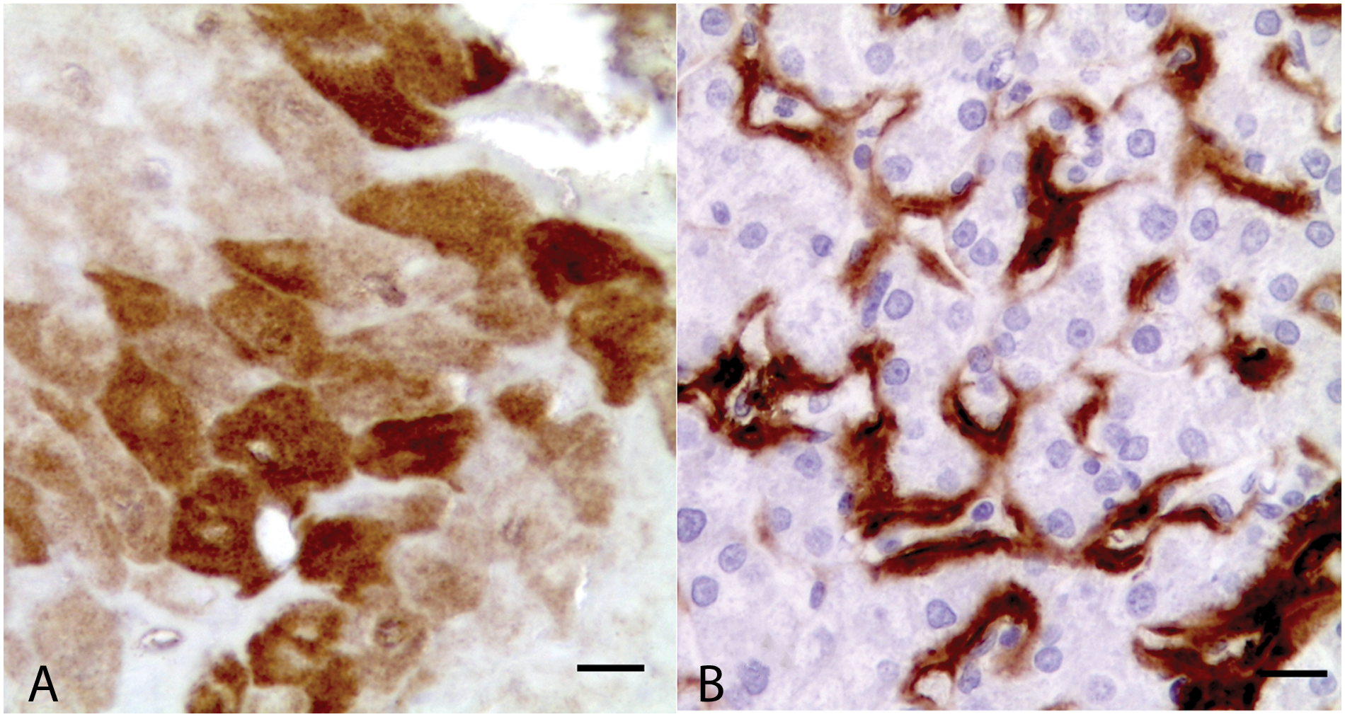
Immunohistochemistry for amyloid A, India-origin rhesus macaque (Macaca mulatta).
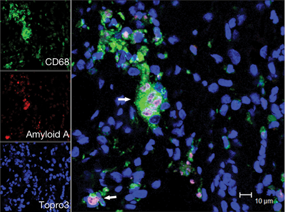
Multi-label confocal microscopy; ischial callosities of India-origin rhesus macaque (Macaca mulatta). Many intralesional cluster of differentiation (CD)68-positive macrophages contain intracytoplasmic amyloid A protein (arrows). Bar = 10 µm. Images for individual channels (macrophages with CD68/Alexa 488, green; amyloid with amyloid A/Fast Red 568, red; nuclei with TO-PRO-3/Alexa 633, blue).
For comparison and assessing the normal histology of ICs, histology was performed on ICs from 5 (3 females and 2 males) 2- to 4-year-old India-origin rhesus macaques as shown in Figure 2B. The epidermis is strikingly thick, having a 3–5 mm layer of well-organized stratum corneum. The stratum granulosum is 1–2 cell layers with abundant granules. The stratum spinosum and basale form serrated rete pegs associated with tall and regular dermal papillae. The subcutaneous tissue is divided into 3 distinct layers. The outer fibrous zone is similar to the superficial dermis in other parts of the body in that it contains a large amount of collagen and many lymphatic and blood vessels. A noteworthy observation is that there are large-sized arteries in this layer. The tunica intima of these arteries is lined by a layer of plump, elongated endothelial cells with a minimal internal elastic lamina. The tunica media is thick with an inner longitudinal, 1–2 cell layer, and an outer circular, 6–10 cell layer of smooth muscle. The tunica adventitia is half the thickness of the media and contains a large amount of collagen and many fibroblasts. The fibro-fatty zone is composed of ovoid adipose lobules circumscribed and separated by connective tissue septa that run from the outer to inner fibrous zone. The inner fibrous zone is formed by dense collagen with fewer blood vessels. The collagen of the inner fibrous zone merges with the underlying perichondrium of the ischial tuberosity.
Reactive amyloidosis, formerly secondary amyloidosis, develops in patients with chronic inflammation or infectious diseases. Chronic inflammation causes increased release of the proinflammatory cytokines IL-1, IL-6, and TNF, which induce a markedly increased synthesis of the acute phase proteins, such as SAA (a 104–amino acid protein), by hepatocytes in liver. The concentration of SAA can be 100- to 1,000-fold higher than normal. 14 The exact function of SAA is unclear, but it has been implicated as an opsonin for Gram-negative bacilli, as a factor in leukocyte chemoattraction, and as a component of cholesterol metabolism. 15 Macrophages rapidly endocytose and transport SAA into lysosomes. Normally, SAA is completely degraded without deposition of amyloid fibrils. However, in some patients, the C-terminal part of SAA is cleaved and then the N-terminal 66–76 amino acid fragments form a β-sheet configuration, which is resistant to proteolysis and accumulates in the extracellular space.13,15 In the current case, there are many CD68-positive macrophages containing intracellular AA protein in the ICs, suggesting an important role of mononuclear phagocytes in the pathogenesis of amyloidosis in nonhuman primates. Similar results have also been reported in human patients with chronic rheumatoid arthritis. 10 Monocyte-macrophage lineage cells play a dual role in the pathogenesis of amyloidosis by initiating amyloid fibril formation 6 and transferring misfolded proteins in blood circulatory system. 12
Reactive amyloidosis has been reported in many domestic and wild animals 18 as well as in nonhuman primates such as rhesus macaques, 2 chimpanzees (Pan troglodytes), 5 and marmosets (Callithrix jacchus). 7 Amyloid deposits are common in gastrointestinal tract, liver, spleen, and lymph nodes in rhesus macaques. 2 Reactive amyloidosis has been found associated with colitis, arthritis, lung mites, strictures of the cecocolic junction, osteoarthritis, and Shigella spp. infection, but no single recognized condition is consistently associated with this syndrome. 2 Reports from both mice and cheetahs suggest that reactive amyloidosis may not be simply a spontaneous disease associated with chronic inflammation but may also be linked to a natural prion-like, transmissible protein misfolding disease.8,19
The morphology of the ICs was first described 67 years ago. 9 Herein that description is expanded by providing distinguishing histologic characteristics of ICs in rhesus macaques using digital imaging. Ischial callosities are bilateral pads of thickened epidermis, located in the gluteal region, that are used for sitting on thin branches during feeding in the peripheral branch zone. Ischial callosities are found in cercopithecoid monkeys, gibbons, and siamangs.16,17 Histologically, the morphology of ICs of normal rhesus macaques is similar to that described previously. 9 However, it was found in the current study that, in addition to deeper large blood vessels, large arteries are also located in the superficial dermis just deep to the epidermis. These arteries have hypertrophic and hyperplastic smooth muscular walls that suggest they are able to withstand pressure during sitting. The thick muscular tunics may also have an important role in regulating local blood flow by active vasoconstriction and vasodilatation with innervation from the vasomotor nerve fibers. 1 The dermis is divided into 3 distinct layers. The outer fibrous zone is similar to the superficial dermis in other areas of skin that contain large amounts of collagen and many lymphatic and blood vessels. The fibro-fatty zone is composed of ovoid adipose lobules vertically circumscribed and separated by connective tissue septa. This structure likely cushions the pressure from the sitting posture.
In summary, this report documented a case of reactive amyloidosis associated with chronic ulcerative ischial callosititis in a rhesus macaque. AA substance was confirmed in intralesional CD68-positive macrophages by confocal microscopy, which suggests that CD68-positive macrophages have an important role in the pathogenesis of amyloidosis in the nonhuman primate. The histology of ICs in rhesus macaques was also reexamined in this report.
Footnotes
Acknowledgements
The authors thank all of the staff of the Tulane National Primate Research Center, especially Dr. Mostafa Bouljihad, Maurice Duplantis, Lifang Li, and Carol Coyne for the technical support.
a.
Dako North America Inc., Carpinteria, CA.
b.
Leica TCS SP2, Leica Microsystems Inc., Buffalo Grove, IL.
d.
Adobe Photoshop version 7.0, Adobe Systems Inc., San Jose, CA.
e.
Roche Diagnostics Corp., Indianapolis, IN.
f.
Molecular Probes, Life Technologies Corp., Carlsbad, CA.
Declaration of conflicting interests
The author(s) declared no potential conflicts of interest with respect to the research, authorship, and/or publication of this article.
Funding
The author(s) disclosed receipt of the following financial support for the research, authorship, and/or publication of this article: This work was supported by the National Center for Research Resources and the Office of Research Infrastructure Programs (ORIP) of the National Institutes of Health through grant number OD011104.