
Abstract
A case of granulocytic sarcoma originating from an eosinophilic lineage is described in a 5-year-old, mixed-breed, female pig. The pig had been originally sent to slaughter in a good plane of nutrition and without displaying clinical disease. At gross examination, green masses were observed in several bones, especially vertebrae, sternum, pelvis, and long bones such as femur and humerus. Similar masses were seen in skeletal muscles, lymph nodes, and kidneys. Cytology revealed large numbers of round cells with round nuclei and scant cytoplasm (myelocytes); some of these cells had a fine eosinophilic granularity to their cytoplasm (eosinophil myelocytes). Histologically, the neoplastic cells formed sheets that completely obliterated the normal architecture of subperiosteal bone marrow. The cytoplasm of the neoplastic cells stained strongly by Sirius red stain of eosinophil and was positively marked by immunohistochemistry using an anti-myeloperoxidase antibody. The association of gross examination, cytology, histology, histochemistry, and immunohistochemistry findings is consistent with a diagnosis of eosinophilic granulocytic sarcoma.
Granulocytic sarcoma is a morphologic presentation of myeloid sarcoma, a hematopoietic neoplasm affecting bones or extramedullary sites 1 ; in this latter case, the growth is also referred to as extramedullary myeloid tumor, myeloblastoma, 7 or myelosarcoma. 2 Granulocytic sarcomas may originate from variably differentiated precursors, from both neutrophilic and eosinophilic lineages. Such sarcomas tend to occur grossly as characteristic green masses; therefore, the sobriquet “chloroma,” derived from the Latin transliteration of the Greek khlorós, meaning green, was given to the neoplasm. 18
In human beings, granulocytic sarcomas precede or occur concomitantly with acute myeloid leukemia, chronic myeloproliferative disorders, or myelodysplastic syndromes but are also described without association with any other hematologic disturbance.1,2,9 In the veterinary literature, granulocytic sarcomas are mentioned affecting dogs, cats, cattle, 19 a rabbit, 15 and a pig. 6 In dogs and cats, the more consistently involved organs are the lungs, intestine, skin, 20 lymph nodes, and liver. 18 In one of the few reports found in the veterinary literature, this tumor is mentioned as a mass in the neck of a Bull Terrier dog. 11 In cattle, skeletal muscle is characteristically affected. 20 In the case reported in a rabbit, the granulocytic sarcoma involved the skin, subcutaneous tissue, and skeletal muscle of the perineum. 15 In the pig, the tumor involved liver, kidneys, and mesenteric lymph nodes. 6 Differently from what occurs in human patients, animals affected by granulocytic sarcomas are almost always aleukemic; however, progression to a leukemic may occur. 20 The current report describes gross findings, cytology, histopathology, histochemistry, and immunohistochemistry of a multicentric eosinophilic granulocytic sarcoma affecting a pig.
Material and methods
A mixed-breed, 5-year-old, female pig in good plane of nutrition and with no detectable clinical manifestation was sent to slaughter. The federal meat inspector detected “areas of green discoloration in the carcass” and sent large tissue samples to be examined at the Veterinary Pathology Laboratory at the Federal University of Santa Maria, Brazil. Cytopathologic slides obtained from the mass were air-dried and stained by rapid panoptic kit, a based on the principle of color hematology established by Romanowsky. Bone samples were decalcified in formic acid prior to processing for histopathology. Multiple samples of bones, skeletal muscles, lymph nodes, and kidneys were fixed in 10% buffered formalin; paraffin embedded, sectioned at 4 µm, and stained by hematoxylin and eosin. Selected sections of bone marrow were stained by Sirius red using the eosinophil technique. For this protocol, the online modification of the original method published by Llewellyn, 10 Sirius red was dissolved in 50 ml of absolute ethanol, 45 ml of double distilled water, and 1 ml of 1% NaOH. NaCl (20%) was added until slight precipitation had occurred. Subsequently, the solution was filtered and sections deparaffinized, stained with hematoxylin for 8 min, differentiated in running tap water, and treated with 70% chloride ethanol for 2 sec. The sections were stained with Sirius red solution at room temperature for 24 hr. After dehydration with increasing concentrations of ethanol, the sections were mounted. The characterization of the eosinophil granulocytes was performed according to its typical morphology and strongly red-stained granules. Sections of eosinophilic meningitis in porcine (salt poisoning) were used as positive control. Such lesions consist of an eosinophilic meningoencephalitis with typical perivascular cuffing of eosinophils around capillaries of cortical parenchyma. 16
To determine the neoplasm origin, different sections from bone marrow were evaluated by immunohistochemistry b for myeloperoxidase of the neutrophil and eosinophil (MPO-7, b 1:400), lysozyme b (polyclonal, 1:100), cluster of differentiation (CD)117 b (polyclonal, 1:50), CD3 (UCHT1, b 1:800), and CD79α (HM57, b 1:200). Serial sections (3 µm) were cut from the tissue blocks for histochemical staining and immunohistochemistry. Sections were incubated at 37°C for 60 min with the primary antibody, diluted briefly, and sections were then dewaxed and rehydrated. Endogenous peroxidase activity was blocked with H2O2 3% in distilled water. Antigen retrieval was by microwaving (10 min at full power) in Tris–ethylenediamine tetra-acetic acid (pH 9.0). Sections were incubated at 37°C for 60 min with the primary antibody. The secondary reagent was biotinylated followed by streptavidin–peroxidase. b Substrate development was with 3,3′-diaminobenzidine c (DAB). Sections were counterstained lightly with Mayer hematoxylin and then coverslipped. Histologic sections of an acute myeloid leukemia in a cat were used as positive controls for the CD117 and MPO-7 antibodies. A normal lymph node was used as positive controls for the CD3 and CD79α antibodies, and a histiocytic sarcoma in a dog as positive control for lysozyme. For negative controls, the same sections were used, utilizing phosphate buffered saline with Tween instead of the primary antibody.
Results
Grossly, over the ribs and beneath the parietal pleura there were green, smooth, and opaque, irregularly contoured, noncircumscribed, soft, homogenous, slightly salient areas. Light green masses that partially or completely obliterated the bone marrow architecture were observed at the cut surfaces of some ribs and several vertebrae and sternebrae. In all lumbosacral vertebrae, typically a subperiosteal presentation was promptly observable (Fig. 1). In the cut surface of long bones (femora and humeri), the same pattern of presentation was observed in the metaphysis (Fig. 2). In the kidneys, there were multiple, 1–3 mm in diameter pink to light green, soft, irregularly shaped nodules. There was a homogeneous aspect to the cut surface of these nodules. At cut surfaces of popliteal and iliac lymph nodes, there were pink or light green areas. In one of the lymph nodes (internal iliac), the cut surface was diffusely light green and crisscrossed by red serpiginous lines (Fig. 3).
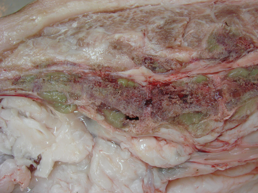
Pig affected by granulocytic sarcoma. Cut surface of a longitudinal section throughout the lumbosacral spine. There are discolored subperiosteal green areas in the vertebral bodies and spinous processes.
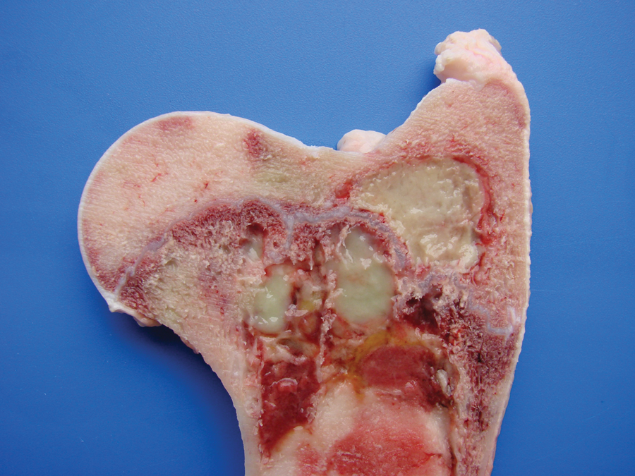
Pig affected by granulocytic sarcoma. Cut surface of a longitudinal through the femur. Green-yellowish soft masses partially obliterate the metaphysis.
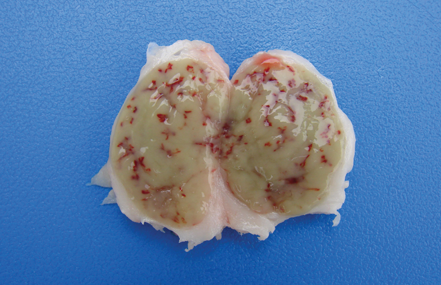
Pig affected by granulocytic sarcoma. The cut surface of internal this iliac lymph node is completely effaced by a homogenous pink to light green mass. Congested blood vessels crisscross the homogenous appearing mass.
Cytological exam of the mass revealed large numbers of round cells approximately 20–30 µm. These round cells had a round, oval, or reniform nuclei that contained slightly clumped chromatin and did not display prominent nucleoli. The cytoplasm was scant, and a fine eosinophilic granularity could be observed in the cytoplasm of some neoplastic cells but not in others. Such cells were interpreted as myelocytes belonging to the eosinophil lineage (Fig. 4A). Histologically, a sheet of round cells with a virtually imperceptible stroma obliterated the bone marrow (Fig. 4B). The bone marrow tissue surrounding these cellular sheets was replaced by fibroblasts and collagen (myelofibrosis). At these sites, there was reabsorption of compact bone and moderate periosteal reaction. Noncircumscribed foci of myeloid cells were observed dissecting, and at times replacing, the renal tubules. Sections of these areas revealed that the neoplastic cell cytoplasm stained red by the Sirius red eosinophil technique (Fig. 4C) and demonstrated strong cytoplasmic positivity for myeloperoxidase by the immunohistochemistry stain (Fig. 4D). All sections were negative for all the other immunohistochemical markers used.
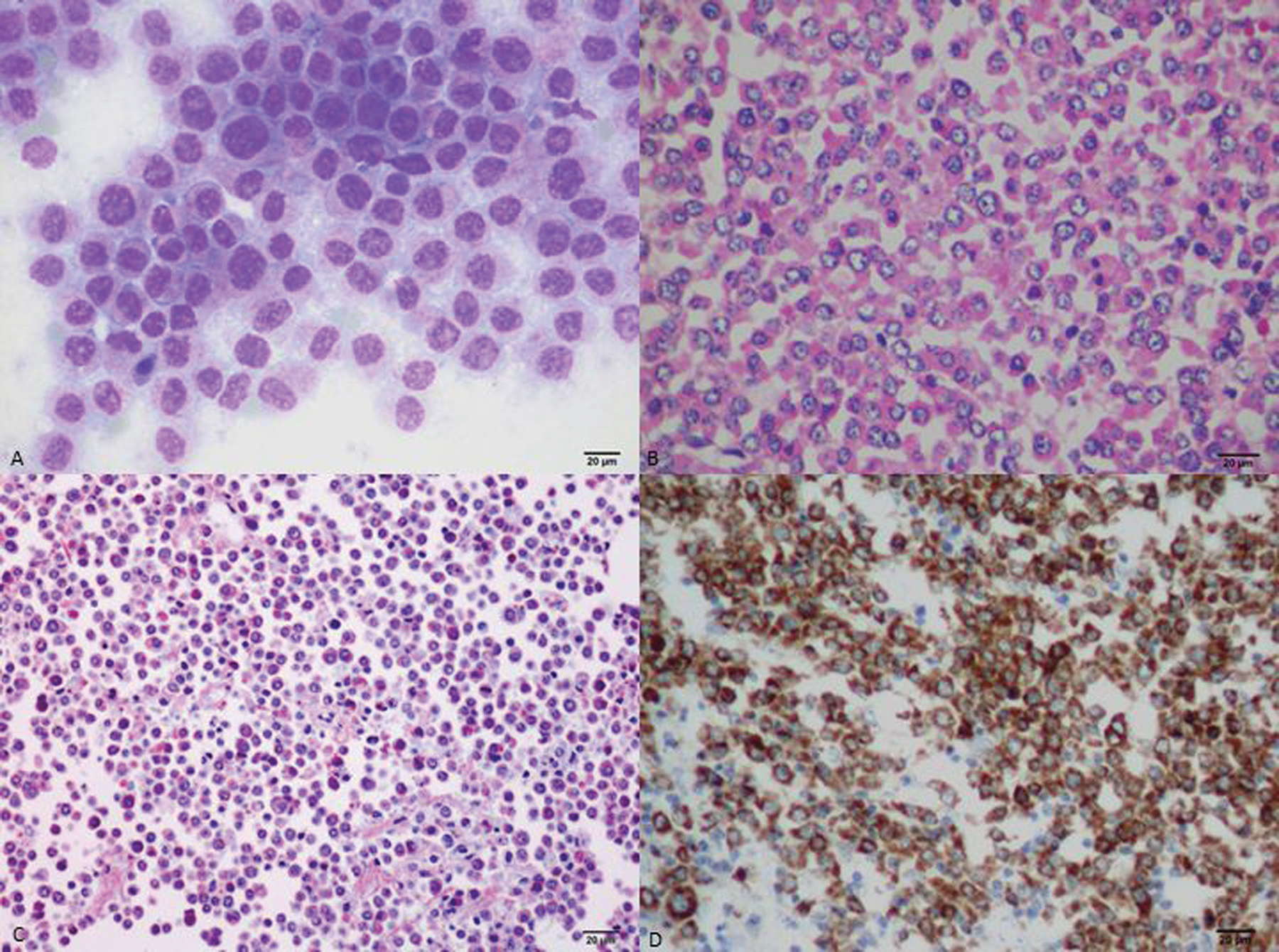
Pig affected by granulocytic sarcoma.
Discussion
Granulocytic sarcoma was suspected in the current case based on the presence of light green masses in the gross inspection of the carcass and viscera, a typical aspect of this tumor. The anatomical distribution of the lesions observed in the current case is quite similar to that described for human granulocytic sarcomas, in which the occurrence is primarily subperiosteal and involves mainly the ribs, sternum, and pelvis. 1 The microscopic presentation pattern, observed both at cytological and histological examination consisting predominantly of precursor with myelocyte differentiation, allowed the presumptive diagnosis of well-differentiated granulocytic sarcoma. Immunohistochemistry results established the cell origin as of the granulocytic lineage, and histochemistry determined that the cells were possibly of eosinophil lineage, definitively confirming the diagnosis suspected at gross examination.
The definitive diagnosis of myeloid sarcomas is based on the association of phenotypic (cytology, histology, cytochemistry, and histochemistry) and immunophenotypic (immunocytochemistry and immunohistochemistry) aspects. 17 An immune phenotype of neoplastic cells positive for myeloperoxidase is the hallmark for granulocytic sarcoma.1,2 Other antibody markers have reportedly yielded positive results when applied to human cases of granulocytic sarcoma, including CD13, CD33, CD117, 1 CD15, CD68, 2 CD43, 12 and lysozyme. 14 However, lysozyme and CD68 are also marked in cases of monoblastic sarcoma, a less common form of myeloid sarcoma. 1
In the porcine tumor described herein, the negative staining for CD117 could be explained by both the predominance of myelocytes and absence of myeloblasts, which are precursor cells that express this antigen. The negative staining for lysozyme was somewhat expected because swine granulocytes have been described as negative for this marker, 3 whereas porcine monocytes and/or macrophages are strongly positive, 5 similar to what is described in human beings, a species in which lysozyme is the choice marker for monocytes and/or macrophages. Based on this species-specific feature, the negative reaction to lysozyme further helps to differentiate granulocytic sarcoma from monoblastic sarcoma.
In human patients, several different forms of lymphoma presentation are often confused with myeloid sarcoma 12 and thus should be the main tumors to be included in the differential diagnosis. 2 In the current case, the differentiation was made based on the following aspects: histological evidence of cytoplasmic eosinophilia, fine granularity observed in the cytoplasm of neoplastic cells when examined in cytological preparations, occurrence within the neoplasm of more mature eosinophil precursors amidst myeloblasts, and the immunohistochemistry marking. Furthermore, the observation of light green masses that partially obliterated the bone marrow from several flat and long bones prompted the suspicion of granulocytic sarcoma, as this is the only hematopoietic neoplasm that expresses this typical color. The green discoloration observed in fresh tissue specimens is due to the presence of myeloperoxidase and substantially helps in the diagnosis of granulocytic sarcoma. 2
Few lesions could grossly resemble sarcoma granulocytic. Such lesions include chlorellosis, a granulomatous inflammatory reaction observed in human beings 13 and animals,8,22 and eosinophilic myositis, which is frequently associated with Sarcocystis spp. infection in cattle 21 and has been described in a pig. 4 The association of gross examination, cytology, histology, histochemistry, and immunohistochemistry findings is consistent with a diagnosis of eosinophilic granulocytic sarcoma.
Footnotes
a.
Interlab LB, Laborclin, Pinhais, PR, Brazil.
b.
LSAB+ System-HRP, Dako North America Inc., Carpinteria, CA.
c.
Sigma-Aldrich, St. Louis, MO.
Declaration of conflicting interests
The author(s) declared no potential conflicts of interest with respect to the research, authorship, and/or publication of this article.
Funding
The author(s) disclosed receipt of the following financial support for the research, authorship, and/or publication of this article: This work was financially supported through a fellowship from the Brazilian Coordination for the Improvement of Post Secondary Education (CAPES) within the National Post-Doctoral Program (PNPD).