
Abstract
Cerebrospinal fluid (CSF) has been examined as a possible source for preclinical diagnosis of prion diseases in hamsters and sheep. The present report describes the detection of chronic wasting disease (CWD) in the CSF of elk and evaluates its usefulness as an antemortem test for CWD. The CSF from 6 captive and 31 free-ranging adult elk was collected at necropsy and evaluated for the presence of the abnormal isoform of the prion protein that has been associated with CWD (PrPCWD) via protein misfolding cyclic amplification. Additionally, the obex from each animal was examined by immunohistochemistry (IHC). Four out of 6 captive animals were CWD-positive and euthanized due to signs of terminal CWD. The remaining 2 were CWD negative. None of the 31 free-range animals showed overt signs of CWD, but 12 out of 31 tested positive for CWD by IHC. Protein misfolding cyclic amplification detected PrPCWD from 3 of the 4 captive animals showing clinical signs of CWD and none of the nonclinical animals that were CWD positive by IHC. The data suggests that CWD prions can be detected in the CSF of elk, but only relatively late in the course of the disease.
Keywords
Chronic wasting disease (CWD) is a fatal, infectious neurodegenerative disease that affects wild and captive cervids. It is a transmissible spongiform encephalopathy, or prion disease. Development of an antemortem test for detecting the misfolded prion protein associated with CWD (PrPCWD) in nonclinical animals would be useful for wildlife and captive population management strategies. To date, preclinical testing for PrPCWD utilizes immunohistochemistry (IHC) of the palatine tonsils or rectal lymphoid tissues in cervids.18,21,24 However, IHC does not routinely detect very early cases of CWD in these tissues.17,20 Various fluids, such as saliva, blood, urine, and cerebrospinal fluid (CSF) have been evaluated in animals for their use in preclinical antemortem detection of abnormal prions.4,6,10,22 Due to the minute concentrations of infectious proteins in saliva, blood, and urine, detection of the misfolded infectious prion protein (PrPres) has been limited or unsuccessful without the use of protein misfolding cyclic amplification (PMCA), which increases the concentration to a detectable level, and is orders of magnitude more sensitive for detecting PrPCWD than commonly used methods such as Western blotting and IHC, 7 enabling detection of minute amounts of infectious prions in animal tissues and environmental samples. 9
In the current study, the usefulness of PMCA to detect PrPCWD in CSF from wild and captive elk (Cervus canadensis nelsoni) was evaluated as a potential tool for early detection of PrPCWD. The PMCA in vitro technique amplifies minute amounts of infectious prions by rapidly converting normal cellular prion protein (PrPc) to PrPres.11,16 Cerebrospinal fluid (5–15 ml) was collected at necropsy from 6 captive and 31 free-ranging, adult elk. Four of the 6 captive elk were euthanized due to positive CWD rectal biopsy results and terminal clinical signs. Cerebrospinal fluid was collected during the postmortem examination just prior to removal of the head. The head was flexed dorsally, the ventral muscles severed, the ventral aspect of the foramen magnum was exposed, and CSF collected with an 18-gauge needle attached to a 12-cc syringe. Great care was taken to ensure that the extraction site of the CSF was clean and without blood contamination. Cerebrospinal fluid was frozen at –80ºC until samples were analyzed using PMCA.
The CWD status of each elk was determined by examination of the obex at the level of the dorsal motor nucleus of the vagus nerve by using previously described IHC techniques. 18 The obex from all CWD-positive elk was scored as previously reported. 19 Briefly, nuclear regions and axonal tracts were evaluated and scored 0–10 for the severity of spongiform degeneration and accumulation of PrPCWD immunoreactivity (IR). Tissue samples showing no detectable IR received a score of zero, and samples showing heavy immunoreactivity in all nuclei and axons received a score of 10.
Brain homogenate from transgenic Tg5037 mice, which express a 5-fold increase in elk PrPc, were used as a source of PrPc substrate in the PMCA. reactions. 2 A 10% (wt/vol) normal brain homogenate (NBH) was prepared as previously described, utilizing a homogenizer a at setting 6 for 5 min. 9 Equivalent volumes of CSF and NBH (25 µl of each) were added to individual 200-µl PCR tubes sealed with plastic paraffin film with a paper backing and sonicated b with 40-sec pulses at 37°C, power setting 7, every 30 min for 24 hr. After each 24-hr round, 25 µl of each sample was mixed with 25 µl of fresh NBH and transferred to a fresh tube. A positive amplification control of 1:100,000 CWD-positive brain homogenate was also amplified with each sample group as were NBH-negative controls. Duplicate samples were amplified in 3 independent replicate experiments for a total of 6 samples. Upon completion of 6 rounds, amplified samples were digested with proteinase K c and immunoblotted as previously described. 9 The proteinase K–resistant core of PrPres appears on Western blots as bands shifted below those seen for full-length PrP. Only samples in which signal was detected in 2 of the 3 replicate experiments were considered positive.
The misfolded prion protein associated with CWD was detected in 3 out of 4 captive, IHC-positive clinical elk (Fig. 1, Table 1). However, the nonclinical, wild CWD IHC-positive elk did not yield detectable CWD in the CSF (Table 1). These animals were in generally good health, as indicated by body and coat condition.
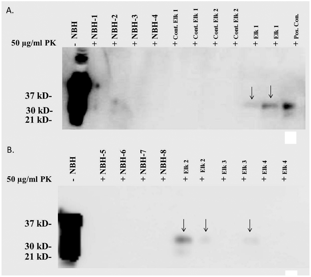
Western blots of captive elk cerebrospinal fluid after 6 rounds of protein misfolding cyclic amplification. NBH = normal brain homogenate negative controls. Positive control 1:100,000 dilution of chronic wasting disease–positive brain homogenate. Chronic wasting disease was detected in captive elk 1–3. Representative of 3 replicates.
Source, and protein misfolding cyclic amplification (PMCA) and immunohistochemistry (IHC) results for 6 captive and 31 free-ranging adult elk (Cervus canadensis nelsoni).
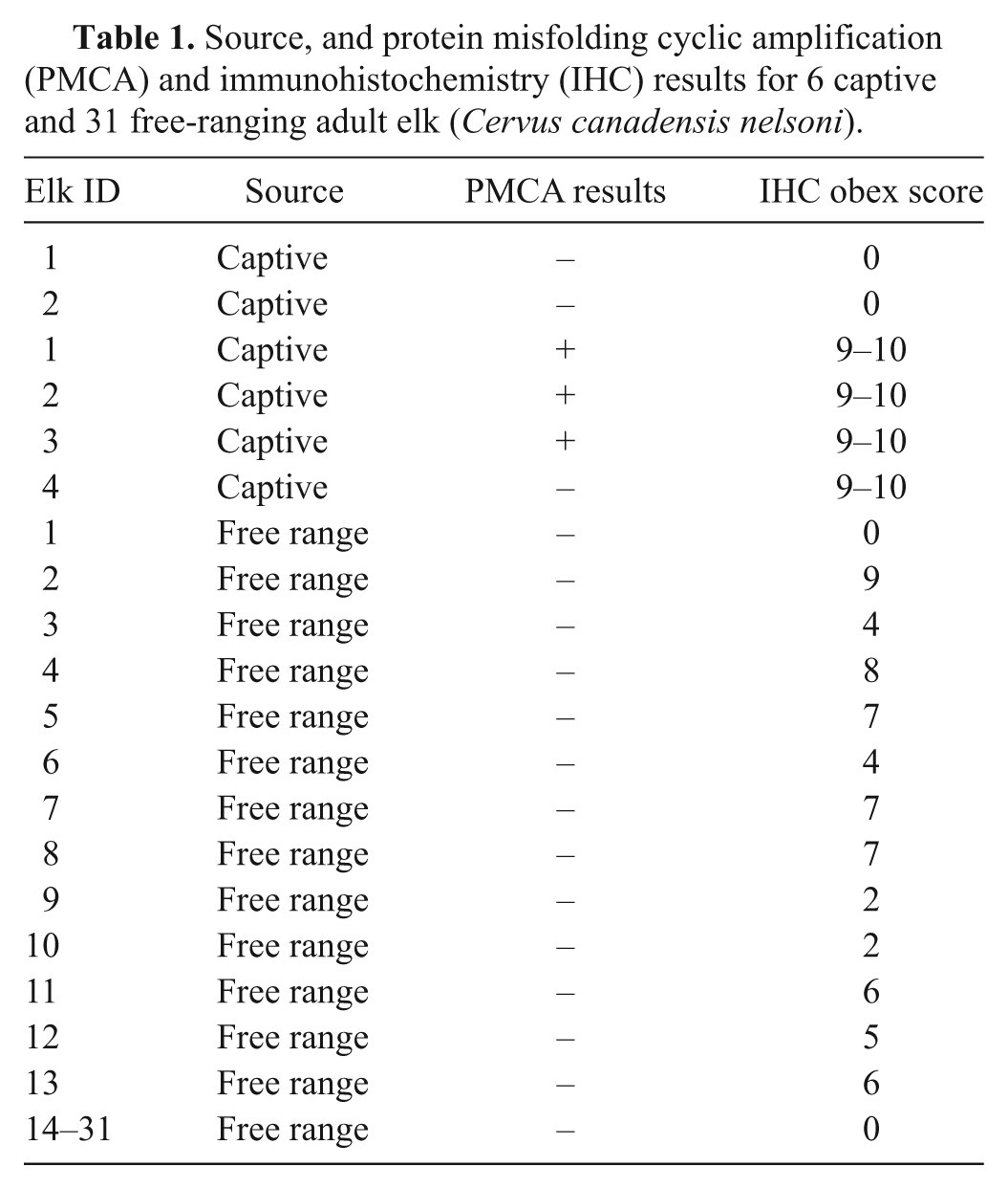
Although it is unknown how long the 3 PMCA-positive elk had been infected, their obex scores ranged from 9 to 10 (Table 1), whereas the obex scores of 12 IHC-positive but PMCA-negative nonclinical elk ranged from 2 to 9. Negative captive elk had no detectable PrPCWD and therefore had scores of 0. Previous studies involving sheep and hamsters demonstrated that PrPres could be detected in the CSF.3,10 However, the present findings are not readily compared to those previous results, which were derived exclusively from terminal, clinical animals.3,10 Transport of prions to the brain from distal regions such as the gut has been shown to occur via movement along peripheral nerves and via the blood. 14 Studies conducted in naturally and orally exposed sheep to scrapie suggest that prions migrate from the gut-associated lymph tissue to the central nervous system along nerve tracts to the spinal cord and brain. 1 A similar pattern of PrPCWD deposition has been observed in deer.8,12,13,23 Chronic wasting disease has been detected in blood, 6 and studies done in sheep demonstrate that intravenous inoculation with scrapie results in neuroinvasion and disease indistinguishable from other routes of infection. 15 It has been postulated in the literature that the circumventricular organs (CVO) in the brain could be a site of entry for prions from the blood into the brain. 15 The CVO are specialized structures that line the third and fourth ventricles of the brain and are considered to be absent of the “blood-brain barrier.” 5 As has been seen with scrapie in sheep, 15 PrPCWD can be detected in the CVO, with the exception of the choroid plexus, in nonclinical deer and elk with obex scores greater than 9 (with the obex scoring system, clinical signs are usually not observed until obex scores of 9 or 10 are achieved (unpublished data, 2005). Early accumulation of scrapie in the CVO has prompted some researchers to propose that because the CVO is devoid of the blood-brain barrier, prions can cross unhampered from the blood to the brain and then on to regional lymph nodes. 15 The data suggests that in the earlier stages of disease, the intact ependymal and CVO appear to sequester PrPCWD and prevent infiltration of PrPCWD into the CSF. It is therefore reasonable to propose that in the latter stages of disease, with obex scores greater than 9, the PrPCWD begins to leak through the epemdyma, allowing PrPCWD access to the CSF. Leakage is not thought to be caused by autolysis at necropsy, as the postmortem interval of the positive wild elk was 3–4 hr and only 1 hr for the captive elk. In conclusion, the present data suggests that PrPCWD infiltrates the CSF at relatively late stages of CWD and that PMCA of CSF is not useful as a diagnostic tool in nonclinical elk. Work needs to continue to investigate other tissues such as saliva and lymph node biopsies as PMCA antemortem diagnostic specimens.
Footnotes
Acknowledgements
The authors thank the North American Deer Farmers Association for research support.
a.
Bullet Blender® homogenizer, Next Advance Inc., Averill Park, NY.
b.
Model 3000MP, Misonix Inc., Farmingdale, NY.
c.
Roche, Madison, WI.
Declaration of conflicting interests
The author(s) declared no potential conflicts of interest with respect to the research, authorship, and/or publication of this article.
Funding
The author(s) received no financial support for the research, authorship, and/or publication of this article.