
Abstract
A captive, 20-year-old female elk (Cervus elaphus nelsoni) euthanized due to progressive lameness and weight loss was presented to Colorado State University Veterinary Diagnostic Laboratory for postmortem examination. Within the uterus there was a poorly demarcated, multilobulated mass measuring 10 cm in diameter. Histologically the tumor was an adenocarcinoma. Histologic examination of the ovaries revealed unilateral metastasis. A focal, 1-cm diameter adenocarcinoma was identified within the abomasum; this tumor was histologically distinct from the neoplasm found in the uterus and ovary. Although this elk had a history of experimental reproductive treatments, including leuprolide, gonadotropin-releasing hormone vaccine, and Brucella abortus vaccination, it was most likely that both tumors represent spontaneous, independent neoplastic transformations and were unrelated.
A captive, 20-year-old female Rocky Mountain elk (Cervus elaphus nelsoni) was euthanized due to progressive lameness and weight loss of 18 to 24 months’ duration. The elk had been wild caught as a calf in 1986 and hand raised at the Colorado Division of Wildlife, Foothills Wildlife Research Facility in Fort Collins, Colorado, where it had been used in multiple contraceptive studies as an adult. From 2000 to 2006 an enlarged cervix and a uterine mass were noted on annual rectal palpation of the reproductive tract. The size of the mass did not change over this period. No antemortem diagnostics were conducted on the cervix/ uterus because the mass was assumed to be scar tissue secondary to a severe dystocia that occurred in 1990; ultrasonography and palpation were not conducted until 2000.
At necropsy, the cow was lean (body condition score = 2.5/57). Severe, chronic osteoarthritis was found within both stifle joints. Degenerative joint disease was less severe in the right tarsal joint. Over the left hip there was a multilobulated, well-encapsulated abscess approximately 6 cm in diameter at the widest point. The left deep inguinal lymph node was approximately 3 times normal size and edematous. A multilobulated mass in the uterine body extended into the proximal portion of both horns. The mass expanded the uterine submucosa, and the enlarged uterus measured up to 10 cm in diameter at the widest point. On cut surface the mass was firm and tan and had foci of necrosis centrally. Both uterine horns were patent; however, the lumen of the left horn was constricted by the mass to approximately one fifth of its original diameter. A nodule, approximately 1 cm in diameter, was in the abomasal submucosa. On section the mass was pale tan, firm, and homogenous.
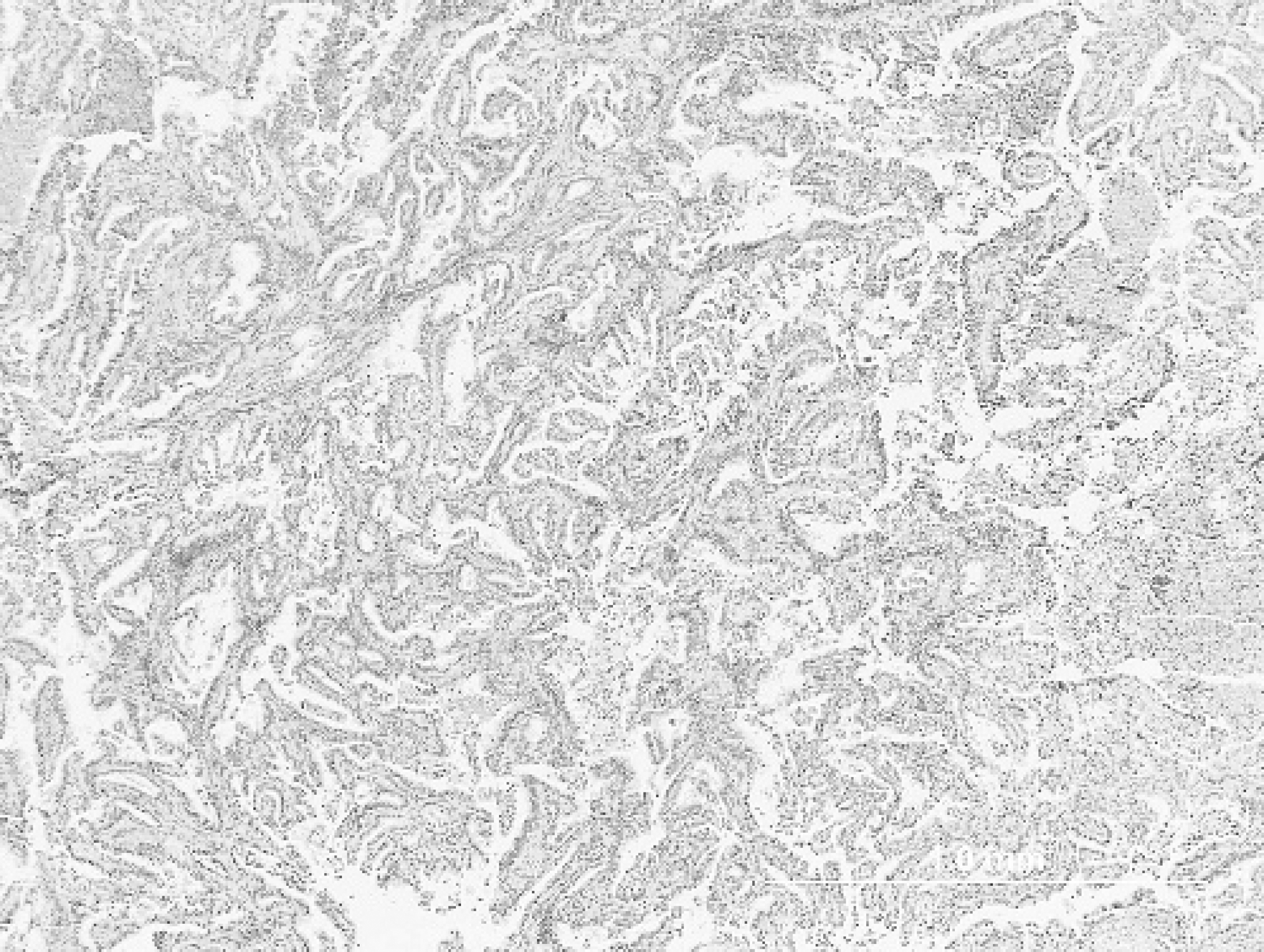
Uterus; elk. Adenocarcinoma forming papillary projections that extend into the uterine lumen. Hematoxylin and eosin. Bar =1.0 mm.
Histologically, the uterine mass was composed of epithelial cells forming fronds and papillary projections (Fig. 1). Neoplastic cells were polygonal but most often cuboidal to short columnar with distinct cell borders and a moderate amount of eosinophilic cytoplasm (Fig. 2). Nuclei were round to oval with coarse clumped chromatin. Neoplastic cells showed marked anisocytosis and anisokaryosis. The numbers of mitotic figures ranged from 2 to 6 per 400− field. The neoplastic cell population was supported by variable amounts of fibrovascular connective tissue stroma. Random foci of necrosis were found throughout the tumor. The right ovary had multifocal to coalescing aggregates of neoplastic cells like those present in the uterine mass (Fig. 3). Neoplastic cells and associated scirrhous response effaced much of the ovarian parenchyma, and no follicles were present. The left ovary was devoid of neoplastic cells, but primordial and atretic follicles were observed.
The abomasal mass was composed of nests of neoplastic epithelial cells that extended from the mucosa, through the basement membrane, and into the submucosa (Fig. 4). Cells were predominantly columnar with abundant basophilic cytoplasm (Fig. 5). Nuclei were round to oval, basally oriented, with coarse clumped chromatin. There was mild to moderate anisocytosis and anisokaryosis. Mitotic figures were uncommon, ranging from 0 to 1 per 400− field. A marked scirrhous response surrounded the neoplastic cells. Relatively normal-appearing parietal cells were scattered among the population of neoplastic cells; however, chief cells were largely absent.
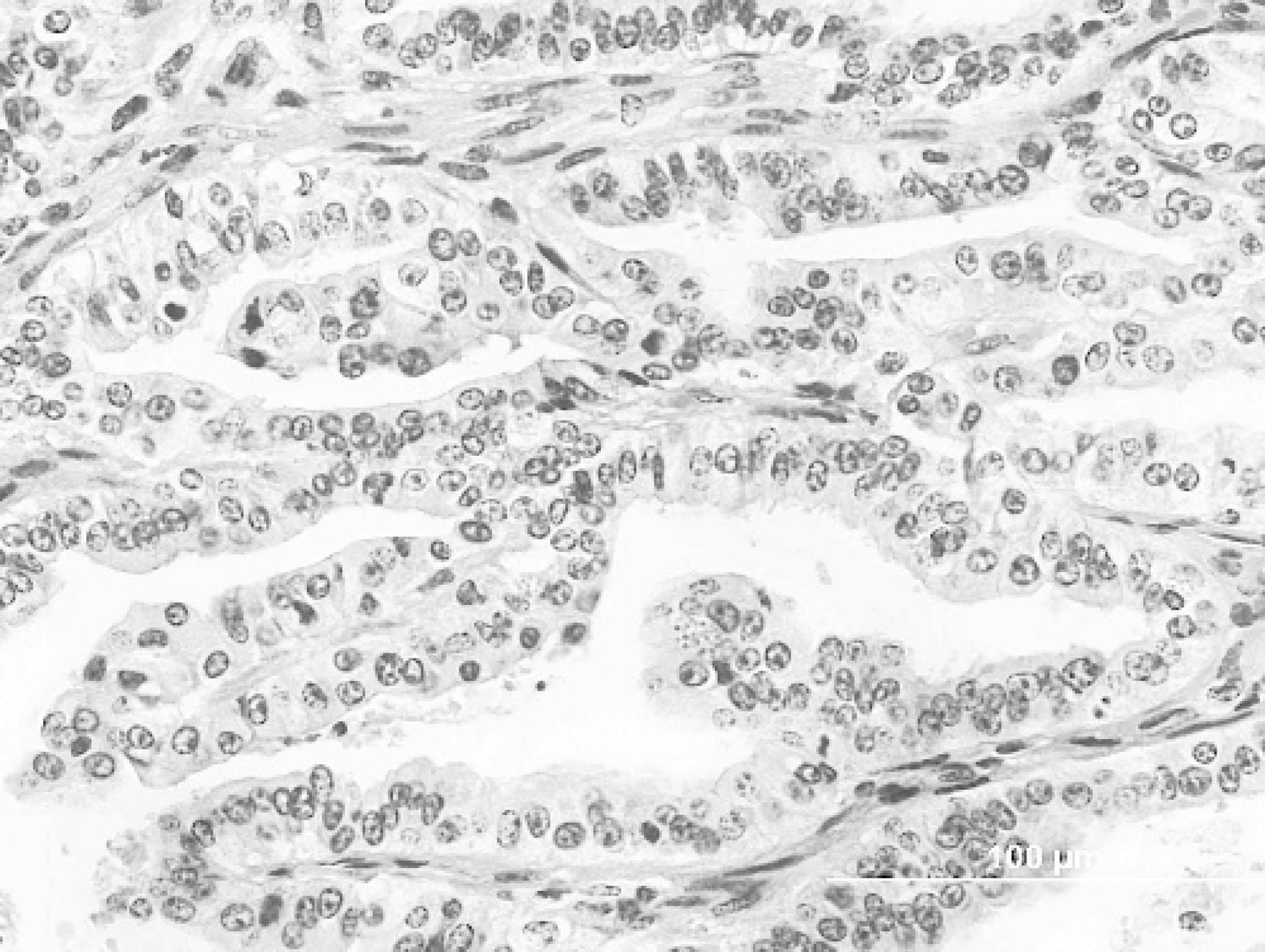
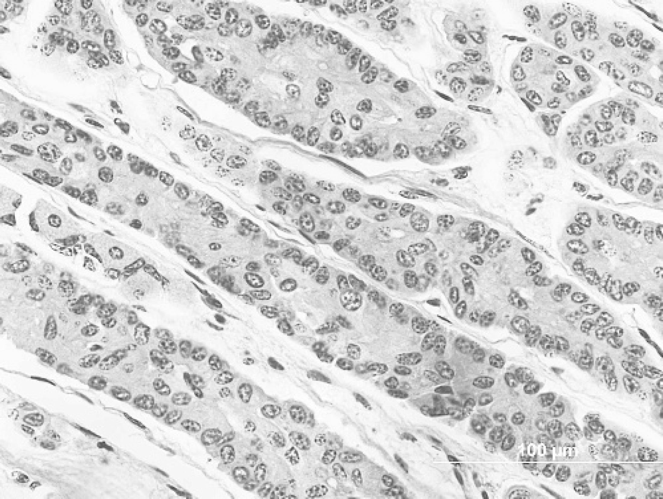
Uterus; elk. Neoplastic cells are cuboidal to columnar and often pile. Hematoxylin and eosin. Bar = 100 μm.
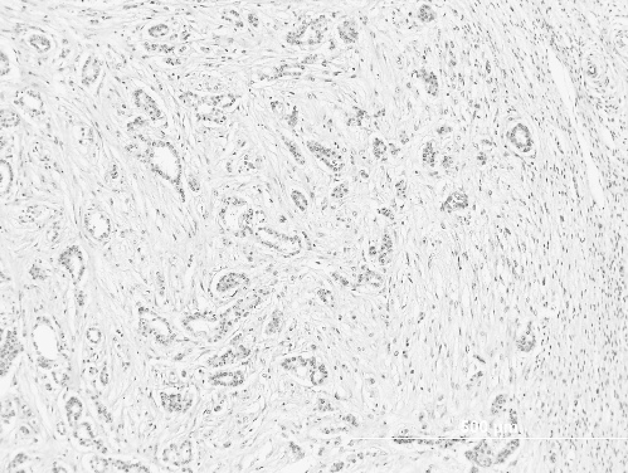
Ovary; elk. Metastases from the uterine adenocarcinoma efface much of the ovarian parenchyma and exhibit a marked scirrhous response. Hematoxylin and eosin. Bar = 500 μm.
Given the history oí a palpable uterine mass oí approximately 6 years’ duration, it was postulated that both the uterine and the ovarian tumors originated in the uterus. Uterine adenocarcinomas have been reported in numerous domestic species, including horses, 3 cattle, 15 dogs, 26 cats, 16 sheep, 25 New World camelids, 18 potbellied pigs, 11 and nonhuman primates. 24 The incidence of uterine tumors in most domestic species is low. 14 In contrast, uterine adenocarcinomas are common in rabbits, with 79% prevalence in animals older than 5 years of age. 9 It has been proposed that this may be due to prolonged estrogen stimulation in these induced ovulators. Likewise, rats and humans with endogenous or exogenous estrogen excess were reportedly at increased risk of developing the tumor. 17,21 Organochlorines were proposed as a risk factor for neoplasia, including uterine adenocarcinomas, in beluga whales of the St. Lawrence estuarv. 12
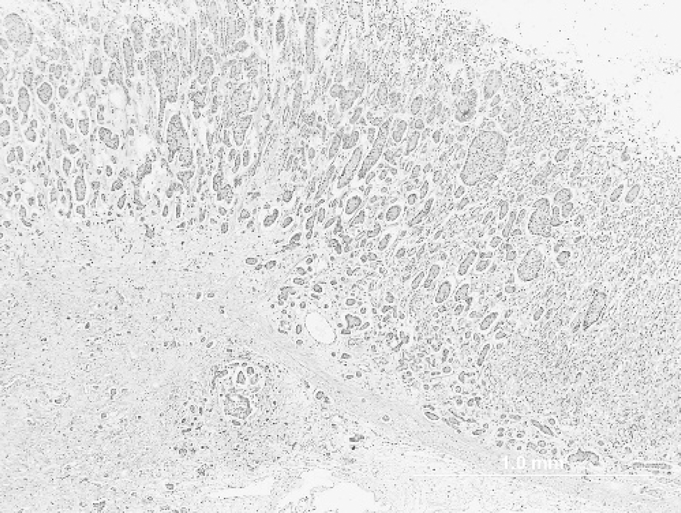
Abomasum; elk. Adenocarcinoma effaces normal mucosa and extends into underlying submucosa. Hematoxylin and eosin. Bar = 1.0 mm.
Between 1990 and 2004, this elk received multiple injections of the short-acting gonadotropin-releasing hormone (GnRH) agonist leuprolide in variable time-release formulations. Treatment with leuprolide suppressed luteinizing hormone (LH) release from the anterior pituitary for 3 to 6 months and prevented pregnancy for 1 breeding season in elk. 1 This elk responded similarly to her cohorts with respect to endocrine hormone profiles but was deemed unfit for pregnancy trials due to the abnormal cervix and uterus. In addition to the GnRH agonist studies, in 1998 it was injected with Brucella abortus strain 19 in a vaccination study but not challenged with wild-type B. abortus. At necropsy the elk still had a positive titer for B. abortus. In 2005 this cow was vaccinated against GnRH with an experimental contraceptive vaccine, resulting in high anti-GnRH antibody titers, which presumably decreased LH release; however, the levels of LH were not measured. The vaccine was given intramuscularly over the left hip, resulting in the abscess found at necropsy.
Despite its being subjected to various experimental reproductive treatments, there was little evidence to suggest that these protocols were related to the development of uterine adenocarcinoma in this elk. Both the GnRH agonist, leuprolide, and the GnRH vaccine decrease gonadotropin secretion from the anterior pituitary gland and have been used to suppress steroidal hormones and treat various estrogen- and androgen-sensitive adenocarcinomas in humans. 6,23 There are, however, sporadic reports of endometrial adenocarcinoma developing shortly after suspending GnRH agonist treatment in humans, 8 as occurred in this elk. It is unlikely that suppressing the action of GnRH either with an agonist or via vaccination could increase reproductive steroid hormone levels, which were associated with uterine adenocarcinomas. 5
Abomasum; elk. Individual neoplastic cells within the abomasal mass are most consistent with chief cells. Hematoxylin and eosin. Bar = 100 μm.
In free-ranging wildlife uterine adenocarcinomas have been described in a beluga whale 12 and a bottlenose dolphin. 22 In both of these marine mammals, marked peritoneal carcinomatosis was also noted. The first report of this tumor in cervids was in a 4-year-old captive sika deer that had a focal uterine mass with peritoneal carcinomatosis and metastases to the lung, liver, spleen, urinary bladder, lymph nodes, diaphragm, and muscular layers of the forestomachs. 20 Uterine adenocarcinomas were often multicentric in rats and rabbits but solitary in cattle and other species. 9 In this elk the uterine adenocarcinoma was focal, and evidence of carcinomatosis was not noted on gross or histologic examination. Metastases to the ovaries were not identified in the sika deer; however, they have been reported in cattle. 10
Histologically the abomasal adenocarcinoma was distinct from the ovarian and uterine tumors; therefore, it was considered to be a second, independent neoplasm. There was progressive loss of chief cells at the transition of normal to neoplastic tissue. Cytoplasmic basophilia of the abomasal carcinoma cells suggested possible chief cell origin. Abomasal adenocarcinomas are rare in cattle 2 and associated with a marked scirrhous response, 4,19 as seen in this case. Tumors of the glandular stomach, including carcinomas, are common in the dog 14 and have been reported in other species, including cats, horses, and pigs. 13 Animals with gastric carcinomas usually show clinical signs late in the course of disease as a result of severe carcinomatosis, metastases, or clinical disease secondary to the size and obstructive nature of the mass. 14,19 In this elk, the mass was small and considered an incidental finding.
Uterine and gastric adenocarcinomas are relatively uncommon tumors of domestic ruminants and previously unreported in elk. The incidence of these tumors in wild or captive elk and their biological significance are unknown.