
Abstract
In 2008, a northwest Texas feedlot underwent an outbreak of Bovine viral diarrhea virus (BVDV) causing high morbidity and mortality involving 2 lots of calves (lots A and B). Severe mucosal surface lesions were observed grossly in the oral cavity, larynx, and esophagus. Mucosal lesions varied from small (1–3 mm) infrequent mucosal ulcerations to large (5 mm to 1 cm) and coalescing ulcerations. Necrotic debris was present in ulcerations of some mortalities with some having plaque-like debris, but other mortalities presented more proliferative lesions. A calf persistently infected with BVDV arrived with one lot and the isolated virus was genotyped as BVDV-1b. Identical BVDV-1b strains were isolated from 2 other mortalities. A BVDV-2a genotype was also isolated in this outbreak. This genotype was identical to all BVDV-2a strains isolated in both lots. Serum samples were collected from exposed and unexposed animals and tested for antibodies for multiple viral pathogens. Seropositivity ranged from zero percent for calicivirus to 100% positive to Pseudocowpox virusx. At the end of the feeding period, the morbidity and mortality for the 2 lots involved was 76.2% and 30.8%, respectively, for lot A, and 49.0% and 5.6%, respectively, for lot B. Differential diagnoses included vesicular stomatitis viruses, Bovine papular stomatitis virus, and Foot-and-mouth disease virus. Based on the present case, acute BVDV should be considered when mucosal lesions are observed grossly.
Keywords
Bovine viral diarrhea virus (BVDV; family Flaviviridae, genus Pestivirus) causes economically important diseases worldwide in cattle. While BVDV infections present in a wide variety of clinical diseases of varying severity, most infections result in subclinical disease.1,9 Several different clinical presentations associated with BVDV infection can involve single or multiple organ systems. The reproductive, gastrointestinal, integumentary, respiratory, immune, and cardiovascular systems can all have pathology individually or in combination from BVDV infections.2,6 A condition of BVDV is persistent infection (PI) resulting from immunotolerance to the virus. Persistent infection occurs from in utero infection of a dam that becomes viremic from BVDV between approximately 42 and 125 days of gestation. If the pregnancy survives, the calf will be born immunotolerant to the specific exposing virus and will be a lifelong shedder of the virus. An outcome of PI is a condition called mucosal disease, resulting from superinfection with a second BVDV, which carries a 100% mortality rate. Mucosal disease presents typical pathology with mucosal ulcerations in segments of or throughout the entire gastrointestinal system. 15
Bovine viral diarrhea virus may also cause acute or transient infections (TIs), which as the name implies, lasts only for a short duration (7–10 days), in contrast to PI, which is lifelong. While it has been reported that TIs are generally subclinical, infections can result in clinically notable disease including enteritis, pneumonia, immunosuppression, and hemorrhagic syndrome. 1 Symptoms include depression, transient fever, leucopenia, anorexia, oculonasal discharge, oral erosions and ulcerations, diarrhea, pneumonia, and production losses such as decreased milk production.2,12,13,15
While gross mucosal lesions presented in mucosal disease are a consistent finding, mucosal lesions from TI tend to be infrequent and less dramatic. Reports of mucosal ulcerations and/or necrosis due to TI are infrequent in the literature.4,7,8,10,11 The present study describes a disease outbreak, associated with acute BVDV infection, that resulted in high morbidity and mortality and extensive mucosal pathology.
In the fall of 2008, a northwest Texas feedlot near Dalhart underwent a disease outbreak associated with BVDV infection and potentially other infectious agents in 2 pens (lots 7587 and 7590; hereafter, lots A and B, respectively) of cattle. Lot A consisted of 159 heifer calves that arrived on December 4, 2008 (day 0; Table 1). The calves averaged 266 kg and originated from auction markets in Tennessee. This lot was purchased by a cattle buyer and included animals from multiple markets. Calves were shipped to a common site after purchase and were sorted for size, sex, and type prior to final shipment to the purchasing feedlots. On day 0, the calves received a vaccine containing modified-live virus (MLV) Bovine herpesvirus 1 (BHV-1), Bovine respiratory syncytial virus (BRSV), Bovine parainfluenza virus 3 (BPIV-3), and killed BVDV-1a and BVDV-2a immunogens. a Other standard arrival procedures were clostridial vaccination, deworming, growth promoting implant, and individual identification tags. Individual ear notch samples were taken for antigen-capture enzyme-linked immunosorbent assay (ELISA; ACE testing) for PI utilizing an NS-3 dual antibody sandwich ELISA protocol. 3 One animal was identified as PI by ACE testing in this lot. It was removed from the pen and placed in an isolated quarantine pen on day 2. This animal (no. 8925) was later confirmed as PI by reverse transcription polymerase chain reaction on serum, ACE, and immunohistochemistry (IHC) on ear notch samples and genotyped as BVDV-1b by protocols previously described.5Also upon initial processing of lot A, rectal temperatures were taken. Any calf with a rectal temperature greater than 40°C was treated with tulathromycin b injection. Animals with rectal temperatures less than 40°C were treated metaphylactically with tilmicosin. c A 5-day treatment moratorium was utilized in that no animals were pulled for sickness and treated until day 5. Thirty-two of the 159 head (20.1%) had rectal temperatures greater than 40°C upon entry into the feedlot (day 0). This lot (A) was administered a second vaccination on day 11 containing MLV BHV-1, BRSV, BPIV-3, BVDV-1a, and BVDV-2a. d Due to higher than average morbidity and mortality experienced in this lot, the calves were vaccinated for a third time at 21 days on feed (DOF) with a MLV vaccine containing BHV-1, BVDV-1a, and BVDV-2a antigens e and again at 33 DOF with a MLV vaccine containing BHV-1, BRSV, BPIV-3, BVDV-1a, and BVDV-2a antigens. f
Acute Bovine viral diarrhea virus infection in a commercial feedlot: lot A (159 head of cattle, day 0, arrival and processing, December 4, 2008).*
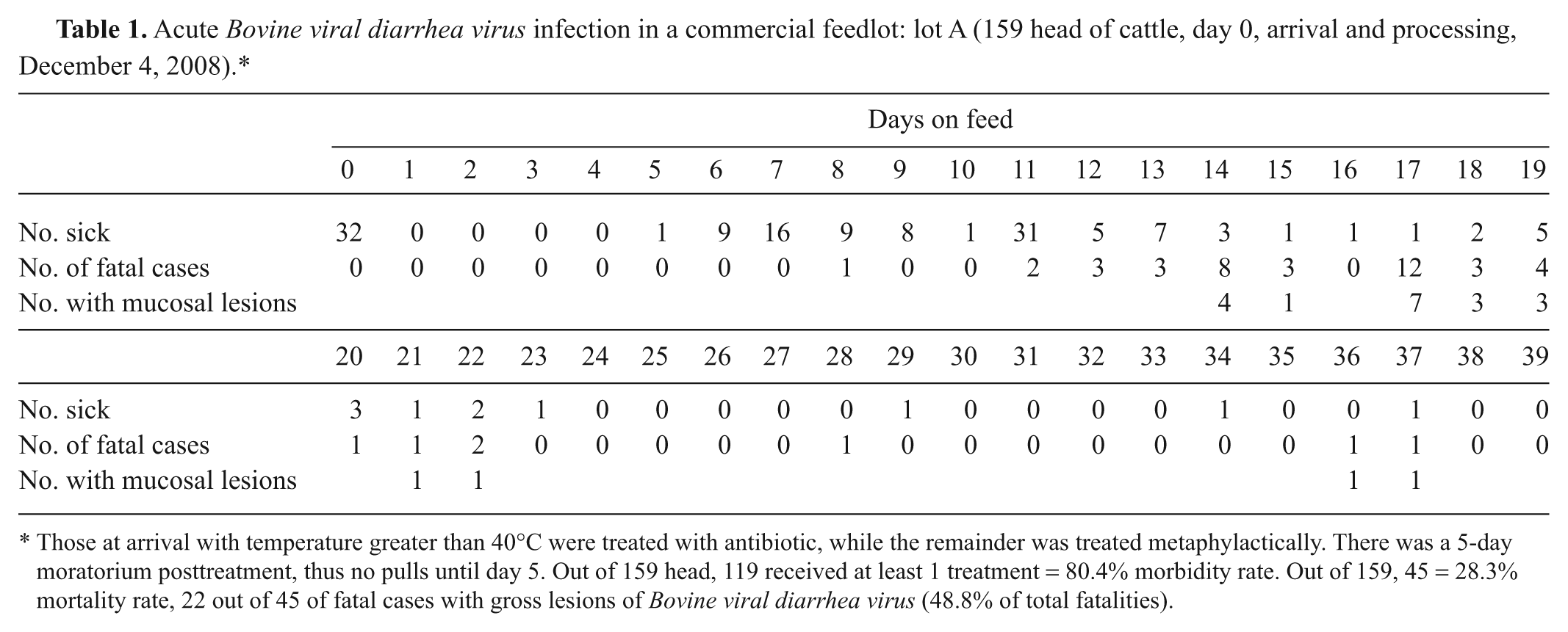
Those at arrival with temperature greater than 40°C were treated with antibiotic, while the remainder was treated metaphylactically. There was a 5-day moratorium posttreatment, thus no pulls until day 5. Out of 159 head, 119 received at least 1 treatment = 80.4% morbidity rate. Out of 159, 45 = 28.3% mortality rate, 22 out of 45 of fatal cases with gross lesions of Bovine viral diarrhea virus (48.8% of total fatalities).
Between 0 and 39 DOF, several animals were identified as sick and were treated according to standard feedyard antimicrobial treatment protocols for bovine respiratory disease (BRD) based on signs of listlessness, depression, increased respiratory rate, pyrexia, anorexia, and ocular and/or nasal discharge. The morbidity of lot A based on DOF is shown in Table 1.
Lot B consisted of 160 heifers with an average weight of 273 kg and originated from multiple auction markets in Texas and Oklahoma. This lot arrived at the feedlot on December 5, 2008 (day 0). The calves received their initial processing on day 1. Ear notch samples were also taken and tested for PI by ACE testing. There were no PI animals identified in this lot. Processing was identical to that of lot A with the exception of the day 21 and 33 vaccinations. Rectal temperatures were also evaluated in lot B at initial processing. In this lot, 43 of 160 animals (26.9%) had temperatures greater than 40°C and received tulathromycin b injections. The remaining 117 animals were given tilmicosin c injections prophylactically. A 5-day treatment moratorium was also followed with this lot. Morbidity for the first 39 DOF for lot B is shown in Table 2.
Acute Bovine viral diarrhea virus infection in a commercial feedlot: lot B (160 head of cattle, day 0, arrival on December 5, and processing on day 1, December 6, 2008).*
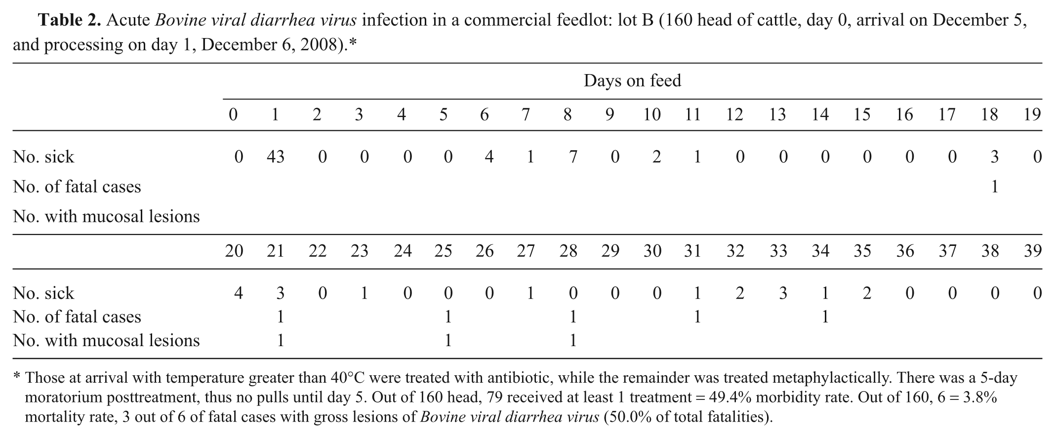
Those at arrival with temperature greater than 40°C were treated with antibiotic, while the remainder was treated metaphylactically. There was a 5-day moratorium posttreatment, thus no pulls until day 5. Out of 160 head, 79 received at least 1 treatment = 49.4% morbidity rate. Out of 160, 6 = 3.8% mortality rate, 3 out of 6 of fatal cases with gross lesions of Bovine viral diarrhea virus (50.0% of total fatalities).
Calves in both lots were treated for BRD according to feedlot protocols as described above. Mortalities for both lots of calves by DOF are shown in Tables 1 and 2. In lot A, 4 out of 8 mortalities on day 14 showed esophageal ulcerations. Samples from 2 of these animals were collected for laboratory analysis. An additional animal died on day 15, and also showed mucosal lesions; samples were taken for laboratory analysis. Samples from these 3 mortalities were sent to the Texas Veterinary Medical Diagnostic Laboratory (Amarillo, Texas) for analysis on December 19, 2008. Table 1 shows fatal cases by DOF from lot A as well as those mortalities showing mucosal lesions. In the first 39 DOF, 119 of the 159 calves received at least 1 therapy (74.8%). There were 45 out of 159 (28.3%) mortalities in the first 39 DOF with 22 out of 45 (48.8%) showing mucosal lesions. Table 2 shows treatments and fatal cases by DOF of lot B. Seventy-nine out of 160 (49.4%) of lot B received at least 1 therapy in the first 39 DOF, and lot B experienced 6 fatal cases. Three of the 6 fatal cases (50.0%) showed gross lesions on mucosal surfaces.
The morbidity and mortality for lot A at the end of the finishing period was 79.2% and 30.8%, respectively. Lot B had a final mortality of 5.6% and a morbidity of 49% at the end of the finishing period.
Multiple samples were taken from fatalities and serums from animals surviving BRD in both lots in addition to the initial 3 mortalities described above. Samples from these animals were submitted to the Oklahoma Animal Disease Diagnostic Laboratory (Oklahoma State University, Stillwater, Oklahoma) for histopathology, virus identification by fluorescent antibody testing, viral isolation, and IHC. 5 In addition, fresh tissues that had gross lesions suggestive of BVDV were swabbed using sterile culture swabs. The swabs were then tested for BVDV using a reverse transcription polymerase chain reaction test. 5 Genomic sequences from the 5′-nontranslated region of BVDV isolates were generated and subgenotypes determined as described previously. 14
Virus isolations were conducted on tissue samples submitted from 11 fatalities. Isolations on cell culture were negative for all viruses with the exception of 4 of 11 being positive for BVDV (Tables 3, 4); no cytopathic agents were isolated. Fluorescent antibody tests for BHV-1, BVDV, and coronavirus were conducted on the tissue samples (8 fatalities) submitted to the Oklahoma Animal Disease Diagnostic Laboratory. All results for fluorescent antibodies were negative. The IHC for BHV-1 was also conducted on these 8 fatalities and returned negative. The IHC for BVDV was evaluated on all 11 fatalities submitted, and all fatalities returned positive results on at least 1 tissue sample (Tables 3, 4). Outcomes from the BVDV laboratory testing performed at Texas Veterinary Medical Diagnostic Laboratory and Oklahoma Animal Disease Diagnostic Laboratory for lots A and B, as well as the genotyping, are shown in Tables 3 and 4, respectively.
Acute Bovine viral diarrhea virus (BVDV) infection in a commercial feedlot: lot A (samples collected from cattle and laboratory test results).*
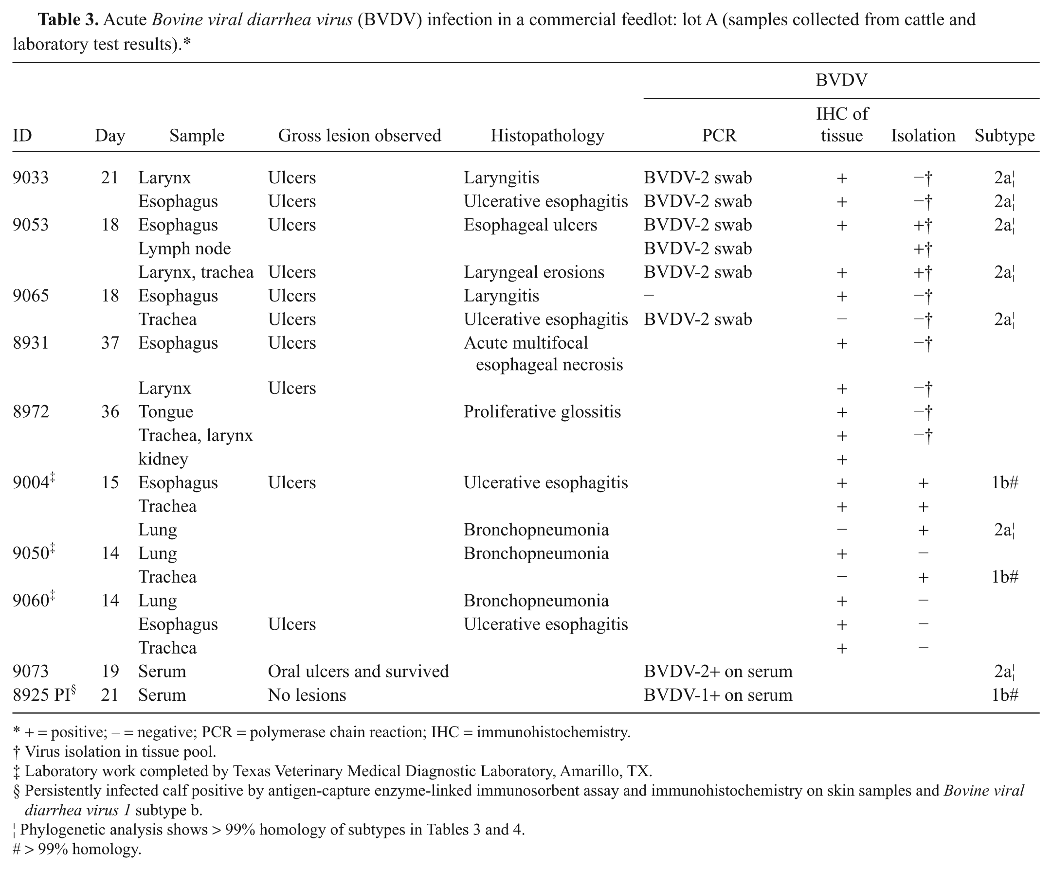
+ = positive; − = negative; PCR = polymerase chain reaction; IHC = immunohistochemistry.
Virus isolation in tissue pool.
Laboratory work completed by Texas Veterinary Medical Diagnostic Laboratory, Amarillo, TX.
Persistently infected calf positive by antigen-capture enzyme-linked immunosorbent assay and immunohistochemistry on skin samples and Bovine viral diarrhea virus 1 subtype b.
Phylogenetic analysis shows > 99% homology of subtypes in Tables 3 and 4.
> 99% homology.
Acute Bovine viral diarrhea virus (BVDV) infection in a commercial feedlot: lot B (samples collected from cattle and laboratory test results).*
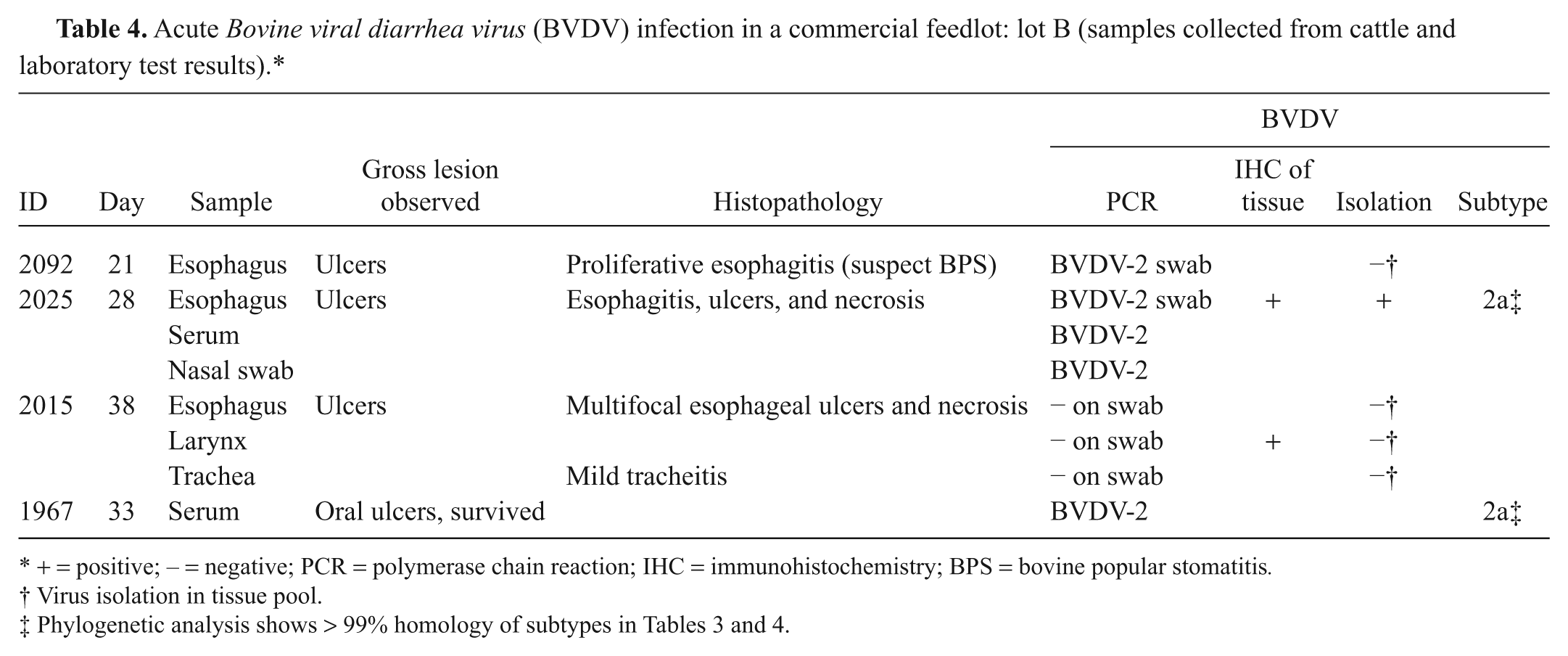
+ = positive; − = negative; PCR = polymerase chain reaction; IHC = immunohistochemistry; BPS = bovine popular stomatitis.
Virus isolation in tissue pool.
It is possible that an infection besides BVDV contributed to the development of the lesions observed. For this reason, serums from animals recovering from oral lesions and other animals not exposed to the affected animals were submitted to the U.S. Department of Agriculture, Animal and Plant Health Inspection Service, National Veterinary Services Laboratory (Ames, Iowa) for testing for antibodies to other viral pathogens. Results are shown in Table 5. Seropositivity ranged from zero for calicivirus to 100% positive by serology to Pseudocowpox virus.
Antibody levels to several viruses in serums from cattle recovering from disease or unexposed to disease.*
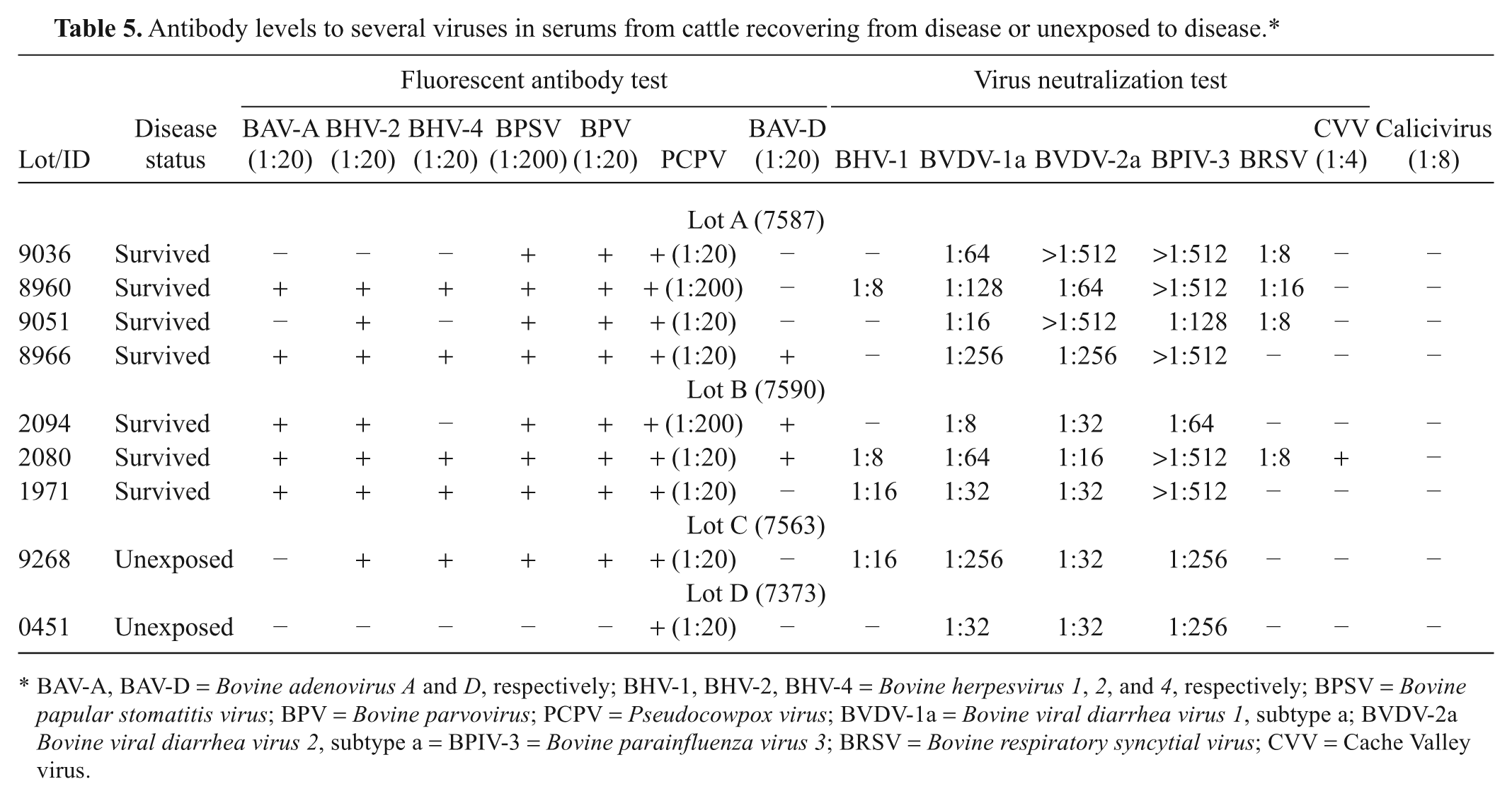
BAV-A, BAV-D = Bovine adenovirus A and D, respectively; BHV-1, BHV-2, BHV-4 = Bovine herpesvirus 1, 2, and 4, respectively; BPSV = Bovine papular stomatitis virus; BPV = Bovine parvovirus; PCPV = Pseudocowpox virus; BVDV-1a = Bovine viral diarrhea virus 1, subtype a; BVDV-2a Bovine viral diarrhea virus 2, subtype a = BPIV-3 = Bovine parainfluenza virus 3; BRSV = Bovine respiratory syncytial virus; CVV = Cache Valley virus.
Grossly, the mucosal lesions observed on necropsy varied dramatically. Lesions were observed in the oral cavity on buccal surfaces and tongue, and in the larynx, trachea, and esophagus. Small infrequent, 1–3 mm, irregular but sharp-edged ulcerations without gross necrotic material were observed in the esophagus of some animals but others had extensive, multifocal, 1mm to 1 cm, circular to ovoid mucosal ulcerations involving the majority of the esophagus with and without necrotic debris. Yet other animals with esophageal involvement showed more proliferative, raised, necrotic lesions. The lesions varied in size from 1–5 mm, circular, raised lesions to up to 1 cm wide and 4 cm long coalescing, raised, proliferative lesions. Some animals showed extensive, coalescing ulcerations involving greater than 50% of the upper esophageal mucosa, which contained firm, yellowish, plaque-like necrotic debris that was not proliferative. Mucosal lesions of the larynx showed large, 1 cm, circular to ovoid mucosal ulcerations with necrotic material but in other animals the lesions were small circular (3–5 mm), raised, proliferative lesions. Gross lesions of the tongue were observed in 2 animals. One animal showed circular, 3–5 mm ulcerations but the other animal had circular, raised, proliferative, 3–8 mm lesions.
Figure 1 shows postmortem lesions from animals in both lot A and lot B. Lesions shown are consistent with lesions observed in other postmortems preformed. These lesions were seen in the oral cavity, esophagus, and larynx and were reported positive for BVDV by IHC and/or PCR testing of lesion swabs.
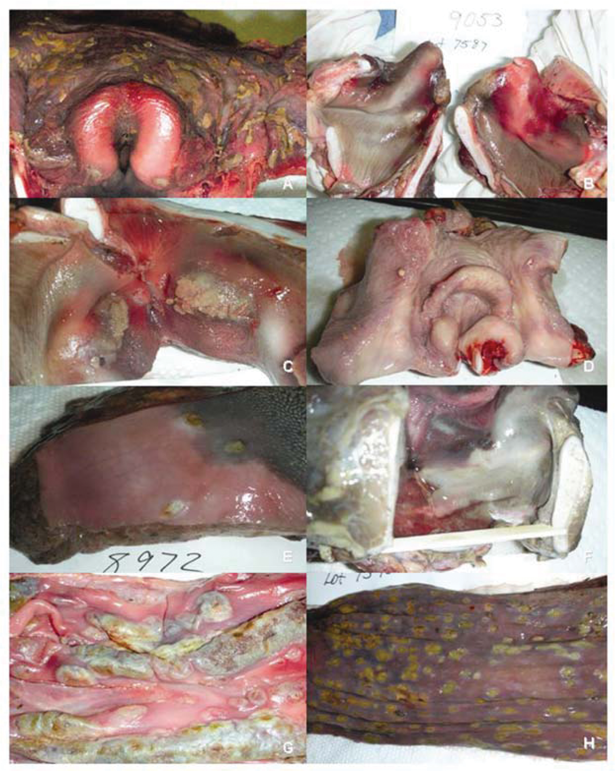
Postmortem lesions testing positive for Bovine viral diarrhea virus.
The BVDV isolates were sequenced for phylogenetic analysis, and results are shown in Figure 2. All BVDV-2a strains isolated from both lots of calves that were sequenced showed 100% homology. The BVDV-1b strain found in the PI animal (no. 8925) of lot A was found to be identical to the strain recovered from the esophagus of animal no. 9004 and from the trachea of animal no. 9050, both from lot A.
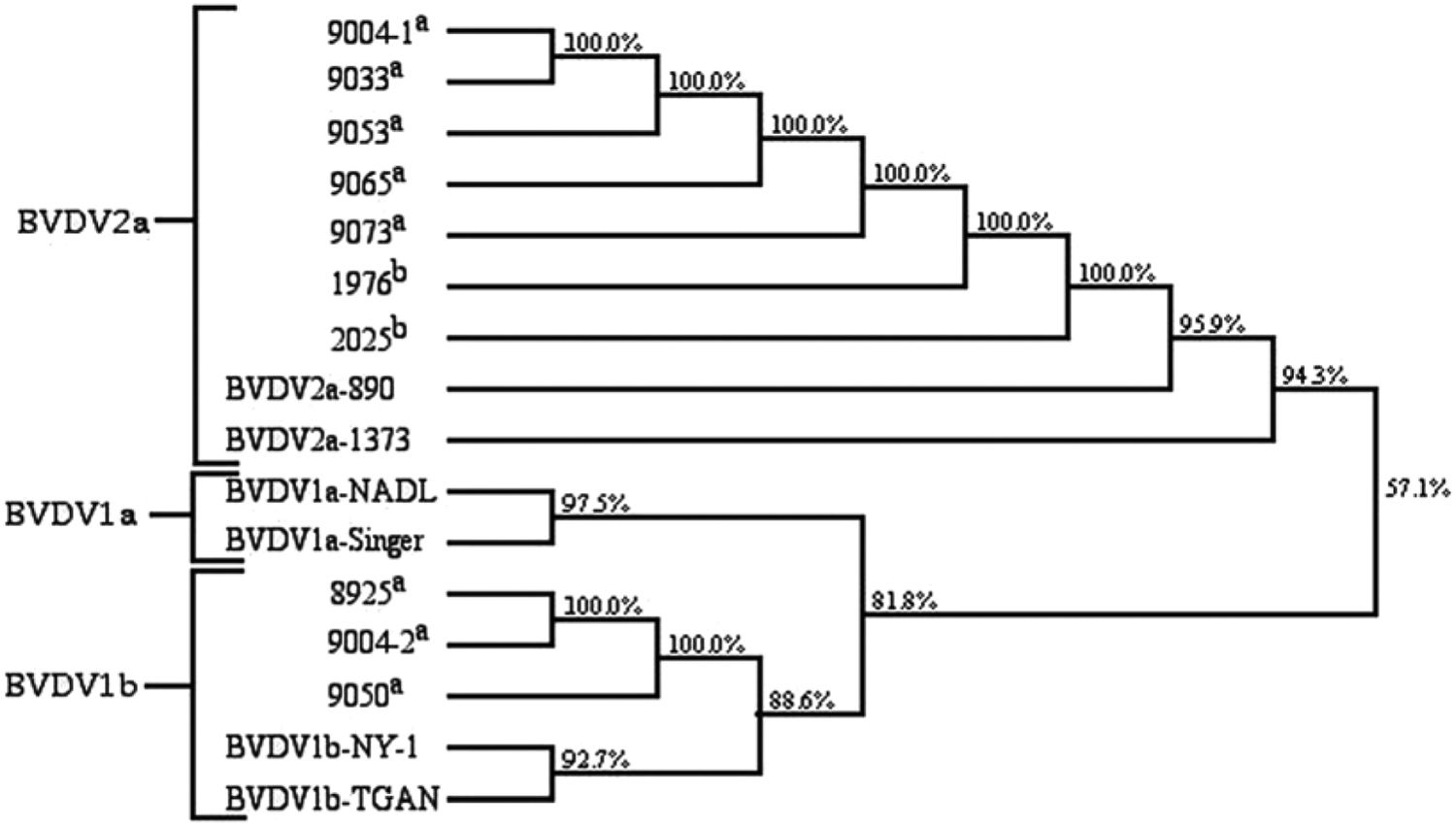
Phylogenetic analysis.
The first laboratory diagnosis of the BVDV-2a strain was from lung tissue of animal no. 9004 (lot A). This animal was 15 DOF when samples were collected. Interestingly, a second BVDV-1b strain was isolated from the esophagus of animal 9004. The second BVDV-1b strain was identical to the 1b strain of the PI animal (no. 8925) identified on arrival by ACE testing. Animal 9004 was the fifth mortality showing gross mucosal lesions. Four out of the 8 calves of lot A, dying at 14 DOF, also had similar lesions. Two of these animals had samples sent in for laboratory diagnosis (nos. 9050 and 9060) and both returned positive results for BVDV. The BVDV strain recovered from the lung of animal no. 9050 was determined to belong to the BVDV-1b genotype, and its sequence was identical to the sequence of the PI strain recovered from animal no. 8925.
The first identical BVDV-2a strain recovered from lot B was on day 21 and was from animal no. 2092, which had a rectal temperature greater than 40°C at initial processing (1 DOF) and was treated with tulathromycin b injection. This animal (no. 2092) was given a second therapy at 13 DOF for lameness, and died at day 21. Postmortem evaluation revealed esophageal mucosal lesions (Fig. 1) as well as pneumonia. The first observation of mucosal lesions upon necropsy in lot B occurred on day 18, although no laboratory analysis was done. A second identical BVDV-2a strain was recovered from an animal (no. 2025) dying at 28 DOF (Fig. 1).
Due to the timing of the mortalities where identical BVDV-2a strains were recovered, it appears that the acute BVD infection originated in lot A and then spread to lot B. The route of transmission from lot A to lot B is unclear as these 2 lots were separated by an empty pen from the time they arrived at the feed yard. Also, none of the hospitalized animals was returned to their original pens due to the severity of the outbreak. Animals pulled from their pen from lot A for treatment of sickness had to pass by lot B via a cattle alley behind the pen housing lot B. This may have provided an opportunity for transmission of the BVDV-2a strain from lot A to lot B.
Postmortem lesions observed in this outbreak were aggressive, well developed, and locally extensive. Ulcerative mucosal lesions were seen in the larynx, trachea, and esophagus in animals where the BVDV-2a strain was recovered. Because severe oral lesion and mucosal ulcerations were observed in this case, differential diagnosis included vesicular stomatitis, papular stomatitis, and foot-and-mouth disease. It should also be considered that given the higher titers for Pseudocowpox virus that previous exposure to pseudocowpox may have played a role or provided conditions for BVDV to produce the severe mucosal lesions seen in this case. Typically, acute BVDV infections result in subclinical or mild infections resulting in immune suppression. The current study describes an aggressive, pronounced clinical disease syndrome and, therefore, acute BVDV infection should be considered as an etiology when severe oral or mucosal lesions are observed.
Footnotes
Acknowledgements
The authors would like to thank the management and staff of Nortex Feeders of Dalhart, Texas for their assistance with this case presentation, as well as the U.S. Department of Agriculture, Animal and Plant Health Inspection Service, Diagnostic Virology Laboratory for their prompt action and support.
a.
Prism 5, Ft. Dodge Animal Health, Ft. Dodge, IA.
b.
Draxxin, Pfizer Animal Health, New York, NY.
c.
Micotil, Elanco, Indianapolis, IN.
d.
Express 5, Boehringer Ingelheim, St. Joseph, MO.
e.
Express 3, Boehringer Ingelheim, St. Joseph, MO.
f.
Pyramid 5, Ft. Dodge Animal Health, Ft. Dodge, IA.
The author(s) declared no potential conflicts of interest with respect to the research, authorship, and/or publication of this article.
The author(s) received no financial support for the research, authorship, and/or publication of this article.