
Abstract
Disseminated lymphoma was diagnosed in an 8-year-old male bonobo (Pan paniscus). The male bonobo presented with a 4–6 week history of dyspnea and facial swelling around the eyes; thoracic radiographs and computed tomography scan indicated a craniodorsal mediastinal soft tissue mass. Upon gross examination, there was a large, cream to white mass expanding the mediastinum and pericardial sac. The mass extended along the thoracic aorta and cranial vena cava, through the thoracic inlet, along and encircling the trachea, and bilaterally into the thyroid glands. Microscopically, neoplastic lymphocytes were present in the thymus, trachea, lungs, kidney, heart, and numerous other tissues. Immunohistochemical staining of neoplastic lymphocytes revealed diffuse immunoreactivity for cluster of differentiation (CD)3 indicating T-cell lymphoma. Routine viral screening was negative via polymerase chain reaction.
The bonobo (pygmy, dwarf, or gracile chimpanzee; Pan paniscus) is a great ape and 1 of the 2 species (the other being P. troglodytes, 6 or common chimpanzee) making up the genus Pan. Endangered in the wild, bonobos are only found in the Democratic Republic of Congo and are among the world’s rarest animals, with approximately 70 in captivity in the United States. 4 These great apes are the closest-living relatives to human beings with 98.5% similar genetic makeup and are used in captivity for behavioral and language development research. 5 Lymphoid neoplasia in chimpanzees is rare. The most common forms of neoplasia are in the female urogenital system and are usually benign. 2 There is a single case of large-cell lymphoma, documented in a 35-year-old male chimpanzee, that expressed cluster of differentiation (CD)4+. 1 The current study describes a disseminated, naturally occurring T-cell lymphoma in a bonobo.
An 8-year-old captive-born male bonobo presented to Iowa State University College of Veterinary Medicine in May 2009 following a 4–6 week history of dyspnea and facial swelling around the eyes. Thoracic radiographs indicated a craniodorsal mediastinal soft tissue mass compressing the entire intra-thoracic trachea. Computed tomography scan revealed the soft tissue mediastinal mass circumferentially compressed the intra-thoracic trachea and was most consistent with neoplasia originating from the mediastinal lymph nodes. Fine-needle aspirates of the mass revealed lymphoma, and there were numerous, large, blastic lymphocytes detected on a peripheral blood smear. The bonobo died, and a necropsy was performed.
Grossly, the cranial mediastinum and cranial pericardial sac were diffusely and markedly thickened by a cream to white colored, poorly demarcated mass. On cut surface, the mass was solid and white (Fig. 1). The mass expanded along the thoracic aorta and its branches (the cranial vena cava), through the thoracic inlet, along and encircling the trachea, and bilaterally into the thyroid glands. The distal 50% of the tracheal lumen was also moderately narrowed. The mass primarily surrounded the thoracic trachea and the distal 30% of the cervical trachea. The epicardium had multifocal pale tan to white discolorations, which did not extend into the underlying myocardium, but the aortic arch and the cranial vena cava were diffusely surrounded. The bronchi and bronchioles were diffusely thickened with prominent walls, and lymph nodes were diffusely enlarged, including the cervical, submandibular, tracheobronchial, inguinal, axillary, and mesenteric nodes. Both kidneys were diffusely enlarged by approximately 50%. There were numerous multifocal pale tan to white nodules throughout the renal cortices, which varied in diameter from 1 mm to 5 mm, were solid and firm on cut surface, and bulged on the capsular surface.
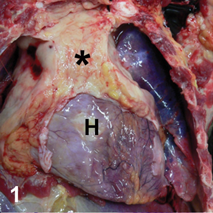
Thoracic cavity; bonobo (Pan paniscus); mediastinal mass. Cranial mediastinum and cranial pericardial sac are diffusely and markedly thickened by a cream to white colored, poorly demarcated mass (*). H = heart.
Tissue samples were fixed in 10% neutral buffered formalin, processed routinely for paraffin embedding, sectioned at 4 μm, and stained with hematoxylin and eosin. Immunohistochemistry was performed on tissue sections with the appropriate controls and standard indirect peroxidase-based methods a for the detection of CD3 and CD79a were utilized.
Neoplastic cells were individualized, round, and compressed into sheets (Fig. 2). These cells were round to oval, had irregularly shaped hyperchromatic nuclei, indistinct nucleoli, a scant to moderate rim of eosinophilic cytoplasm, and distinct cell borders. Anisokaryosis was moderate to marked, and nuclear shape varied from round or oval to reniform, cleaved or multilobular (Fig 3). The mitotic rate was 1–2 per 400× field with scattered apoptotic cells throughout. Throughout the thoracic mass were multifocal zones of necrosis, characterized by loss of cellular detail, accumulation of pyknotic and karyorrhectic cellular debris, and nuclear smudging. Similar neoplastic cells were present in the peri-tracheal connective tissue, thyroid glands, parathyroid glands, pituitary gland, gasserian ganglia, peripheral nerves, bone marrow, lungs (Fig. 4), kidney, heart, spleen, lymph nodes, brain, testicles, urinary bladder, tonsils, aorta and peri-aortic tissue, eyes, stomach, small intestine, and colon. Immunohistochemical staining of the thoracic mass and numerous other tissues revealed intense, diffuse immunoreactivity for CD3 (neoplastic cells were negative for CD79a, a B-cell marker), indicating T-cell lymphoma (Fig. 5). Virologic testing via polymerase chain reaction (PCR) on frozen liver and testis was negative for Simian immunodeficiency virus, Simian retrovirus, Primate T-lymphotropic virus 1–3, Simian foamy virus, and Rhesus monkey rhadinovirus, a gamma-2 herpesvirus related to Human herpesvirus 8. Tissues also showed no evidence of a detectible lymphocryptovirus via PCR.
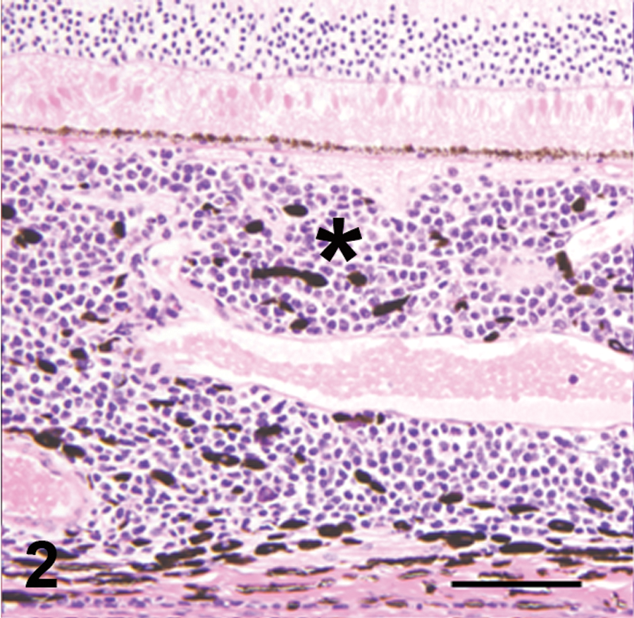
Choroid and retina; bonobo (Pan paniscus); malignant lymphoma. The choroid (*) is diffusely expanded to 2–4 times normal thickness by an infiltrate of individualized neoplastic lymphocytes. Bar = 100 µm.
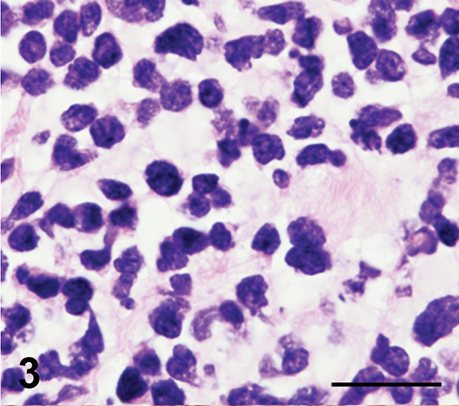
Mediastinal mass; bonobo (Pan paniscus); malignant lymphoma. Neoplastic cells were round to oval, had irregularly shaped hyperchromatic nuclei, indistinct nucleoli, a scant to moderate rim of eosinophilic cytoplasm, and distinct cell borders. Bar = 50 µm.

Lung; bonobo (Pan paniscus); malignant lymphoma. Focus of neoplastic lymphocytes replacing normal architecture. L = normal lung. Bar = 100 µm.
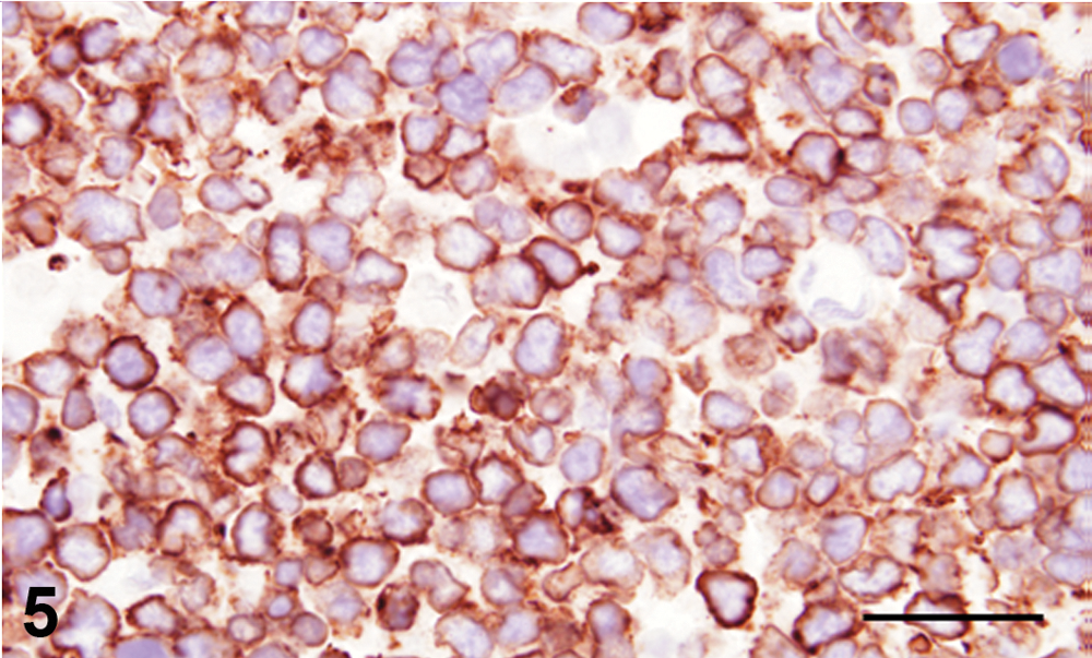
Mediastinal lymph node; bonobo (Pan paniscus). Intense cytoplasmic immunohistochemical staining for cluster of differentiation (CD)3 in neoplastic lymphocytes. Bar = 50 µm.
The findings are consistent with widely disseminated, malignant T-cell lymphoma. Anaplastic, large T-cell lymphoma with disseminated abdominal metastases was reported in a 35-year-old male chimpanzee and was negative for retroviral infection. 1 More commonly, lymphoid malignancies are associated with viral infection in nonhuman primates. Primate T-lymphotropic virus 1, 2, and 3 have been isolated from bonobos and have been associated with T-cell lymphoma in other primates.3,4 In the current case, tissues were negative for this virus.
Footnotes
Acknowledgements
The authors would like to thank the Great Ape Trust of Iowa and Dr. Brigetta Hughes for submitting this case, Deb Moore for immunohistochemical assistance, and Toni Christofferson, Diane Gerjets, and Jenny Groeltz-Thrush for histology assistance. The authors thank Dr. Andrew D. Miller at Harvard Medical School, Division of Comparative Pathology and the Pathogen Detection Laboratory at the California National Primate Research Center, Davis, CA for viral testing.
a.
Dako North America Inc., Carpinteria, CA.
The author(s) declared no potential conflicts of interest with respect to the research, authorship, and/or publication of this article.
The author(s) received no financial support for the research, authorship, and/or publication of this article.