
Abstract
In the current report, a case in Italy of disseminated Mycobacterium avium subsp. hominissuis infection in a dog from an American lineage of Basset Hounds is described. A 2-year-old intact female Basset Hound presented with persistent lymphadenopathy, lameness, and a history characterized by coccidiosis, bacterial gastroenteritis, and alopecia. Lymphadenitis, with macrophages containing a few intracytoplasmic, negative staining, Ziehl–Neelsen-positive bacilli, was detected by a popliteal fine-needle aspirate leading to the diagnosis of mycobacteriosis. Ultrasound and X-ray examinations revealed visceral and mediastinal lymphadenopathy. Because of the extent of the disease, the dog was humanely euthanized. Significant gross abnormalities, such as enlargement of the cranial mediastinal lymph nodes with encapsulated areas of caseous necrosis and generalized lymphadenopathy, were observed at necropsy. Granulomatous lesions were histopathologically detected in the liver and spleen. Ziehl–Neelsen-positive bacilli were observed in all examined lymph node, liver, spleen, lung, and bone marrow smears. Lymph nodes and liver were collected in order to pursue speciation by bacterial culture and molecular biology; multiplex polymerase chain reaction results classified the pathogen as M. avium subsp. hominissuis. Although an immune system deficiency was not investigated, anamnesis suggests that the dog was immunocompromised. Furthermore, the dog came from an American stock of Basset Hound, and for some of this breed, a predisposition to this infection has been hypothesized.
A 2-year-old intact female Basset Hound dog was presented for evaluation of persistent lymphadenopathy and lameness. From its birth by Caesarian section, which gave rise to 4 empty amniotic sacs, the history of the dog had been characterized by episodes of coccidiosis and severe bacterial gastroenteritis responsive to anticoccidial and antibiotic therapy. At 4 months of age, the dog spent a short period in a breeding house in northern Italy, and soon after its return, the dog showed periodic lameness, without a detectable neurologic defect, and diffuse alopecia. A systemic form of lupus was suspected, and an anti-nuclear antibody test was positive. Continuous treatments with prednisone (almost 6 cycles) were necessary to prevent recurrence of lameness.
At 2 years of age, a popliteal fine-needle aspirate was performed and sent to the Department of Animal Pathology of the University of Pisa (Pisa, Italy) for cytopathological evaluation. One of the 2 obtained slides was stained with a Romanowsky stain, and low cellularity was noted with prevalent macrophages and small lymphocytes. A few rod-shaped, nonstaining organisms were seen within the cytoplasm of 1 macrophage (Fig. 1A). A Ziehl–Neelsen (ZN) stain was performed on the remaining unstained slide and revealed a few acid-fast bacilli morphologically consistent with Mycobacterium species (Fig. 1B). Since mycobacterial infection had been previously diagnosed by one of the authors in another Basset Hound dog, and Basset Hounds are known to be predisposed to Mycobacterium avium intracellulare complex (MAIC) infection, 10 further investigation was recommended.
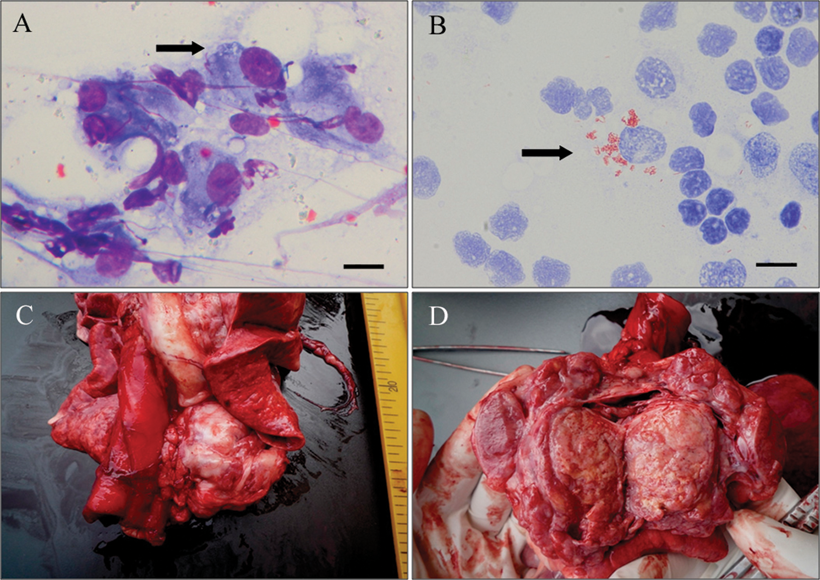
Basset Hound dog. Popliteal lymph node fine-needle aspirate (A, B) and postmortem view (C, D).
The dog was thus referred to the Department of Veterinary Clinics of the University of Pisa. On physical examination, the dog was bright, alert, and responsive, with a body condition score of 3 out of 5, and temperature of 38.9°C. The mucous membranes were pink, and the respiratory and heart rates were within normal limits. The main abnormal findings were generalized lymphadenopathy, with increased consistency of all palpable lymph nodes, and splenomegaly. Abnormal hematological findings were limited to a mildly microcytic, hypochromic anemia, and hypoalbuminemia.
Thoracic radiographs revealed a 3 cm in diameter, mediastinal mass compatible with an enlarged cranial mediastinal lymph node. On abdominal ultrasonography, the spleen appeared enlarged, with scattered, small, round, hypoechoic areas. The liver was mildly enlarged, but had normal echogenicity, and the abdominal lymph nodes were enlarged and hypoechoic, with rounded edges.
New fine-needle aspirates were obtained from prescapular lymph nodes. Some of these samples were stained with a Romanowsky stain and ZN, while others were sent to the Microbiology Laboratory of the Department of Animal Pathology of the University of Pisa for bacterial culture. The cytological findings were characterized by a granulomatous lymphadenitis with a predominance of large macrophages and hypersegmented neutrophils. Within the macrophage cytoplasm and free in the background, nonstaining negative images of bacterial rods were present, which stained brilliant pink with ZN. Morphological changes thus confirmed the presence of mycobacterial infection characterized by high numbers of bacteria within the cytoplasm.
Because of the extent of the disease, the owners opted not to pursue treatment, and the dog was humanely euthanized. At necropsy, the significant gross abnormalities were limited to enlargement of the cranial mediastinal lymph nodes (Fig. 1C), with encapsulated areas of caseous necrosis (Fig. 1D) and enlargement of all peripheral and visceral lymph nodes, with loss of corticomedullary distinction. No gross alterations of the joints that could be considered a source of the lameness were seen. Representative portions of cranial mediastinal, mesenteric, and submandibular lymph nodes, lungs, heart, liver, spleen, stomach, intestine, kidney, brain, and bone marrow were sampled for histologic evaluation.
Microscopically, all examined lymph nodes showed massive infiltration of epithelioid macrophages, leading to complete loss of the normal lymphoid tissue architecture. Furthermore, in the cranial mediastinal lymph node, multiple foci of necrosis were observed (Fig. 2). The lungs were markedly congested. The liver showed sinusoidal ectasia and congestion, and portal fibrosis with mixed infiltration by macrophages, lymphocytes, and neutrophils. Bile ducts were hyperplastic with evidence of cholestasis. Multiple granulomatous foci composed of epithelioid macrophages were randomly distributed throughout the parenchyma (Fig. 3). The splenic red pulp was diffusely infiltrated by epithelioid macrophages; occasional megakaryocytes, extramedullary hematopoietic foci, and hemosiderosis were also present. The bone marrow was partially replaced by epithelioid macrophages with residual aggregates of erythropoietic cells (Fig. 4). No alterations were present in samples examined from other tissues. Ziehl–Neelsen stain was performed on all samples obtained. Numerous epithelioid macrophages containing acid-fast, intracytoplasmic, bacterial rods were seen in all lymph nodes, liver, spleen, bone marrow, and, to a lesser extent, in the lungs.
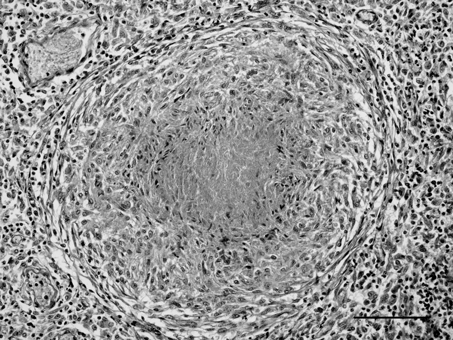
Histology; Basset Hound dog. Mediastinal lymph node with massive infiltration of epithelioid macrophages around an area of caseous necrosis. Hematoxylin and eosin. Bar = 100 µm.
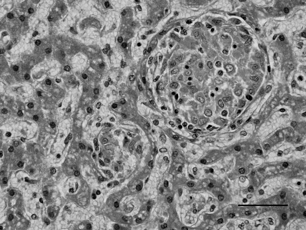
Histology; Basset Hound dog. Granulomatous hepatic lesion with necrosis and epithelioid macrophages. Hematoxylin and eosin. Bar = 50 µm.
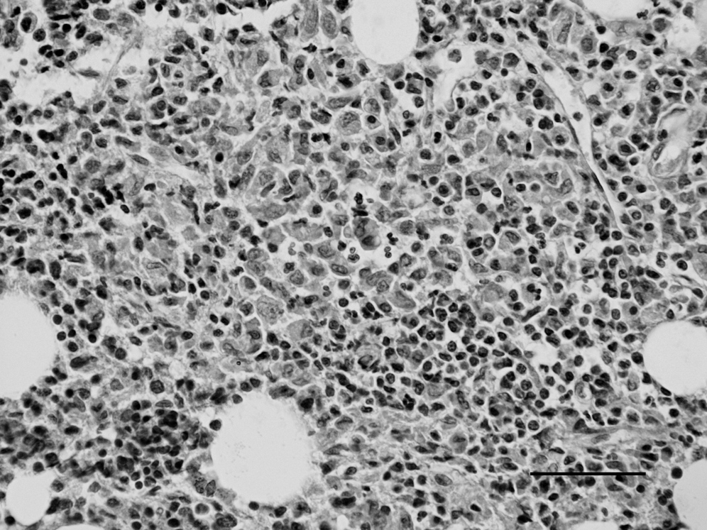
Histology; Basset Hound dog. Bone marrow is partially replaced by macrophages with residual aggregates of erythropoietic cells. Hematoxylin and eosin. Bar = 50 µm.
Lymph node aspirates collected antemortem, and lymph node and liver samples taken at necropsy were cultured in Dubos medium after decontamination with a 1% hexadecylpyridinium chloride solution. For each sample, a single tube was incubated at 37°C while another tube was incubated at 43°C in order to differentiate between M. avium and other Mycobacterium species. Colonies that grew at 43°C after 10 days were submitted for ZN staining. The acid-fast isolates were identified as M. avium by DNA probe hybridization a performed following the manufacturer’s instructions. DNA samples were extracted from the isolates using a commercial kit b according to manufacturer’s recommendations. The samples were successively used in a multiplex polymerase chain reaction (PCR) assay in order to identify the M. avium subspecies. The primers IS901-f (5′-GGATTGCTAACCACGTGGTG- 3′) and IS901-r (5′-GCGAGTTGCTTGATGAGCG-3′) were used in the PCR protocol to amplify a 577-bp fragment, whereas the primers IS1245-f (5′-GAGTTGACCGCGTTCATCG-3′) and IS1245-r (5′-CGTCGAGGAAGACATACGG-3′) were employed to amplify a 385-bp fragment. The fragments correspond to the IS901 and IS1245 genes, respectively, which are both present in M. avium subsp. avium and M. avium subsp. silvaticum, whereas only the IS1245 gene is present in M. avium subsp hominissuis. 17 The PCR amplification was performed in 50 µl total volume containing 200 µM of deoxynucleoside triphosphates, 0.5 µM of each primer, 1.25 U of Taq polymerase, b 5 µl of 10× PCR buffer, b and 2 µl of extracted DNA. The PCR amplifications were performed in an automated thermal cycler c for 40 cycles. Each cycle consisted of a denaturation phase (95°C for 1 min), an annealing phase (58°C for 30 sec), and an extension phase (72°C for 1 min). An initial denaturation of 5 min at 95°C and a final extension of 5 min at 72°C were performed. The PCR products were analyzed by electrophoresis on a 1.5% agarose gel at 100 V for 45 min; the gel was stained with ethidium bromide and observed under ultraviolet light. All isolates showed the 385-bp band, but not the 577-bp band. On the basis of these results, the isolates were identified as M. avium subsp. hominissuis.
In dogs, infections due to M. avium are sporadically reported, and in most of the cases, the subspecies was not identified. * Mycobacterium avium subsp. hominissuis is generally isolated from pigs and human beings,14,15 and cases of infection have been reported in a horse, 13 pet parrots, 21 and 2 dogs. 9 Mycobacterium avium intracellulare complex bacilli are ubiquitous in the environment and can remain viable for over 2 years.3,22 In dogs, infection with M. avium subsp. hominissuis occurs because of direct contact with infected animals or from the environment. It has also been documented that dogs can become infected by their owners, but the opposite situation, namely the dog as a source of infection for immunocompromised human beings, is the higher risk. 13 The source of infection was not determined in the current case; however, the dog lived outdoors for a long time in northern Italy in a rural area, and during that period, it was exposed to caged pet birds and pigeons, and had access to a pond, which was frequented by migrating waterfowl. No known exposure to chicken or swine carcasses, or human beings known to have tuberculosis in that area was suspected.
Mycobacterium avium subsp. hominissuis can invade the body through the skin, or the respiratory or gastrointestinal tracts. Normally, the infected subject develops an early granulomatous lesion at the site of entry and in the regional lymph node (complete primary complex) or only in the regional lymph node (incomplete primary complex), with or without secondary lymphohematogenous dissemination. 8 Mycobacterium avium intracellulare complex infections in dogs are frequently associated with the disseminated form of disease, with multiple organ system involvement.18,22 In the present case, the extensive mediastinal and tracheobronchial lymph node involvement suggests a respiratory route of infection, with formation of an incomplete primary complex and secondary lymphohematogenous dissemination.
Diagnosis is a challenge since clinical signs may be vague. Cytology is very helpful in the early diagnostic work-up, allowing for rapid detection of the rod-shaped bacteria; however, these can be few in number and easily overlooked. In the current case, cytology revealed the infectious nature of the disease, with further methods used to better characterize the involved pathogen. Cytology and histopathology are indeed useful to detect the rod-shaped, acid-fast bacilli, but do not help in differentiating between zoonotic and nonzoonotic mycobacterial species. The lepromatous type of MAIC infection, characterized by florid, monomorphic, epithelioid macrophage responses with numerous intracytoplasmic bacteria, is cytologically and histologically different from the pleocellular granulomatous response characteristic of infection due to Mycobacterium tuberculosis or Mycobacterium bovis. This lepromatous type of infection includes Langhan type, multinucleated giant cells, and few intracytoplasmic bacilli. However, definitive diagnosis relies on the use of supplementary tools such as bacterial culture and molecular genetic techniques such as PCR. Mycobacterial culture requires a long period of time (4–12 weeks) until a definitive diagnosis is provided.
The molecular techniques designed to identify the various subspecies of M. avium are a relatively recent development, and in many published cases, were not used, and therefore the subspecies was not identified. In the present case, multiplex PCR allowed the identification of the pathogen as M. avium subsp. hominissuis, and this is, to the authors’ knowledge, the third case described worldwide in the dog and the first in Italy.
Certain breeds of dog, such as the Basset Hound and Miniature Schnauzer, appear to be predisposed to this disease 10 ; the basis of this breed predisposition is unclear. Only in a few cases has the efficiency of the immune status been investigated. Other authors 4 have suggested that Basset Hounds might be predisposed to a cell-mediated immunodeficiency against intracellular pathogens due to a defect in either T cells or intracellular killing ability of macrophages. In another case, 18 a concurrent infection with 3 types of pathogens, one of which was M. avium, was reported in a Great Pyrenees dog, suggesting that an immunodeficient state also existed for this dog. These examples support the hypothesis that an immunocompromised state might predispose to the sporadic form of the disease. In the case herein described, the breed is one previously reported to be predisposed to the infection, and severity and massive dissemination of the lesions suggest that glucocorticoids might have promoted spread of the infection.
From the study that described the 5 cases of MAIC infection in Basset Hounds, 4 pedigrees were available and studied only in 3 dogs; from these, it emerged that 1 male was the sire of one of the affected dogs and that one of his sons was the sire of the other 2 dogs. For the cases of MAIC described in mixed breeds,2,12 whether the dogs were genetically related to Basset Hounds was not presented. The dog described in the current report was born from an American lineage of Basset Hounds (both mother and father). Direct documentation does not exist on whether the parents were direct descendants of the 5 Basset Hounds described in 1988. 4
Since human beings are relatively resistant to infection with MAIC, unless they are immunocompromised, and zoonotic transmission is no more likely than environmental acquisition, treatment of affected dogs might be considered. However, in most of the reported cases, dogs were euthanized for the progressive and often rapid worsening of clinical signs without any attempt at specific therapy.1,5-7,9,12,18 Only in a few cases has specific antimycobacterial therapy been initiated. One affected dog was treated with isoniazid for 10 months; however, the subject was euthanized because of continuing deterioration. 20 Three of the 5 Basset Hounds with MAIC disseminated infection described in the United States 4 were specifically treated for periods lasting from 7 days to 1 month, and in all 3 cases, a lack of response and progressive course of disease prompted the decision for euthanasia. Antimycobacterial therapy was also attempted in a Schnauzer dog whose condition seemed to initially improve, but the dog rapidly deteriorated. 16 Mycobacterium avium intracellulare complex organisms are resistant to common anti-tuberculosis drugs; however, fluoroquinolones and new quinolones have been shown to be active in vitro against many mycobacterial species. 11 Unfortunately, since clinical signs tend to be vague and often lead to misdiagnosis if rod-shaped organisms are not detected in cytological specimens, diagnosis is not usually achieved in an early, more treatable stage of the disease. In the current case, therapy was not attempted, mostly because of dissemination of the disease with multi-organ involvement.
In conclusion, the authors report a case of disseminated mycobacteriosis in Italy in a Basset Hound of American lineage. The introduction of molecular biology to the diagnostic plan allowed for the identification of the pathogen as M. avium subsp. hominissuis, and to the authors’ knowledge, systemic MAIC infection due to this subspecies is rarely described and has not been reported in Italy.
Footnotes
a.
INNO-LiPA Mycobacteria v2, Innogenetics NV, Ghent, Belgium.
b.
DNeasy®, Qiagen GmBH, Hilden, Germany.
c.
Gene-Amp PCR System 2700, Perkin-Elmer Inc., Waltham, MA.
The author(s) declared no potential conflicts of interest with respect to the research, authorship, and/or publication of this article.
The author(s) received no financial support for the research, authorship, and/or publication of this article.