
Abstract
Ovariohysterectomy in a healthy, nonpregnant, non-pseudopregnant, 9.5-month-old female dog revealed a 3 cm in diameter, swollen segmental portion of the uterus. A large amount of mucoserous fluid was present in the segmental portion, and the mucous membrane was covered by many yellowish-white villi protruding into the lumen. Histopathologic examination revealed endometrial hyperplasia associated with dilatation of the uterine glands in the segmental portion. The morphological findings were quite similar to those of a maternal placenta of pregnancy without the fetus and fetal membrane. Accordingly, this young female dog was diagnosed with spontaneous pseudo-placentational endometrial hyperplasia.
Cystic endometrial hyperplasia (CEH) in female dogs is well known as an important and commonly occurring lesion that can develop into pyometra.1,2,13,14 However, there have been many experimental reports on canine deciduoma, which is defined as a placenta-like endometrial proliferation induced by artificial stimuli to the endometrium during the luteal phase of the ovarian cycle.6-12 Furthermore, a previous study 4 reported for the first time a case of maternal placenta-like endometrial hyperplasia which occurred spontaneously in a female dog. A 2008 study 13 proposed that these endometrial hyperplasias, which differ from CEH, define a condition named pseudo-placentational endometrial hyperplasia (PEH). This report describes a case of spontaneous canine PEH in a young female dog.
This case involves a 9.5-month-old 6.5 kg female Miniature Schnauzer. At 6 months of age, the dog had cystitis and then cystitis associated with urolithiasis at 7 months. The dog was given antibiotics and a pH control diet a for treatment of cystitis and urolithiasis. The bottle-type water container that had been used for the dog was changed to a tray-type container for sufficient water supply. The dog recovered completely with proper medication and improvement of its water supply. The dog showed signs of estrus at 7.5 months, but had no chance of mating during the estrus period because it was always kept indoors. Ovariohysterectomy was performed under inhalation anesthesia when the dog was 9.5 months old.
Macroscopically, the surgically removed uterus was thickened, and many longitudinal folds were present on its surface. A swollen segmental portion, approximately 3 cm in diameter, was observed in the right uterine horn, and many blood vessels were apparent on its surface (Fig. 1). Macroscopically, this segmental portion was quite similar to the placentation site of the uterus in normal pregnant dogs. A large amount of mucoserous fluid, which was semitransparent and greenish in color, was observed in the lumen of the segmental portion, and the mucous membrane was covered by many yellowish-white villi protruding into the lumen (Fig. 2). No embryo or any other appendage was observed in the lumen of the segmental portion. Direct smears of the mucoserous fluid were stained by Gram and Giemsa stains and examined microscopically, but no inflammatory cells or bacteria were observed.
The excised uterus and ovaries were fixed in 15% formalin and stained with hematoxylin and eosin for histopathological examination. Histopathologically, the uterine lumen of the segmental portion was lined by long villi extending from the endometrial surface and contained accumulations of endometrial secretions on the luminal surface. The smooth muscle layer in the segmental portion became thinner than other area of the uterus probably due to expansion of the lumen (Fig. 3). The endometrium of the segmental portion consisted of 3 layers. The inner layer was made up of villous folds lined by endometrial epithelium. The middle layer consisted of a thin band of connective tissue covered by epithelial cells, and the outermost layer contained conspicuously distended endometrial glands (Fig. 4). The epithelium in the inner layer multiplied and protruded into the lumen and formed a long chorion, which was distributed regularly. These properties were extremely similar to those of the endometrium observed in maternal placentation during normal pregnancy. The endometrium of the nonsegmental portion consisted of short folds covered by epithelial cells; however, no glandular dilatation was observed. The smooth muscle layer of the nonsegmental portion was thicker than that of the segmental portion (Fig. 5). No inflammatory cells were observed in the endometrium or smooth muscle layer in any part of the uterus. In addition, well-developed corpus lutea and immature ovarian follicles were observed on the right and left ovaries (Fig. 6).
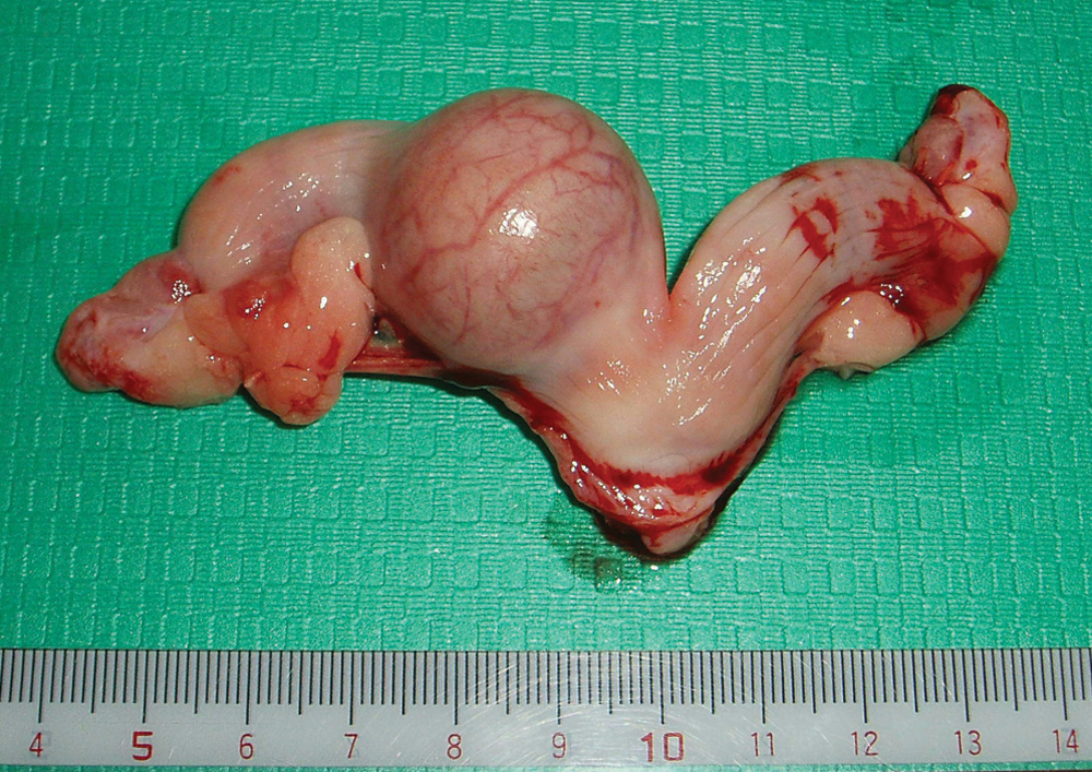
A spherical swollen portion, approximately 3 cm in diameter, was observed in the right uterine horn with many blood vessels were apparent on its surface.
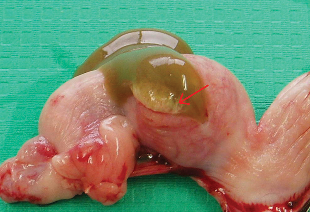
A large amount of mucoserous fluid, semitransparent and greenish in color, was observed in the lumen of the segmental portion. The mucous membrane was covered by many yellowish-white villi protruding into the lumen.
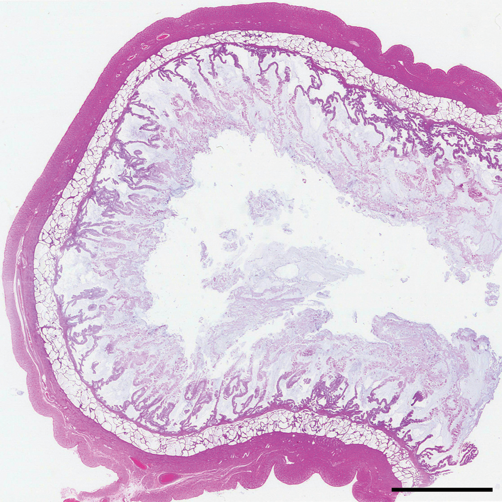
The uterine lumen of the segmental portion was lined by long villi extending from the endometrial surface and contained accumulations of endometrial secretions on the top. Hematoxylin and eosin stain. Bar = 5 mm.
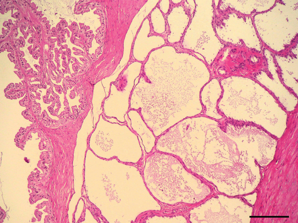
The endometrium of the segmental portion consisted of 3 layers. The inner layer was made up of villous folds lined by endometrial epithelium, the middle layer was a thin band of connective tissue covered by epithelial cells, and the outermost layer contained distended endometrial glands. Hematoxylin and eosin stain. Bar = 200 µm.
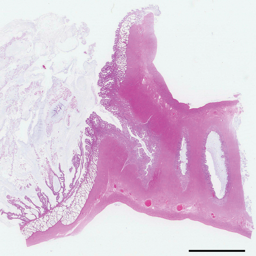
The endometrium of the nonsegmental portion consisted of short folds covered by epithelial cells; however, no glandular dilatation was observed. The smooth muscle layer of the nonsegmental portion was thicker than that of the segmental portion. Hematoxylin and eosin stain. Bar = 5 mm.

Two well-developed corpus lutea and immature follicles were observed in the ovary. Hematoxylin and eosin stain. Bar = 5 mm.
The present report describes the morphological features of the segmental portion of the uterus discovered in a 9.5-month-old female dog during a routine ovariohysterectomy. The dog had a history of both cystitis and urolithiasis between 6 and 7 months of age from which the dog recovered completely and thus was healthy at the time of surgery.
Although the dog showed signs of estrus 2 months before the operation, the dog had no chance of mating and showed no signs of pseudopregnancy.3,5 A large amount of mucoserous fluid was observed in the lumen of the segmental portion; however, no solid material or bacteria were observed. Mature corpus lutea were observed in the ovaries, which is typical of the luteal phase of the ovarian cycle. The segmental portion was located at the posterior part of the right uterine horn and was confined to one location.
Initially, pregnancy or pyometra was suspected from the above-mentioned findings. 3 Because the dog was kept indoors, it had no chance to mate. Furthermore, no inflammatory cells or bacteria were detected in the uterus. The findings indicated that the dog was neither pregnant nor had pyometra. Histopathological examination revealed that the segmental portion of the uterus was equivalent to the structure of the maternal placenta except for the embryo and fetal membrane. The observations suggest that unknown factors caused the decidual reaction followed by maternal placenta-like endometrial proliferation in the absence of an embryo in the uterus.6-12
A previous study 13 proposed the division of uterine lesions associated with endometrial hyperplasia into 2 forms, namely CEH and PEH. Cystic endometrial hyperplasia is the most common form and frequently develops into pyometra in older dogs. Pseudo-placentational endometrial hyperplasia is very different from CEH and is not widely recognized by clinicians or diagnostic pathologists. 13 Pseudo-placentational endometrial hyperplasia has also been referred to as deciduoma, 6 endometrial hyperplasia in pseudocyesis,3,5 maternal placenta-like endometrial hyperplasia, 4 and segmental endometrial hyperplasia. 14 The present case was ultimately diagnosed as spontaneously occurring PEH.
Although the cause of canine PEH has not been elucidated, bacteria invading the uterus from the vagina during estrus is believed to induce this condition. 11 The dog described herein had a history of lower urinary tract diseases including cystitis and urolithiasis between 6 and 7 months of age. Immediately after the dog recovered from the lower urinary tract diseases, it showed signs of estrus. Two months later, ovariohysterectomy was performed during the luteal phase. The clinical findings suggest that the bacteria, which contaminated the vulva, invaded the uterus and stimulated the endometrium, as would a fertile ovum, and subsequently induced PEH. It appears important to note that the condition occurs during the luteal phase and may be induced by a variety of materials including bacteria as reported previously.6-12,13 More case reports are required to clarify the pathogenesis and incidence of canine PEH.
Footnotes
a.
Royal Canin SA, Aimargues, France.
The author declared no potential conflicts of interest with respect to the research, authorship, and/or publication of this article.
The author received no financial support for the research, authorship, and/or publication of this article.