
Abstract
A 6-month-old male mixed-breed dog weighing 12.6 kg weight was presented for evaluation of a subcutaneous nodule on the dorsum. The medical history indicated trimethoprim–sulfamethoxazole treatment 2 months before presentation at the veterinary hospital. The initial complete blood cell count (CBC) results included an apparent left shift. Microscopic examination of a blood smear (Panoptic stain) revealed granulocytes with hyposegmented nuclei, coarse mature chromatin, and a nuclear shape varying from round to bilobed (pince-nez) or slightly indented. Occasional neutrophils and eosinophils had typical segmentation of nuclei. Abnormalities were not present in limited serum biochemical testing. The CBC was repeated 17 and 120 days later, and the results were similar to those observed in the first examination. The parents of the patient were located, and a CBC was performed on both animals. The dam, but not the sire, had nuclear hyposegmentation of granulocytes, confirming the diagnosis of Pelger-Huët anomaly.
Pelger-Huët anomaly is a hereditary disorder of the leukocytes characterized by granulocytes with hyposegmented nuclei and a coarse, mature pattern of chromatin. The morphology of the granulocyte nuclei may be round, oval, dumbbell-shaped, peanut-shaped, or bilobulate, or they may appear as a band. 15 The present report describes cases of Pelger-Huët anomaly in 2 related mixed-breed dogs.
A 6-month-old male mixed-breed dog weighing 12.6 kg was presented to the veterinary hospital at the Universidade Federal Rural do Semi-Árido, Mossoró, Rio Grande do Norte, Brazil, for evaluation of a subcutaneous nodule on the dorsum. The medical history disclosed parvovirus infection 2 months before presentation, which was treated with trimethoprim–sulfamethoxazole a (30 mg/kg intramuscular twice daily for 5 days) to prevent secondary bacterial infection.
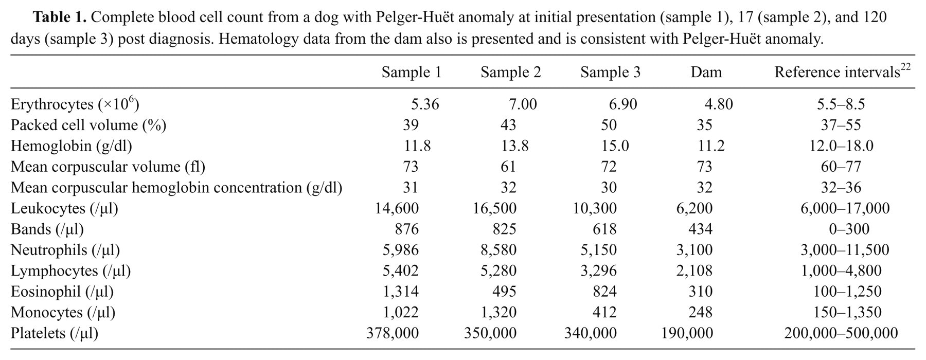
The subcutaneous, 5.0 cm × 6.0 cm, round nodule was diagnosed as an injection granulomas by fine-needle aspiration cytology. No other remarkable findings were found on physical examination. An ethylenediamine tetra-acetic acid–anticoagulated blood sample was collected for a complete blood cell count (CBC). Red blood cell, white blood cell, and platelet counts were determined using an improved Neubauer-ruled hematocytometer. The leukocyte differential count was performed using a stained blood smear. The packed cell volume was measured by microhematocrit centrifugation and the hemoglobin concentration was determined by the cyanmethemoglobin method. The serum alanine aminotransferase, aspartate aminotransferase, and alkaline phosphatase activities and urea nitrogen, creatinine, and total protein concentrations were also determined. b Relevant hematologic findings (Table 1) were suggestive of Pelger-Huët anomaly. Microscopic examination of the blood smear (Panoptic stain) revealed that most granulocytes (neutrophils, eosinophils, and basophils) had hyposegmented nuclei with a coarse, mature chromatin pattern. The nuclear shape varied from round to bilobed (pince-nez) to slightly indented (Fig. 1). Occasional neutrophils and eosinophils had typical segmentation of nuclei. No abnormalities were found in limited biochemical testing.
Complete blood cell count from a dog with Pelger-Huët anomaly at initial presentation (sample 1), 17 (sample 2), and 120 days (sample 3) post diagnosis. Hematology data from the dam also is presented and is consistent with Pelger-Huët anomaly.

Blood smear from a mixed-breed dog with Pelger-Huët anomaly. Note the nuclear hyposegmentation of neutrophils (A–D) and eosinophils (E, F). Panoptic stain. 100× objective.
Blood sample collection was repeated 17 days later for assessment of the CBC and serum biochemical tests. The apparent left shift was still present (Table 1), and blood smear examination confirmed continued morphological alterations of the granulocytes. Again, no abnormalities were found in the serum biochemical test results.
After enquiring about the relatives of the dog, the parents were located 120 days after the first examination of the initial patient. Blood samples were collected from both parents and again from the original patient. Hematological analysis revealed the presence of Pelger-Huët anomaly in the original patient and its dam, but not in the sire.
Pelger-Huët anomaly has been reported in human beings, rabbits, 24 cats, 20 dogs, 7,16,17,19 horses, 8,10 and mice. 26 In dogs, the condition has been described in a number of breeds, including the Cocker Spaniel, 7 Basenji, 19 Border Collie, English and American Foxhounds, Samoyed, 17 Australian Shepherd, 16 Australian Cattle Dog, Boston Terrier, German Shepherd Dog, coonhound, 15 and mixed-breed dogs. 14 Pelger-Huët anomaly is usually a rare disease but is more common in some breeds such as Australian Shepherds where an estimated incidence of 9.8% has been reported. 16 To the authors’ knowledge, there has only been 1 previous report of Pelger-Huët anomaly in mixed-breed dogs. 14
Pelger-Huët anomaly is an autosomal dominant hereditary anomaly, except in Australian Shepherds where incomplete penetrance has been reported. 16 In human beings, the anomaly has been attributed to a mutation in the lamin B receptor gene. 1 This receptor is an inner nuclear membrane protein responsible for the traffic of heterochromatin and lamins to the nuclear membrane. 30 Its disruption causes abnormalities in nuclear heterochromatin and, consequently, in nuclear morphology as described previously.
Acquired Pelger-Huët anomaly, also known as pseudo–Pelger-Huët anomaly, has morphological features that resemble the congenital anomaly. 15 The acquired anomaly is sporadic, but has been observed in human patients with hematological diseases such as myelodysplastic syndrome, 27,29 and has been associated with the administration of drugs such as sulfisoxazole, 13 sulfadiazine–trimethoprim, 28 valproate, 21 taxoid, 12 immunosuppressive agents, 4,6,9 and ibuprofen. 23 Acquired Pelger-Huët anomaly has also been associated with infections and inflammatory diseases in human beings 5 and animals. 2,15,25,28 The dog in the present report had been treated with trimethoprim–sulfamethoxazole 2 months before first hematological analysis, but this treatment was not responsible for the anomaly. Blood smear examinations were repeated 17 and 120 days after initial assessment and the morphologic changes of the granulocytes persisted. The time interval between administration of trimethoprim–sulfamethoxazole and the first hemogram should have been sufficient for recovery from pseudo–Pelger-Huët anomaly, if present. Furthermore, these test intervals also excluded the possibility of infectious or neoplastic diseases.
It is generally accepted that immunodeficiency and predisposition to infection do not occur in patients with Pelger-Huët anomaly. 15 The comparison of the function of Pelger-Huët neutrophils with normal neutrophils of dogs revealed no significant differences in neutrophil adherence, random movement, chemotaxis, phagocytosis, and bactericidal activity. 18 Other studies also demonstrated that Pelger-Huët leukocytes function similarly to normal cells. 3,10,11
The practical significance of recognizing Pelger-Huët anomaly is to distinguish it from severe infection and to thereby avoid unnecessary treatment, further diagnostic testing, and associated medical expenses. This differentiation can be made through blood smear evaluation by observation of the appearance of the nuclear lobes, nuclear size, and chromatin pattern. Demonstrating similar nuclear hyposegmentation in relatives such as the sire, dam, and/or siblings is crucial for a definitive diagnosis of Pelger-Huët anomaly. 15 In the present report, the dam, but not the sire, was found to have the same condition, which provided the genetic predisposition for the diagnosis of Pelger-Huët anomaly.
Footnotes
a.
Sultrinjex, Biovet, Cotia, São Paulo, Brazil.
b.
Bioplus BIO-2000, Barueri, São Paulo, Brazil.
The author(s) declared no potential conflicts of interest with respect to the research, authorship, and/or publication of this article.
The author(s) received no financial support for the research, authorship, and/or publication of this article.