
Abstract
A male German Shepherd Dog mix was presented for chronic non–weight-bearing lameness of the left hind limb. At clinical examination, the dog’s left hind limb had severe joint contractures, with the presence of what appeared to be a second paw. The dog also had 2 sets of external genitalia of opposite phenotypic sex; a complete male reproductive tract with a left retained testicle and a right descended testicle, as well as rudimentary female external genitalia including a hypoplastic vulva with a blind-end vagina and a hypertrophied clitoris. The female genitalia were located on the proximal posterior third of the deformed limb. Following amputation of the hind limb, gross pathologic analysis revealed a duplication of the fibula, tarsal, and metatarsal bones, digits, and appendices. The supernumerary structures and female genitalia were concluded to represent a parasitic twin. As conjoined or parasitic twinning of non-identical twins is thought to be impossible, the presence of genitalia of opposite phenotypic sex appeared paradoxical. Polymerase chain reaction analyses were therefore performed to determine the genotypic sex of both animals, which revealed the presence of the Y chromosome in all tissues, including the female genitalia. The non-masculinization of the external genitalia in the parasitic twin was presumed to be the result of an embryonic developmental defect. On this basis, a diagnosis of atypical caudal duplication (parasitic twinning) with phenotypic sex reversal was made.
A parasitic twin is a conjoined twin in which one embryo, described as the autosite, maintains a dominant development over the other embryo, referred to as the parasite.2,3,7,8 Whether conjoined and parasitic twins arise from the incomplete fission of a single embryo or the fusion of 2 distinct embryos has not been completely resolved.2,3,7,8 In either event, they are always reported to be genetically identical monozygotic twins, and thus always of the same sex.2,4,5,7 To the authors’ knowledge, no case of conjoined twins or parasitic twinning of opposite phenotypic sex has been reported in the veterinary literature.
A 2-year-old, 24 kg, male, intact mixed-breed German Shepherd Dog was presented to the small animal teaching hospital of the Faculté de Médecine Vétérinaire de l’Université de Montréal (Quebec, Canada) with a history of chronic, non–weight-bearing left pelvic limb lameness. At the physical examination, the affected limb was shorter than its contralateral counterpart and showed severe joint contractures with skeletal muscle atrophy (Fig. 1B). The femorotibial and tibiotarsal joints were ankylosed in a flexed position. The distal segment of the leg was polydactyl with several palpable supernumerary metatarsal bones with articulated phalanges and appendices, forming what appeared to be a complete second paw (Fig. 1B). A brief neurological examination revealed a normal response to superficial pain. The physical examination of the dog also revealed the presence of 2 sets of external genitalia. A complete male reproductive tract was present in the normal anatomical position, and consisted of a normal penis, a right descended testicle, and a left retained (cryptorchid) testicle that was palpable in the left inguinal canal. In addition, rudimentary female external genitalia were localized on the posterior proximal third of the non-bearing leg. These consisted of a small vulva with a blind-end vestibule and a protruding, hypertrophic clitoris (Fig. 1C).
Left lateral and ventrodorsal radiographic projections of the pelvis and left pelvic limb were taken in order to evaluate the relationships between structures. Radiographic findings of the left pelvic limb showed angular deformities of the femur, as well as a shorter tibia. Two fibulas were present, both with severe angular deformities (Fig. 2A). Consistent with the physical examination, supernumerary articulated and non-articulated metatarsal bones with articulated phalanges were detected (Fig. 2B). Abdominal ultrasound examination did not reveal any additional anomalies.
Amputation of the left hind limb at the coxofemoral joint was performed in order to improve the dog’s comfort and mobility. A bilateral castration was also performed. All resected tissues were submitted for macroscopic examination. The female external reproductive tract was located on the proximal third of the leg (Fig. 2C) and was composed of a closed-ended vestibule measuring 2.5 cm long and an enlarged clitoris-like structure measuring 0.5 cm in diameter and approximately 4 cm in length that lacked a urethral opening. The skin and skeletal muscles were removed from the limb, and the examination of the bones revealed a single left femur with an angular deformity at the level of the distal metaphysis/epiphysis. The femur rested on an enlarged and deformed tibial plateau. The tibia itself was abnormally thick, measuring 7 cm in length, and was centered between 2 deformed and enlarged fibulas (described herein as “lateral” and “medial”; Fig. 2D). Each fibula was 13 cm long and was bent at a 90º angle at the level of the proximal diaphysis (Fig. 2E). Whereas the lateral fibula was fused with the tibia both proximally and distally, the medial fibula was articulated to the tibia at both ends. The tarsus was composed of 2 fused tibial tarsal bones centered between 2 calcanea (medial and lateral). The duplicated tarsus gave rise to 8 metatarsal bones. Medially, there were 4, full-length metatarsi (II–V), extending from the tarsus to the phalanges. These metatarsal long bones were fully formed, normal in appearance, and were articulated to the first phalanges to give rise to fully formed digits and appendices. Laterally, there was a second, distinct set of 4 metatarsi. Of these, metatarsi III and IV extended from the tarsus to the phalanges and were of a normal length. However, metatarsi II and V were one-third of the normal length and were not articulated with the tarsus. These 4 additional metatarsi were articulated to the first phalanges and gave rise to a second set of fully formed digits with appendices. This second set of metatarsi and phalanges formed the rudimentary second paw described at the physical examination. Both surgically removed testes were macroscopically smaller than normal (bilateral testicular atrophy). Histopathologically, the descended right testis showed normal spermatogenesis in 25% of the seminiferous tubules. Most of the seminiferous tubules (75%) were lined with Sertoli cells only. The retained left testis presented a Sertoli cell–only phenotype (not shown). The histopathologic examination of the vulva and vestibule demonstrated a normal vestibular mucosa and submucosa characterized by a stratified squamous epithelium on an abundant propria submucosa composed of fibrovascular connective tissues (Fig. 2F). The clitoris-like structure lacked a centrally located urethra surrounded by corpus spongiosum and was characterized histologically by a central core that contained large irregular vascular spaces lined by endothelial cells (corpora cavernosa) surrounded by fibrovascular connective tissue with nerve bundles (not shown).
Results indicated a parasitic twin, characterized by the duplication of the hind leg as well as an additional set of external genitalia (monocephalus tripus dibrachius). The presence of female external genitalia in the parasitic twin represented an intriguing paradox, as this seemed to indicate that the animals were non-identical twins, which may be a biological impossibility.2,7 It was therefore decided to determine the genetic sex of the parasitic twin. Polymerase chain reaction (PCR) was performed to detect the Y chromosome gene SRY in tissue samples taken from both the autosite and the parasitic twin. Results showed the presence of the SRY gene in all samples, notably including the clitoris and both fibulas (Fig. 3). Although the analysis was not truly quantitative, DNA bands were of comparable strength in all samples, suggesting that false-positive results due to minor contamination with XY cells (i.e., leukocytes) from the autosite were unlikely. These results strongly support the notion that the parasitic twin was indeed an identical twin, and that the female genitalia were the result of a phenotypic sexual development defect that occurred during embryogenesis.
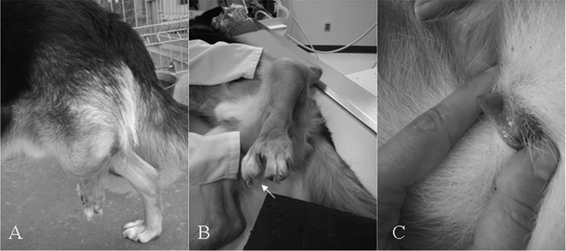
Photographs of the left hind leg of a male German Shepherd Dog mix showing (A) limb deformities and joint contractures, (B) supernumerary metatarsi and phalanges (arrow), and (C) ectopic vulva and hypertrophic clitoris.
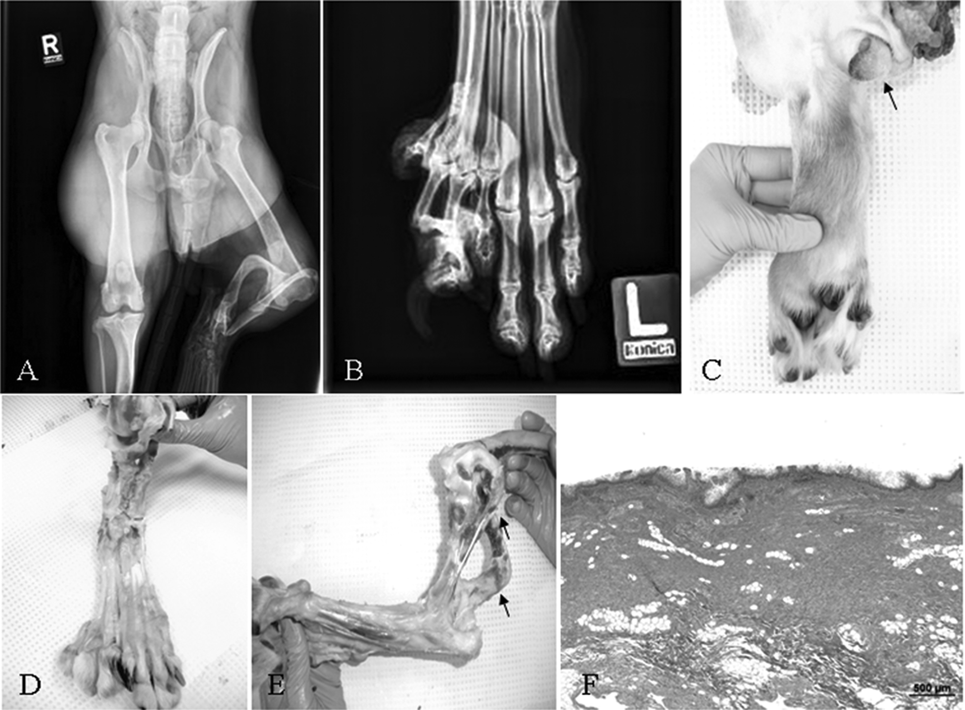
Radiographic, macroscopic, and histopathological analyses. A, ventrodorsal radiographic view of the coxofemoral, femorotibial and tibiotarsal joints showing the misshapen femorotibial and tibiotarsal joints with skeletal muscle atrophy. B, rostrocaudal radiographic view of the left hind paw showing the supranumeral metatarsi and phalanges. C, picture of the resected left hind leg showing the location of the rudimentary female external genitalia (arrow) and the duplication of the lower limb with 2 paws. D, rostrocaudal view of the left hind leg demonstrating the supranumeral (medial) fibula, enlarged tarsus with supranumeral tarsal bones, and supranumeral metatarsies associated with complete digits and appendices. Findings suggest the fusion of 2 lower limbs. E, lateral view of the resected left hind leg demonstrating a dysmorphic femorotibial joint, 2 fibulas (lateral and medial) bent at a 90-degree angle (arrows), and a short, stubby tibia. F, photomicrograph (hematoxylin and eosin stain) of the vaginal mucosa and submucosa characterized by a stratified squamous epithelium on a thick fibrovascular propria-submucosa. Bar = 500 µm.
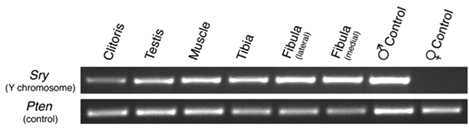
Polymerase chain reaction (PCR) genotype analysis. The PCR was conducted on genomic DNA from tissues from the parasitic twin (clitoris), the male dog (testis), and tissues that may have originated from either animal (muscle, tibia, medial, and lateral fibulas). Control tissues were uterus and testis taken from unrelated dogs following routine elective sterilization procedures. The PCR procedures amplified specific regions of the Y chromosome gene SRY (positive result is indicative of XY genotype), or the unrelated control gene Pten to control for DNA sample integrity. The PCR samples were separated by 2% agarose gel electrophoresis, stained with ethidium bromide, and photographed under ultraviolet light.
The embryonic development of the male gonad and reproductive tract is triggered by the activation of the SRY gene. This in turn activates a male-specific genetic program that culminates in the production of testosterone by the Leydig cells, which supports the differentiation of the Wolffian ducts into male sex organs, as well as the production of anti- Müllerian hormone (AMH) by the Sertoli cells, which activates the degradation of the Müllerian ducts. 1 Androgen and AMH production by the gonad also appears essential for the proper development of male external genitalia, as defects in the production or action of either hormone in XY mammals results in ambiguous or female external genitalia.1,6 In the current case, it therefore seems likely that the genitalia of the parasitic twin did not receive the required developmental signals, presumably due to the loss or non-development of its own gonads. The developmental signals originating from the gonads of the autosite were presumably not sufficient to ensure proper masculinization of the parasitic twin, or were not available to it during the critical developmental period during which the development of the external genitalia normally occurs.
Footnotes
The author(s) declared no potential conflicts of interest with respect to the research, authorship, and/or publication of this article.
The author(s) received no financial support for the research, authorship, and/or publication of this article.