
Abstract
A 2-year-old Pekinese dog was diagnosed with hepatic yersiniosis. Grossly, white-to-yellow nodules consisting of degenerated inflammatory cells, cell debris, and bacterial clumps were scattered throughout the liver. Histopathologically, suppurative and necrotizing hepatitis was apparent. Yersinia enterocolitica biotype 4, serotype O3 (4:O3) was identified and confirmed in the liver immunohistochemically, using a monoclonal antibody. The virulence genes ystA and ail were detected, but the isolate was negative for autoagglutination and calcium-dependent growth. To confirm systemic yersiniosis in animals, it is imperative that the organism(s) be identified because the hepatic lesions are similar to those of Y. pseudotuberculosis and other diseases, including plague, which is also a zoonotic pathogen.
Keywords
Yersinia enterocolitica has been isolated from mammals, birds, and rodents, as well as from the environment, such as soil and water. 1,3,11 It is also a well-known food-borne pathogen in human beings. 1 In domestic animals, yersiniosis, caused by Yersinia enterocolitica, is usually sporadic and limited to the gastrointestinal system, such as enteritis. 3 In dogs, it is considered commensal, because the organism is recovered from the feces of healthy dogs, which are a known reservoir. 6,7 It is sometimes associated with enteritis in young dogs. 5,14 Although myocardial yersiniosis caused by Y. enterocolitica O9 has been reported in a Rottweiler puppy, 17 systemic Y. enterocolitica infection in an adult dog is very rare. The current report describes hepatic yersiniosis caused by Y. enterocolitica 4:O3 in an adult dog.
A 2-year-old Pekinese dog was rescued by a local shelter in Incheon, western Korea, in late March 2006. The dog developed lethargy and anorexia 2 days after its rescue. Although the dog was treated with antimicrobials and critical care, its condition deteriorated, and it was euthanized. The carcass was referred to the Animal Disease Diagnostic Center of the National Veterinary Research and Quarantine Service (Anyang, Republic of Korea), where a necropsy was performed. The brain, lung, liver, heart, spleen, and kidney were fixed in 10% buffered formalin for histopathology. The fixed tissues were processed routinely and stained with hematoxylin and eosin. The liver was aseptically inoculated on sheep's blood and MacConkey agar and aerobically incubated at 37°C for 48 hr. Nonhemolytic and lactose-negative colonies were seen on the blood and MacConkey agar after incubation for 24 hr. Gram-negative coccobacilli were identified using an automated system. a The bacilli were biotyped using standard methods, as described previously, 1 and serotyped using commercial rabbit O antisera. b Yersinia enterocolitica ATCC9610 was used as a positive control. To determine the virulence of the organism, virulence genes including ail, ystA, ystB, inv, yadA, and virF were amplified by polymerase chain reaction (PCR). 16 Briefly, all the reactions were separately carried out in 50-μl reaction volumes containing 5 μl of DNA template, 25 μl of PCR mix, c and 2 μM of concentration of each forward and reverse primer. The PCR was performed with a thermal cycler d under the same conditions described previously. 16 Afterward, 10 μl of amplicon was analyzed by electrophoresis on a 2.5% agarose gel. Phenotypic features such as autoagglutination and calcium-dependent growth were also determined by the previously described methods. 9,12 With a mouse monoclonal antibody against Y. enterocolitica O3, e immunohistochemistry confirmed the existence of the organism in tissue.
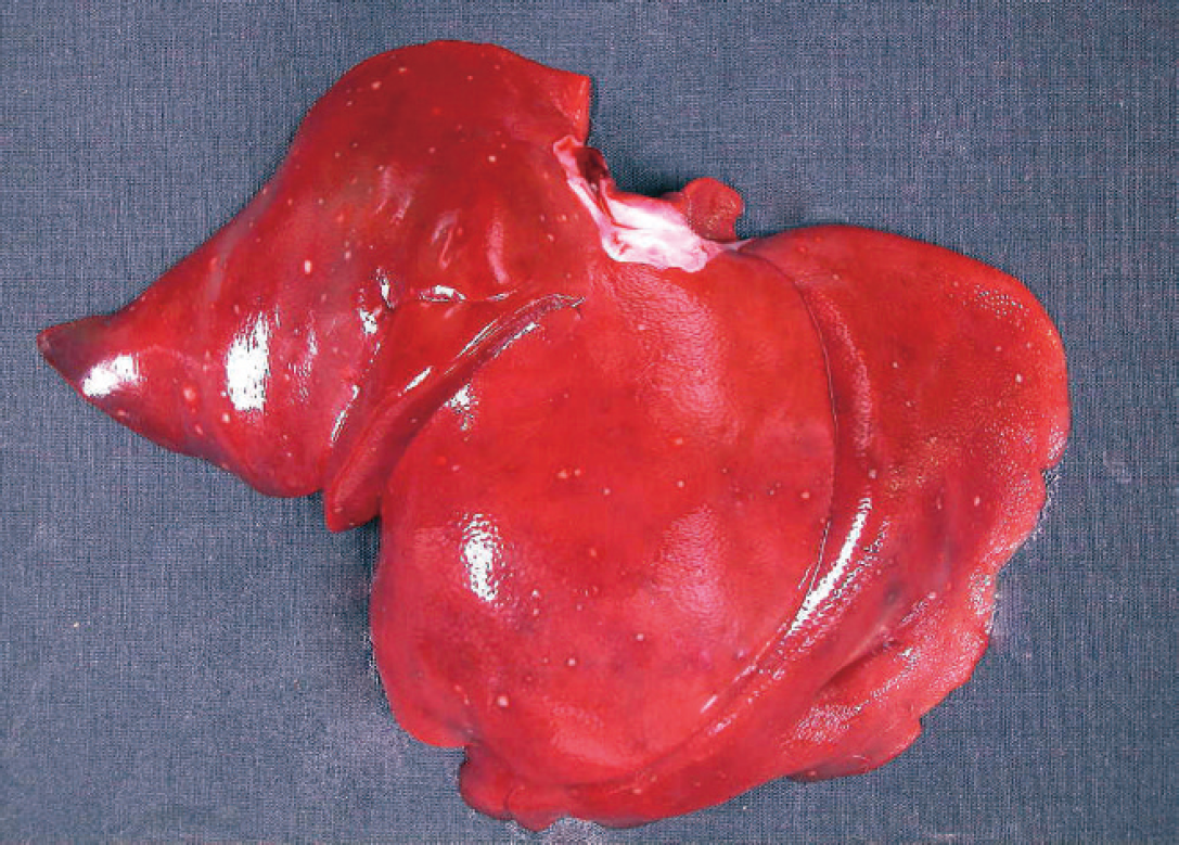
White-to-yellow foci are scattered diffusely throughout the liver.
Grossly, white-to-yellow foci were scattered diffusely throughout the liver (Fig. 1). The liver was the only organ that showed gross changes. Histopathologically, the necrotic nodules were mixed with inflammatory cells, cell debris, and bacterial clumps (Fig. 2). The inflammatory cells were primarily neutrophils and macrophages, although lymphocytes and plasma cells were also observed. Necrotizing hepatocytes and bacterial colonies were seen at the outer margin of the lesion. The bacterial colonies reacted strongly to a monoclonal antibody for Y. enterocolitica O3. Some of the necrotic lesions were surrounded by a thin connective tissue layer. Lymphocytes and macrophages were also infiltrated in the portal area. The hepatic cords were collapsed in the necrotic area; however, the hepatic capsule was not affected. Hepatic yersiniosis was confirmed by bacterial isolation and immunohistochemistry. Of the virulence genes tested, the ail and ystA genes were detected in the isolate. The isolate did not show autoagglutination or calcium-dependent growth.
Yersinia enterocolitica is part of the normal intestinal flora of dogs and is associated with enteritis in young dogs subject to stress, such as cold. 3,11 There are few reports on systemic infection caused by Y. enterocolitica. In the present case, hepatic yersiniosis was confirmed by isolating Y. enterocolitica 4:O3 and immunohistochemistry of the lesion. The lesions were similar to those of systemic Y. pseudotuberculosis infection in cats and wild animals. 10,13 Virulence genes and phenotypical tests, such as autoagglutination, have been used to identify the pathogenic isolates. 9,12,16 The isolate was positive for ail and ystA but negative for autoagglutination and calcium-dependent growth. The ail and ystA genes are responsible for attachment and invasion locus and the production of heat-stable enterotoxin, respectively. 1,16 Immunohistochemistry is widely used to identify the causative agents in diagnostic laboratories. The bacteria seen in the liver reacted positively with a monoclonal antibody for Y. enterocolitica O3. It would be difficult to confirm the source and route of the infection due to the limited history available. Generally, stray dogs are likely to contact materials contaminated with pathogenic organisms due to their free-ranging nature. The consumption of infected animals and food contributes to Yersinia spp. infection in domestic animals. 3 It is possible that the dog in the current study ate pork or by-products contaminated with Y. enterocolitica. This hypothesis is supported by the fact that the area where the dog was rescued was located between a large abattoir for pigs and a conventional market with a large number of butchers. 7 Several studies 4,8,11 have suggested that pork is associated with the transmission of Y. enterocolitica 4:O3 to dogs. In addition, in Korea, Y. enterocolitica O3 has predominantly been isolated from pigs and dogs. 15 The dog was thought to be starving and stressed, which would make it more susceptible to pathogens. 2 Accordingly, an intestinal infection could have developed opportunistically and caused the lesions in the liver. To diagnose yersiniosis, it is important to identify the organism(s) because the hepatic lesions are similar to those of Y. pseudotuberculosis and other diseases, including plague, which is also a zoonotic pathogen. To the authors' knowledge, this is the first report of hepatic yersiniosis caused by Y. enterocolitica 4:O3 in an adult dog.
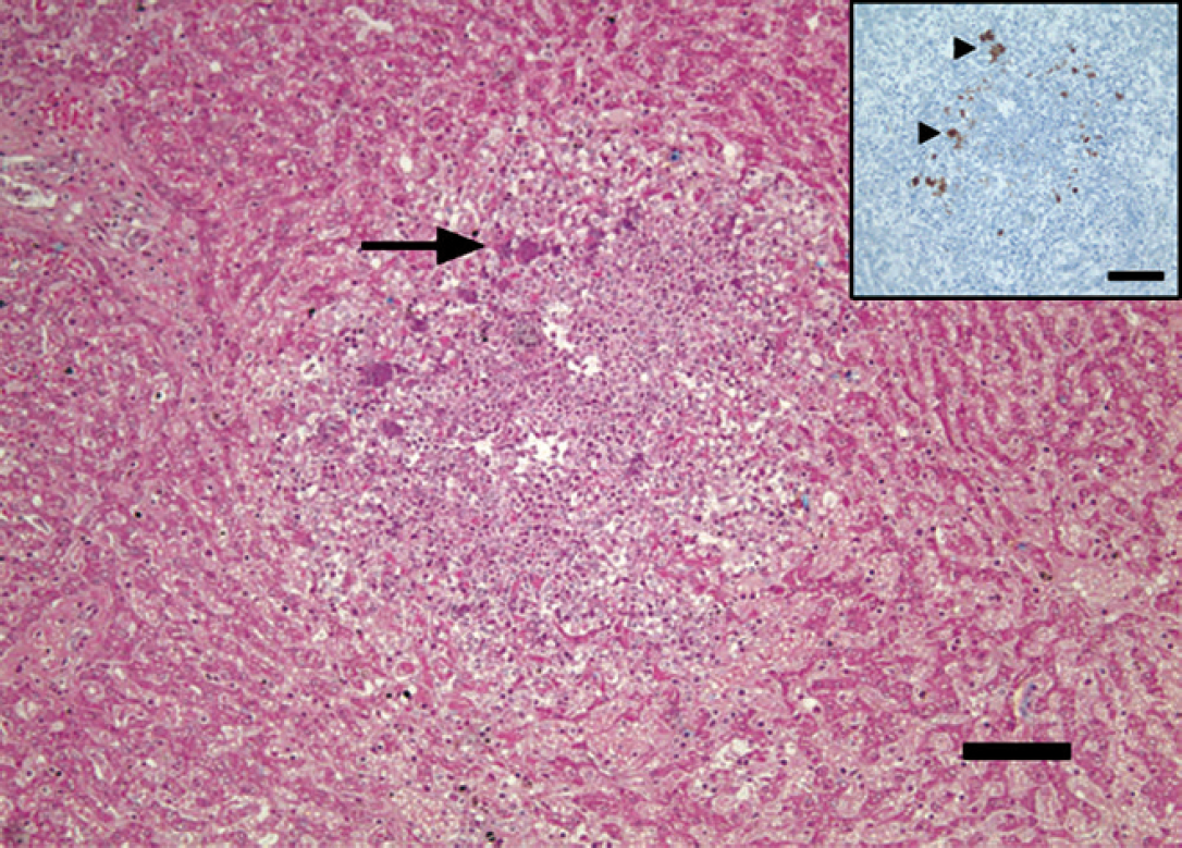
The necrotic area of liver contains inflammatory cells, primarily neutrophils, degenerative hepatocytes, and bacterial colonies (arrow). Hematoxylin and eosin. Bar = 100 μm.
Footnotes
a.
VITEK® 2, bioMérieux Inc., Durham, NC.
b.
Denka Seiken Co. Ltd., Tokyo, Japan.
c.
EmeraldAmp®, TaKaRa Bio Inc., Otsu, Shiga, Japan.
d.
GeneAmp® PCR System 9700, Applied Biosystems, Foster City, CA.
e.
PROGEN Biotechnik GmbH, Heidelberg, Germany.