
Abstract
Bacteria of the genus Helicobacter are associated with disease in humans and animals. Reports of infection in reptiles are very limited. In the present study, pathological findings and molecular characterization are reported for a Helicobacter species associated with septicemia in a pancake tortoise (Malacochersus tornieri). Disseminated infection resulted in regional cellulitis and edema of the head and neck, and pericarditis. Spiral bacteria were identified in cytological preparations and Warthin-Starry-stained sections of pathological lesions. Amplification of partial sequence of the 16S ribosomal gene using polymerase chain reaction identified the organism as Helicobacter and suggest that it is a novel species.
Keywords
Helicobacter (phylum Proteobacteria, class Epsilonprote-bacteria, order Campylobacterales, family Helicobacteraceae) are a genus of spiral-shaped, Gram-negative bacteria that are phylogenetically distinct from bacteria within the phylum Spirochaetes. In mammals and birds, Helicobacter have been associated with gastric ulcers, ulcerative colitis, cholangiohepatitis, cellulitis, and bacteremia. 5,7,10
Reported cases of infection by spiral bacteria in reptiles are very limited. The first documented case described the detection and ultrastructural characterization of spiral-shaped bacteria associated with septicemia in a moribund rhinoceros iguana (Cyclura cornuta). 6,9 This bacterium was not cultured or otherwise further identified. To the authors' knowledge, the only other relevant report in reptiles is a brief abstract describing gastritis in chelonians associated with Helicobacter species (Busch MD, Terio KA, Low-enstine L, et al.: 2002, Gastritis associated with Helicobacter infections in chelonians (order Testudines). In: Proceedings of the American Association of Zoo Veterinarians Annual Conference, pp. 82-83, Milwaukee, WI). The apparent rarity of documented cases may be caused in part by fastidious growth requirements of Helicobacter, which limits detection by routine culture techniques. The present report describes a case of septicemia caused by a probable novel Helicobacter species in a pancake tortoise (Malacochersus tornieri) resulting in regional cellulitis and pericarditis. Pathologic findings are described, and initial genetic characterization is presented.
An adult male pancake tortoise was among a large group of tortoises confiscated from smugglers (6 months prior to the tortoise's death) and was subsequently transferred to a zoological institution where it was housed for 8 days. The tortoise drank, but would not eat, and became progressively weaker and lethargic over the 24 hr preceding death. Severe edema of the head and neck, and weakness of the forelimbs were noted near the time of death.
On gross examination, the tortoise was found to be in good nutritional condition. External abnormalities included marked edema of entire neck region and eyelids (Fig. 1). Upon examination of the coelomic viscera, the pericardial sac appeared thickened by edema and contained 2.0 ml of cloudy, serous fluid. Thin strands of fibrin were diffusely adhered to the epicardium, and multifocal white foci were observed on the surface of the ventricle. Cytological examination of the pericardial fluid revealed myriad spiral bacteria 6.0–10.0 μm in length that were distributed within a granular, proteinaceous background (Fig. 2). Spiral bacteria also were observed in organ cytological impressions of lung, liver, spleen, and kidney and were interpreted to reflect organisms in circulation. Tissues were fixed in 10% neutral buffered formalin and processed by routine histological methods.
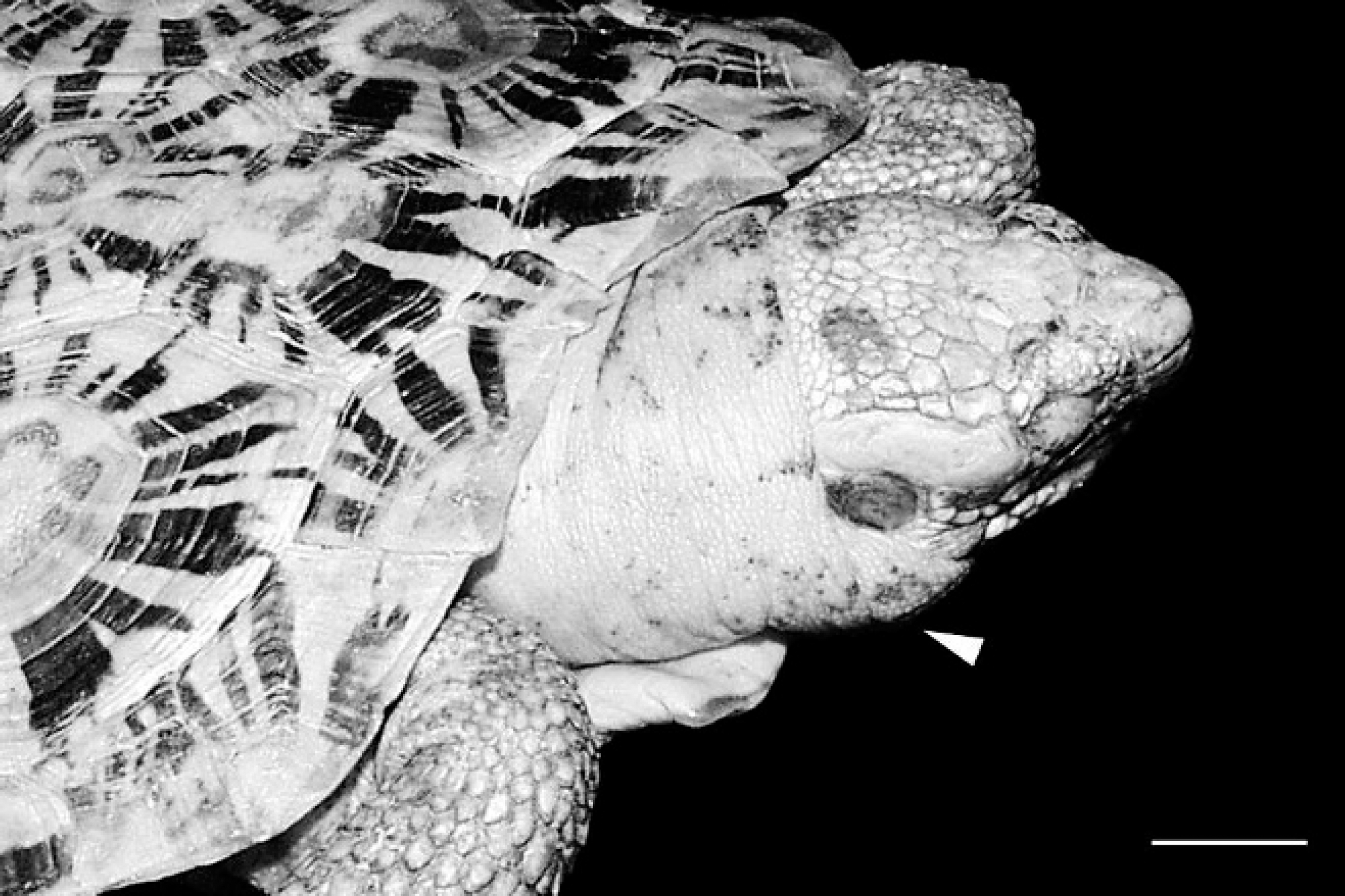
Pancake tortoise (Malacochersus tornieri). The neck region and eyelids are markedly edematous because of regionally extensive cellulitis. Bar = 11 cm.
Histological examination of the integument and underlying soft tissue of the neck region revealed severe edema and diffuse infiltration of deep tissues by heterophils and macrophages. Lymphatic vessels contained coagula of fibrin and leukocytes interspersed with spiral bacteria (Fig. 3). Acute degeneration of associated myofibers also was observed. Similar findings were noted in the tongue and pharyngeal region. In addition, large numbers of intact and degranulated heterophils and fewer macrophages infiltrated the epicardium and multifocally extended into the subjacent myocardium (Fig. 2). Many cardiac myofibers were undergoing degeneration and necrosis within areas of myocardial infiltration. Abundant spiral bacteria were interspersed with the inflammation and were intensely stained with Warthin-Starry silver stain. A similar, mild to moderate inflammatory infiltrate expanded the pulmonary interstitium, and edema of various organs and tissues was observed. No hepatic changes were present. Lastly, there was moderate to severe hyperplasia of the mucosa of the small intestine associated with numerous apical round protozoa (1.0–5.0 μm in diameter), consistent with Cryptosporidium species. No spiral bacteria were observed in Warthin-Starry-stained sections of stomach (body and pylorus), small intestine, and colon. No bacterial growth was obtained from a postmortem heart blood sample. Culture media used included Columbia blood agar with 5% sheep blood, MacConkey agar, and colistin nalidixic acid agar. All plates were incubated under aerobic conditions at 25°C.
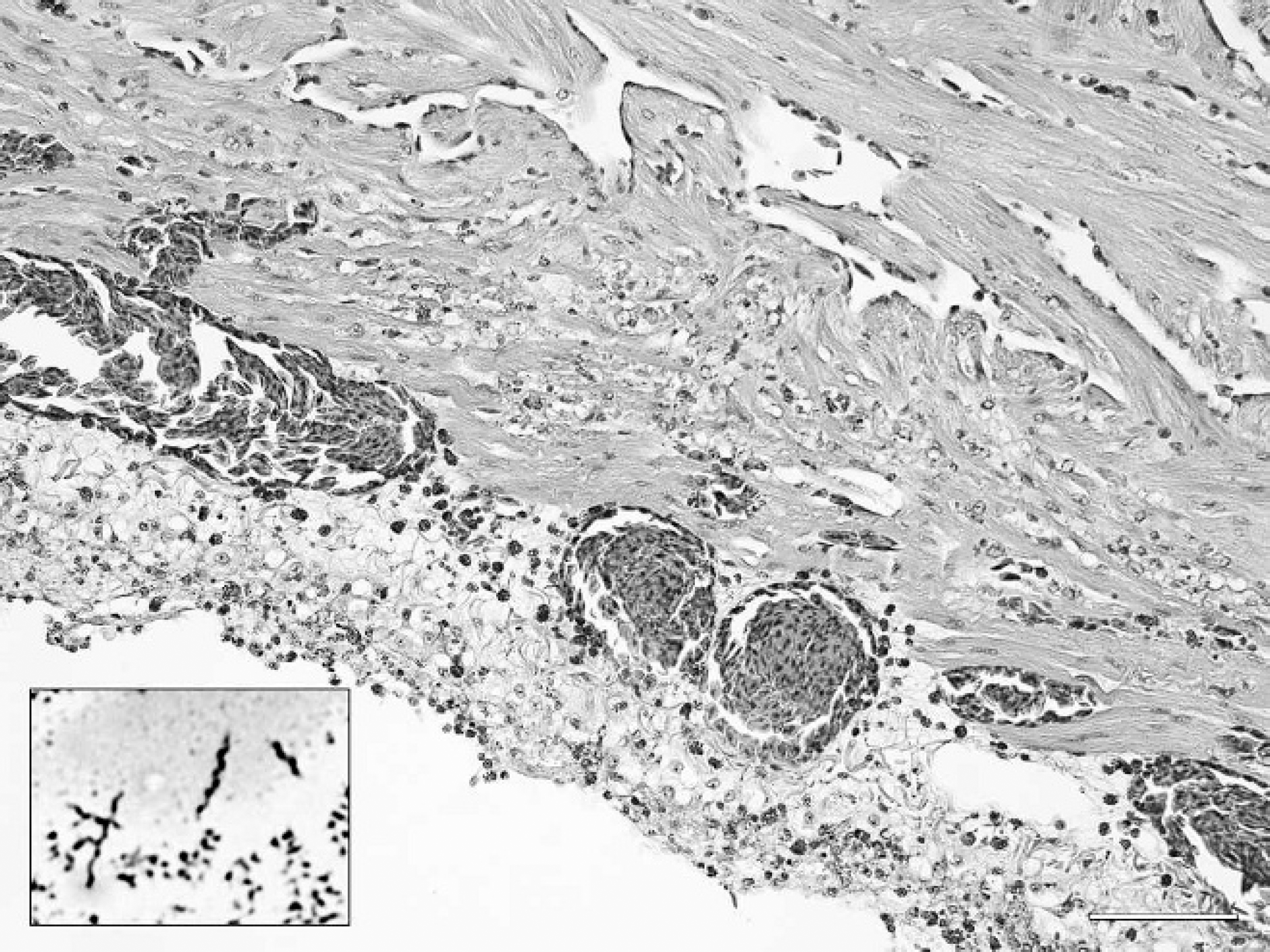
Pancake tortoise (Malacochersus tornieri); epicardium. The epicardium and subjacent myocardium are infiltrated by large numbers of heterophils and macrophages. Hematoxylin and eosin. Bar = 100 μm. Inset: A cytological preparation of pericardial fluid containing numerous spiral bacteria (Helicobacter sp.) is shown. Wright-Giemsa stain. Bar = 15 μm.
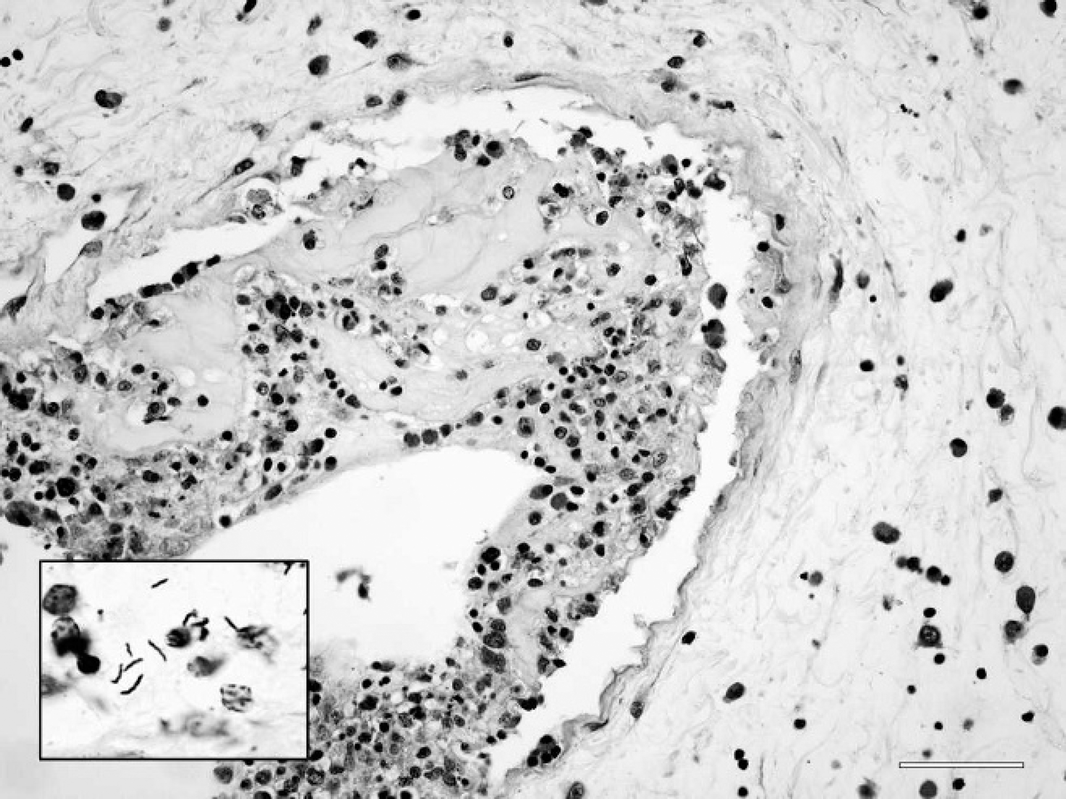
Pancake tortoise (Malacochersus tornieri); deep lymphatic vessel, neck region. Inset: The lymphatic vessel contains a coagulum of leukocytes and fibrin interspersed with spiral bacteria (Helicobacter sp.). Warthin-Starry stain. Bar = 30 μm. The surrounding stromal elements are separated by edema in infiltrated by heterophils and fewer macrophages. Hematoxylin and eosin. Bar = 50 μm.
To identify the spiral bacteria, DNA was extracted from pericardial fluid using a commercial tissue kit. a Previously described polymerase chain reaction (PCR) methods 8 were used to amplify a 1,370-bp region of the bacterial 16S ribosomal RNA (rRNA) gene. Direct sequencing was performed using a commercial kit b and analyzed on automated DNA sequencers. c Sequence from the pancake tortoise bacteria was submitted to GenBank (accession no. FJ667779) and was compared with other sequences in GenBank (National Center for Biotechnology Information, Bethesda, MD), EMBL (Cambridge, United Kingdom), and Data Bank of Japan (Mishima, Shizuoka, Japan) databases using BLASTN. 1 BLASTN results indicated that the sequence obtained from the pancake tortoise was that of a Helicobacter sp., and the highest score was obtained for Helicobacter fastidiosus sp. nov. 16S small subunit rRNA gene, with which there was 94% identity (GenBank accession no. AM992061). Thus, a diagnosis of disseminated Helicobacter infection was based on the spiral morphology of bacteria in pathologic lesions, abundance of these bacteria in the pericardial fluid used for PCR, and high degree of genetic homology between the tortoise bacteria and characterized Helicobacter species.
Although Helicobacter appear to be common in many clinically normal individuals of multiple species, changing environmental factors may result in significant disease. For example, both wild and captive cheetahs (Acinonyx jubatus) are colonized by the same Helicobacter species; however, associated disease is prevalent in captive cheetahs with higher stress levels and is virtually absent in wild cheetahs. A similar host-pathogen relationship is plausible in chelonians. Furthermore, animals seized from wildlife smuggling operations, as occurred in the present case, undoubtedly are exposed to densities of individuals and other species that would not be encountered under natural conditions. Exposure of immunologically naïve animals, unsanitary conditions, stress of capture and transport, and other suboptimal environmental conditions provide ample opportunity for transmission of pathogens and manifestation of disease. This scenario is of critical importance to the ultimate disposition of confiscated animals, which may pose a significant health risk to both captive populations of receiving institutions and wild populations if release is elected.
Infection in the pancake tortoise manifested as septicemia with regional cellulitis of the head and neck, and pericarditis. Bacteremia, cellulitis, and edema have been reported in humans associated with Helicobacter cinaedi, Helicobacter westmeadi, and Helicobacter rappini infections. 5 Based on the known pathogenesis of other Helicobacter, it is likely that the bacteria in the pancake tortoise originated from the alimentary tract. 5 However, neither gastroenteric colonization nor pathological lesions consistent with Helicobacter infection were observed. The proliferative enteritis seen in the current case was attributed to cryptosporidiosis, and no mucosal-associated spiral bacteria were observed. It is possible that the mucosal injury associated with cryptosporidiosis may have provided the mechanism for translocation of bacteria from the gastroenteric tract and predisposed the tortoise to disseminated infection.
The zoonotic significance of this organism remains to be determined. Some Helicobacter species from diverse hosts may have zoonotic potential and have been associated with disease in both animals and humans. 3,5
Failure to isolate the Helicobacter species in the present report was not surprising given the standard methods utilized for diagnostic culture and the difficulties in isolating these fastidious organisms. 5 Molecular detection and characterization were very helpful in identifying the organism and provided a timely and relatively cost effective means of obtaining a diagnosis.
Standards for describing new species of Helicobacter have been reported, and involve several biochemical tests, sequence data from the 16S rRNA, DNA-DNA hybridization, and the examination of a minimum of 5 strains for morphology, staining behavior, and motility. 4 Thus, characterization of the organism given in the present report does meet the criteria for designation of bacterial species status. Nonetheless, comparison across a relatively conserved region of the 16S RNA gene indicated that genetic distances between the tortoise Helicobacter sp. and other Helicobacter species were comparable with interspecific differences between recognized species of Helicobacter. This difference suggests that the bacterium detected in the pancake tortoise is a novel species.
The present report expands upon the only other previously documented cases of Helicobacter infection in chelonians, which were all limited to associated gastritis, and the range of species known to become infected. The prevalence and association with disease in pancake tortoises and other chelonians requires further study, as does Helicobacter infection in other reptiles.
Footnotes
a.
DNeasy®, Qiagen, Valencia, CA.
b.
BigDye® Terminator Kit, Applied Biosystems, Foster City, CA.
c.
ABI 3130, Applied Biosystems, Foster City, CA.