
Abstract
The present report describes a case of fibroma with osseous metaplasia of the external auditory canal in a 7-year-old male Pomeranian dog. Upon otoscopic examination, the right external auditory canal was almost completely obstructed by a well-circumscribed mass adjacent to the eardrum. The mass was surgically excised. Grossly, it was well demarcated, firm when cut, pink, and measured 0.3 cm × 0.2 cm × 0.7 cm. The cut surface of the mass exhibited a central portion of homogeneously white osseous components surrounded by brown to pink soft tissue. Microscopically, the resected external auditory canal mass mainly consisted of fibroblastic spindle cells showing differentiation to metaplastic osteoblast-like cells. Metaplastic osteoblasts and osteoclasts lining the osteoid bony trabeculae were also observed. Bony trabeculae and spicules were separated by abundant collagen and neoplastic fibroblastic cells. Fibromatous components, irregular formation of woven bone spicules, and the presence of osteoblasts lining bony trabeculae led to a diagnosis of fibroma with osseous metaplasia.
Aural tumors have been reported infrequently in dogs and other animals. 10,11 A study of 81 dogs with tumors of the ear canal reported that most ear canal tumors were of ceruminous glandular epithelial and squamous epithelial origin. 11 Fibroma is a benign tumor composed of mesenchymal fibrocytes with abundant collagenous stroma. 13 In veterinary medicine, fibromas are not common, but they are most often reported in dogs. 13 In dogs, fibromas are reported to be commonly found on the limbs and the head. 13 However, to the authors' knowledge, fibromas with osseous metaplasia have not been reported previously in the external auditory canal of dogs. The present report describes a rare case of fibroma with osseous metaplasia that occurred in the external auditory canal in a dog.
A 3-year-old male Pomeranian dog was presented to a local veterinary hospital because of a hemorrhagic otitis externa of the right ear. On physical examination, the dog appeared to have no signs of disease. Serum biochemical and complete blood count test results were within reference intervals. Upon otoscopic examination, the right external auditory canal was almost completely obstructed by a proliferative and well-circumscribed mass adjacent to the eardrum (Fig. 1). Bloody to crusty discharge also exuded from the ear canal mass. The dog was treated with a topical antibiotic; however, there was no noticeable response. On cytological examination, spindle-shaped stromal cells with oval nuclei and scant cytoplasm were observed. The stromal cells were regular in shape, and a few neutrophils were also observed. Surgically, the ear canal mass was excised, and the mass was sent to the Department of Veterinary Pathology, Kyungpook National University (Daegu City, Republic of Korea), for histopathological examination.
Grossly, the ear canal mass was well demarcated, firm when cut, pink, and measured 0.3 cm × 0.2 cm × 0.7 cm. The cut surface of the mass exhibited a central portion of homogeneous white osseous components surrounded by brown to pink soft tissue (Fig. 1). The resected mass was immediately fixed in 10% neutral buffered formalin, processed routinely, embedded in paraffin wax, and sectioned at 4 μm in thickness. The sections were stained with hematoxylin and eosin for microscopic examination. To identify the collagenous fibers and bony trabeculae, Masson's trichrome stain was used. Microscopically, the resected external auditory canal mass was mainly composed of fibroblastic spindle cells with differentiation to metaplastic osteoblasts-like cells (Fig. 2). The metaplastic osteoblasts formed and surrounded woven bone trabeculae and spicules (Fig. 2). The woven bone trabeculae and spicules were separated by abundant collagen and neoplastic fibroblastic cells (Fig. 3). The fibrous stroma of the mass was moderately vascularized and exhibited moderate infiltrates of neutrophils, lymphocytes, and plasma cells. Neoplastic spindle cells were characterized by elongated ovoid nuclei, scant cytoplasm, and indistinct cell borders. Mitotic figures were not observed. Osteoclasts were adjacent to bony spicules in some parts of the mass. The epidermal layer showed inward growth with mild spongiosis, and most parts of the epidermis were desquamated. The deep part of the mass was connected to the ear canal, and many cholesterol clefts were also observed. In Masson's trichrome stain, the bony trabeculae and spicules were differentiated clearly.
In dogs, aural tumors have been reported to occur uncommonly. 19 In addition, tumors of the ear canal in dogs constituted 2% to 6% of all tumors in dogs admitted for aural surgery. 10 A study reported that 33 benign tumors of the ear canal in dogs consisted of 8 polyps, 6 papillomas, 5 sebaceous gland adenomas, 5 basal cell tumors, 4 ceruminous gland adenomas, 2 histiocytomas, 1 plasmacytoma, 1 benign melanoma, and 1 fibroma. 11 Moreover, in that study, 48 malignant tumors of the ear canal in dogs consisted of 23 ceruminous gland adenocarcinomas, 9 carcinomas of undetermined origin, 8 squamous cell carcinomas, 3 round cell tumors, 2 sarcomas, 2 malignant melanomas, and 1 hemangiosarcoma. 11 These results indicate that most ear canal tumors are derived from ceruminous glandular epithelium and squamous epithelium lining the auditory canal.
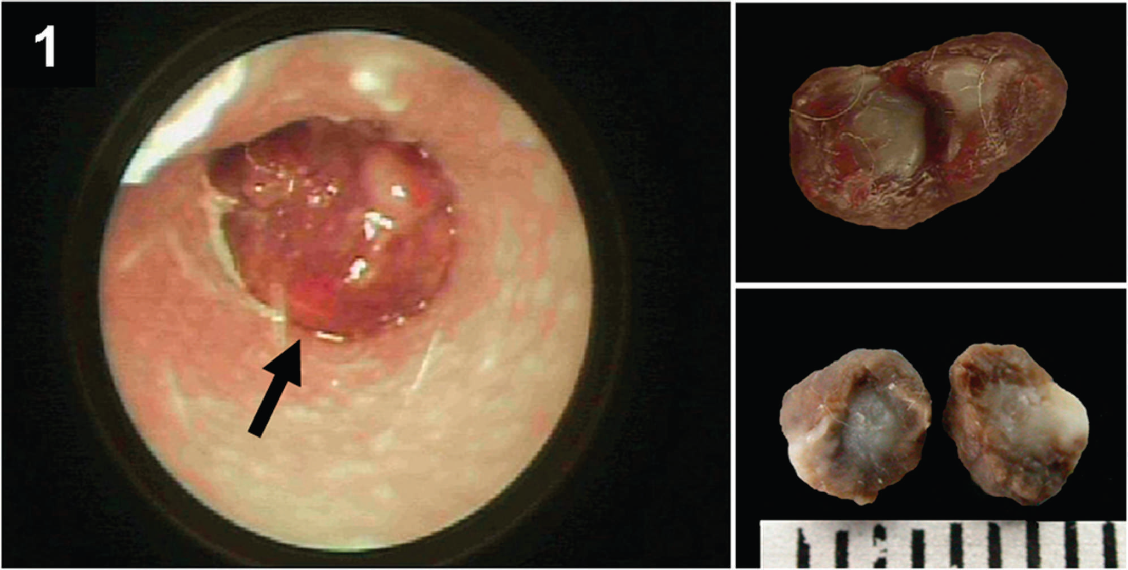
Otoscopic examination indicates a mass (arrow) obstructing the external auditory canal. Top inset, well-demarcated mass measuring 0.3 cm × 0.2 cm × 0.7 cm, and Bottom inset, a cross section of the mass showing a homogeneous white component in the center.
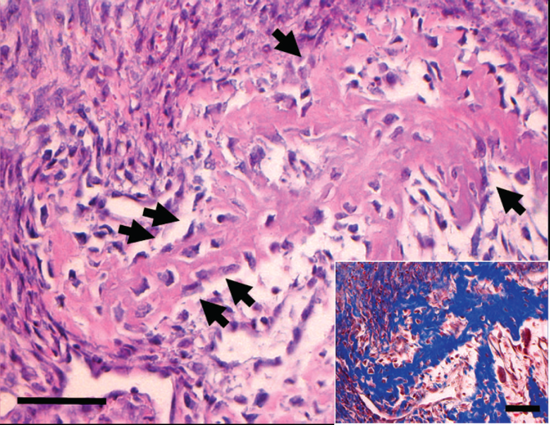
Irregular woven bone trabeculae rimmed by a row of fibroblasts undergoing transformation into osteoblasts (arrows). Hematoxylin and eosin stain. Bar = 50 μm. Inset, Masson's trichrome stain. Bar = 50 μm.
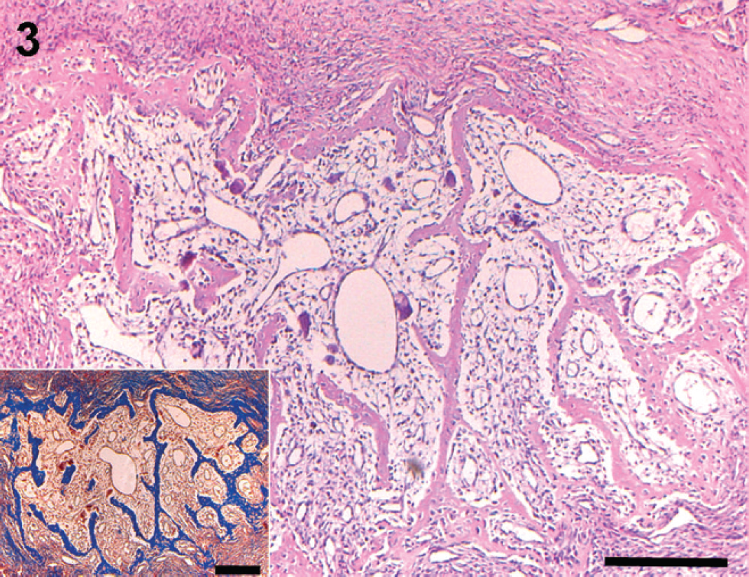
Ossifying component in the central part surrounded by cellular fibrous stroma. Hematoxylin and eosin stain. Bar = 200 μm. Inset, Masson's trichrome stain. Bar = 200 μm.
Osseous metaplasia is one type of ectopic ossification of fibrous connective tissue. 16 Osseous metaplasia indicates an alternative differentiation of fibroblast-like cells into osteoblasts producing osteoid components. 16 Therefore, the metaplastic bone formed by this process develops directly from soft connective tissue. Although fibromas can occur wherever there is fibrous connective tissue, only 1 case of fibroma of the external auditory ear canal has been reported in the veterinary literature to date to the author's knowledge. 11 Moreover, fibromas with osseous metaplasia have not been reported before in veterinary medicine. In human beings, only a few cases of extraskeletal mesenchymal tumors with osseous metaplasia have been reported, including 6 lipomas, 1 3,5 , 7,18 1 fibromyxoid sarcoma with osseous metaplasia, 20 1 ossifying epithelioid hemangioendothelioma, 8,1 fibroma with osseous metaplasia. 9 Therefore, the present case might represent a highly unusual case of extraskeletal mesenchymal tumors with osseous metaplasia.
Osteoma, ossifying fibroma, and fibrous dysplasia belong to a miscellaneous group of benign lesions found primarily in intramembranous bone, which can be confused with other fibro-osseous lesions. 14 Therefore, ossifying fibroma, osteoma, and fibrous dysplasia should be differentiated from other fibro-osseous lesions. 12,15 The present case exhibited numerous fibroblastic spindle cells that showed differentiation to metaplastic osteoblast-like cells forming bony trabeculae. The collagenous fibers produced by fibroblastic neoplastic cells and bony trabeculae were confirmed through Masson's trichrome stain, which was consistent with previously reported ossifying fibromas in animals. However, ossifying fibroma is an expansile intraosseous lesion that can be a lytic and invasive mass developing in intramembranous bone, especially the mandible. 15 In the present case, the aural mass was well demarcated from adjacent tissues. Moreover, the mass did not appear to arise from intramembranous bone grossly or microscopically. Thus, the present case was differentiated from ossifying fibromas.
Chronic otitis has been a well-known clinical sign of ear canal tumors. 4 Chronic otitis was believed to be a pivotal factor in the development of tumors in human beings. 4,6 A previous veterinary publication has described a close relationship between chronic otitis and the development of ear canal tumors. 19 Aural tumors are often accompanied by bacterial infections due to partial or complete obstruction of the ear canal. 19 Moreover, it has been hypothesized that hyperplasia of epithelial cells and adnexa occurring in chronic otitis progresses to dysplastic changes or neoplastic transformation. 19 Therefore, it is thought that long-standing localized inflammation plays an important role in the development of aural tumors. In the present study, the dog had a hemorrhagic otitis externa of the right ear canal that has previously been reported in cases of ear canal tumor in animals and human beings. Histologically, the resected ear canal tissue had moderate infiltrates of neutrophils, lymphocytes, and plasma cells (chronic active inflammation), suggesting that the formation of fibroma with osseous metaplasia in the external ear canal was associated with chronic otitis. It is possible that chronic otitis led to neoplastic transformation, but it seems equally likely that the presence of a neoplastic process resulted in what could clinically appear to be chronic otitis. Therefore, the precise primary cause of otitis externa in the present case could not be determined. The primary treatment of benign ear canal tumors is surgery in human beings and animals.
Too few cases of tumors of the ear canal have been reported to demonstrate the absolute characteristics of these neoplasms. Thus, many veterinary pathologists may have difficulty distinguishing an aural bony mass with osseous metaplasia from other bone tumors, especially osteoma and ossifying fibroma. Since osseous metaplasia and ossifying fibromas can resemble osteomas, the distinction between them can sometimes be obscure. 17 In the present case, the diagnosis of fibroma with osseous metaplasia rather than ossifying fibroma and osteoma was based on the observation that the mass was not derived from the intramembranous bone tissue but arose from soft tissues.
Acknowledgements
This work was supported by a grant (code: CBM 31-B3003-01-01-00) from the Center for Biological Modulation of the 21st Century Frontier R&D Program, the Ministry of Science and Technology, Republic of Korea.