
Abstract
A 3.5-year-old, male, neutered ferret (Mustela putorius furo) was presented with a 3-day history of lethargy and anorexia. Splenic aspirates revealed high numbers of intermediate-sized lymphocytes and Mott cells interpreted as lymphoma with Mott cells. The ferret was euthanized because of a poor clinical prognosis. Postmortem examination revealed markedly enlarged spleen and lymph nodes, with multifocal white nodules in the liver parenchyma. Histologically, the spleen had multifocal large nodules composed of neoplastic lymphocytes with frequent Mott cells. Similar neoplastic cells were present in the sections of liver, lymph nodes, and bone marrow. These cells were cluster of differentiation (CD)3-negative, CD79α-positive, and lambda light-chain—positive. Electron microscopy revealed that the cytoplasm of the neoplastic Mott cells had increased, disorganized, dilated, rough endoplasmic reticulum containing electron-dense immunoglobulin. On the basis of cytologic, histopathologic, immunohistochemical, and electron microscopic findings, a malignant B-cell lymphoma with Mott cell differentiation was diagnosed.
Keywords
In ferrets, lymphoma is one of the 3 most common neoplasms, after adrenal cortical neoplasms and insulinoma. The prevalence of lymphoma in ferrets ranges from 11.9% to 29.2%. 2,3 Lymphoma in ferrets has been classified into multicentric, gastrointestinal, mediastinal, cutaneous, and extranodal forms according to the site of origin, with no particular age association. 13 Several variants of lymphoma exist in ferrets. The lymphocytic form is most common and occurs in ferrets older than 2 years of age. Affected ferrets typically display a chronic course of disease with involvement of visceral organs, especially the spleen, liver, kidneys, and lymph nodes. 28 Younger ferrets (less than 2 years of age) more commonly develop the lymphoblastic variant, which typically involves mediastinum, thymus, spleen, liver, and bone marrow. An immunoblastic polymorphous type can occur at all ages and have varying degrees of peripheral lymphadenopathy and visceral organ involvement. 28 The current report describes the clinical, microscopic, immunohistochemical, and transmission electron microscopic features of a malignant B-cell lymphoma with Mott cell differentiation in a ferret.
A 3.5-year-old, male, neutered ferret was presented to the exotics medicine service of Louisiana State University Veterinary Teaching Hospital and Clinics (Baton Rouge, LA) with a 3-day history of lethargy and anorexia. On physical examination, the ferret was slightly dehydrated, and other parameters were within the normal limits. No abnormalities could be palpated in the abdomen except for moderate splenomegaly. Results of the complete blood cell count were unremarkable except for hemolyzed plasma, a minimal increase in hematocrit percentage (62.3%; reference interval: 44–61%). The red blood cell count (RBC) and hemoglobin concentration were within reference intervals. The spun packed cell volume (PCV) was 57% (reference interval: 48–59%). The only abnormality noted on the serum biochemical profile was hypokalemia (potassium 3.9 mmol/l; reference interval: 4.5–7.7 mmol/l), which was attributed to anorexia. Urinalysis was unremarkable. An abdominal ultrasound examination revealed a severely enlarged spleen with multiple, ill-defined, hypoechoic nodules. Multiple, irregular, slightly enlarged (6 mm in diameter) lymph nodes having heterogeneous echogenicity were seen at the level of the portal vein and mesentery. Anechoic cysts measuring 4 mm and 1 cm in diameter were present in the right and left kidneys, respectively.
Fine-needle aspirate smears of the spleen were collected by ultrasound guidance and stained with Wright—Giemsa. The smears contained high numbers of nucleated cells with adequate cellular preservation. Numerous erythrocytes were present along with platelets. There were numerous intermediate-sized (15–20 μm in diameter) lymphocytes with round nuclei, finely stippled chromatin, frequent prominent single to multiple nucleoli, and scant to small amounts of mildly basophilic cytoplasm (∼50%). Fewer small lymphocytes were present. There were also numerous (>50% of the nucleated cells) Mott cells of variable size (20–30 μm). The Mott cells had single, round to oval, eccentric nuclei with clumped chromatin and mild anisokaryosis. The cytoplasm of these cells was densely packed with numerous, well-demarcated, round to oval, variably sized, clear to light blue, Russell bodies (Fig. 1A). There were many erythroid and myeloid precursors, low numbers of megakaryocytes, and fewer plasma cells. A cytologic interpretation of lymphoma with Mott cell differentiation was made. Because of the unavailability of a serum sample, a plasma sample was submitted for protein electrophoresis. The results of electrophoresis revealed a total protein of 6.0 g/dl with normal albumin and globulin fractions and no evidence of a monoclonal peak in the globulin fraction. The ferret was euthanized because of a poor clinical prognosis and submitted for postmortem examination.
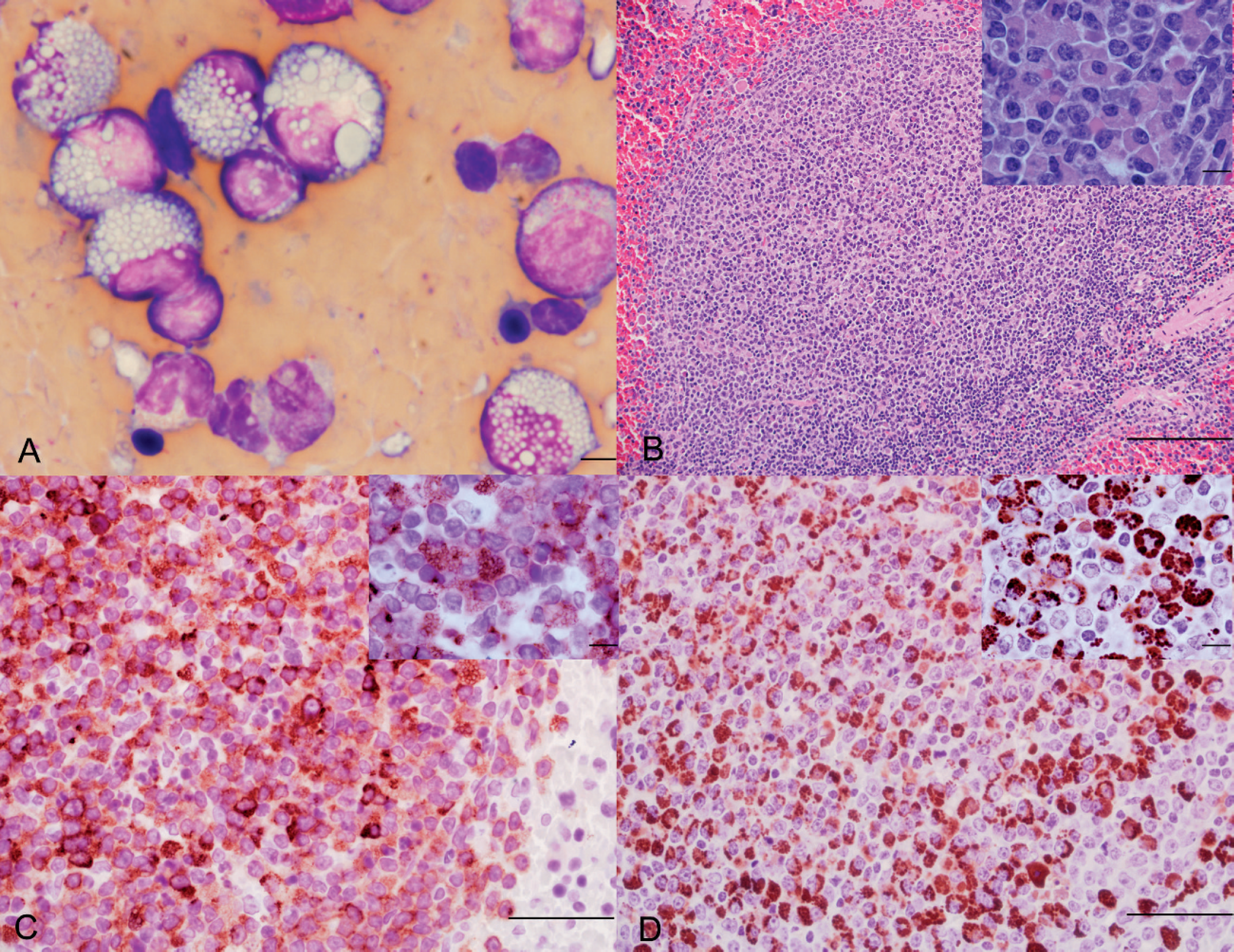
Spleen; ferret (Mustela putorius furo).
Postmortem examination revealed bilaterally enlarged prescapular lymph nodes (3 mm in diameter) and marked enlargement of mediastinal (6 mm in diameter) and mesenteric (2 cm × 2 cm × 3 cm) lymph nodes. The spleen was severely enlarged (4 cm × 10 cm × 1 cm). On cut surface, the liver had multifocal, 1–2-mm-diameter, white nodules in the parenchyma. No other major gross findings were noticed. The tissue samples were fixed in 10% neutral buffered formalin. The samples were routinely processed, paraffin embedded, sectioned at 5 μm, and stained with hematoxylin and eosin. Histologically, the spleen was characterized by multifocal large nodules composed of neoplastic lymphocytes. Approximately, 50–60% of the periarteriolar lymphoid sheaths were expanded with unencapsulated, infiltrative, moderately cellular, well-demarcated nodules of neoplastic lymphocytes arranged in sheets. The neoplastic cells had variably distinct borders and contained minimal amounts of eosinophilic cytoplasm. Nuclei were round to oval with vesiculated chromatin and 1 or 2 nucleoli. Mitoses averaged 1 per 10 high-power fields of view. The neoplastic cell population frequently contained plasmacytoid cells containing large numbers of eosinophilic, intracytoplasmic globules consistent with Russell bodies (Mott cells; Fig. 1B). The globules in the Mott cells were periodic acid—Schiff (PAS) positive. In liver sections, the neoplastic lymphocytes predominately infiltrated portal areas but also surrounded central veins and replaced the hepatic parenchyma with nodular aggregates of neoplastic lymphocytes containing small numbers of Mott cells. The architecture of lymph nodes (mesenteric, prescapular, mediastinal) and bone marrow were severely and diffusely effaced by the lymphoma. Only 1 section of bone marrow was examined, but the tissue was effaced with neoplastic lymphocytes. Bone marrow from different locations was not examined.
Immunohistochemical staining of formalin-fixed, paraffin-embedded replicate tissue sections was performed as described by the manufacturer with the use of rabbit polyclonal anti-human cluster of differentiation (CD)3 antibody (code no. A0452), mouse monoclonal anti-human CD79α antibody (clone no. HM57), and rabbit polyclonal anti-human kappa and lambda light-chain antibodies (code nos. A0191 and A0193). a The positive control tissue was a section of lymph node and spleen from a healthy rabbit. The negative control tissue was identical but lacked application of the primary monoclonal antibody. Almost all neoplastic lymphocytes were positive for CD79α (Fig. 1C) and were negative for CD3. The Mott cells stained strongly positive for λ light chain (Fig. 1D) and negative for κ light chain.
For transmission electron microscopic examination, 1-mm 3 fragments of fresh spleen were collected at postmortem examination and fixed in 3% glutaraldehyde, postfixed in 1% osmium tetroxide, dehydrated in ethanol, and embedded in liquid epoxy resin. b The ultrathin sections (70–90 nm) were stained with lead citrate and uranyl acetate and examined with an electron microscope. c Ultrastructurally, many Mott cells were identified throughout the tissue sections (Fig. 2). Mott cells had a single, round to oval, eccentric nucleus that contained prominent heterochromatin with rare prominent nucleoli. The cytoplasm of these cells contained differing amounts of variably dilated, disorganized cisternae of endoplasmic reticulum containing electron-dense, mostly homogeneous to rarely flocculent proteinaceous material consistent with immunoglobulin. No Golgi apparatuses were seen in the neoplastic cells.
The cytologic, histologic, immunohistochemical staining (CD3—, CD79α+) and electron microscopic morphology of the neoplastic cells in the current case were indicative of B-cell lymphoma with Mott cell differentiation. Most lymphomas in ferrets have been T cell in origin, and most commonly affect lymph nodes, spleen, liver, small intestine, mediastinum, bone marrow, lungs, and kidney. 18 Splenomegaly, a common finding observed in ferrets with lymphoma, 7 was also observed by abdominal ultrasonography and postmortem examination in the present case. The lymphoma in this ferret was unusual because of Mott cell proliferation among the neoplastic lymphocytes. Recently, lymphoma with Mott cell differentiation in dogs has been reported 3 times. 6,16,24 These cases primarily involved the gastrointestinal tract and lymph nodes 6,16,24 ; a single case had multiple nodules in spleen, liver, and lung. 6,16,24 In contrast, the gastrointestinal tract was not affected in the ferret in the current report; however, marked splenomegaly, generalized lymphadenomegally, and scattered white nodules in the liver were present. Benign plasma cell neoplasms in dogs and cats are usually irregularly positive for lambda and kappa light chains. 26 However, atypical plasma cell neoplasms are more likely positive only for lambda chain, which was consistent with the present report. The positive PAS staining, positive CD79α and lambda light-chain immunohistochemical staining, and electron microscopic findings were consistent with previous case reports of lymphoma with Mott cells in dogs. 16,24 An earlier report in a 4-year-old pig described follicular lymphoma involving various lymph nodes with Russell body—type inclusions, 14 which was also consistent with the current study.
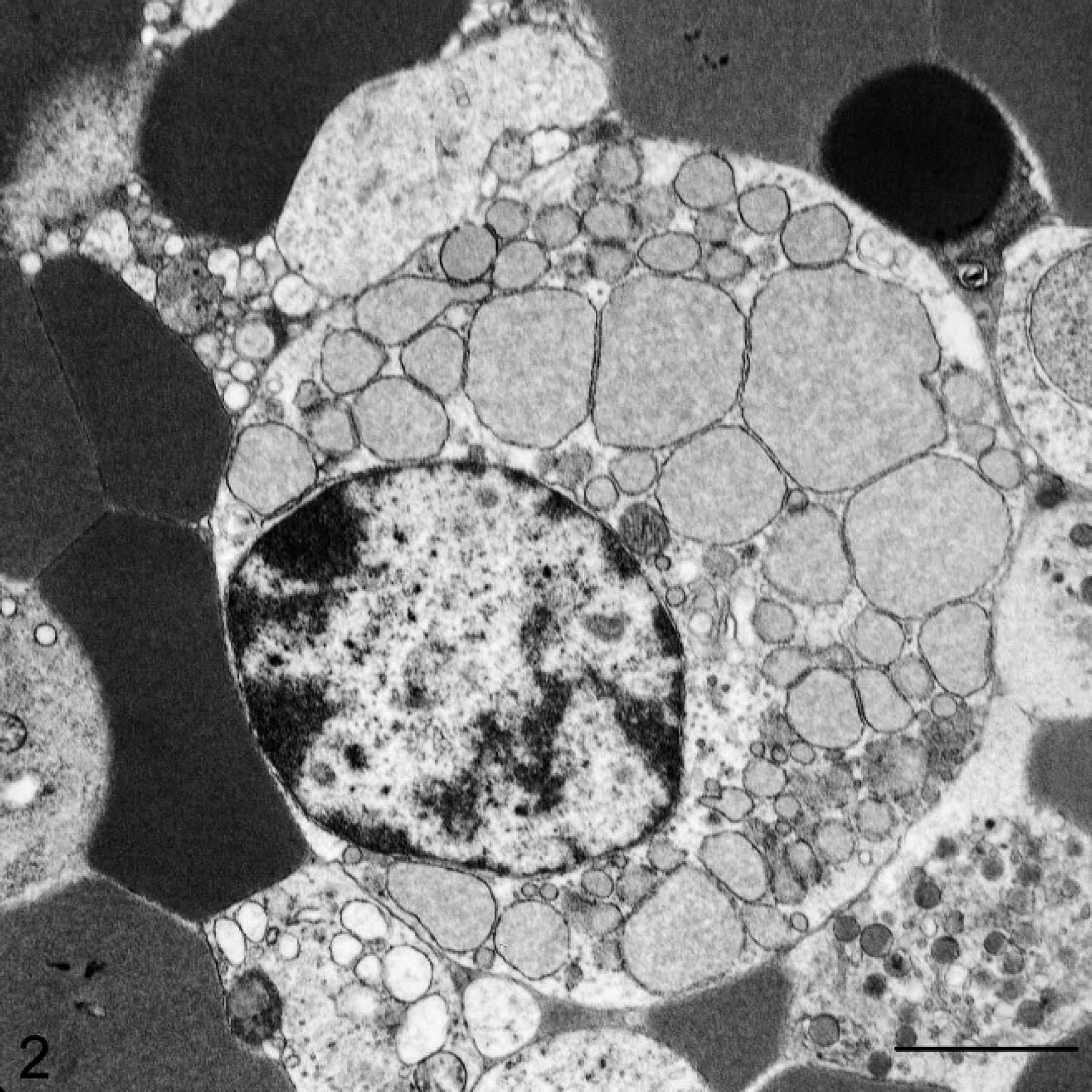
Spleen of a ferret (Mustela putorius furo) with Mott cell lymphoma. An ultrastructural view of a Mott cell reveals an eccentric nucleus, marginated heterochromatin, and dilated cisternae of the endoplasmic reticulum. Electron-dense material within the endoplasmic reticulum is consistent with immunoglobulin (Russell body inclusions). Transmission electron micrograph. Bar = 2 μm.
The ferret in the present study was middle aged (3.5 years old), but acute onset of clinical signs along with rapid progression of disease was more typical for the juvenile form of ferret lymphoma. Lymphopenia has been observed commonly in older ferrets with lymphoma, whereas lymphocytosis has been more common in younger ferrets with lymphoma. 7 Neither change was observed in this ferret, which was consistent with a previous study in ferrets with lymphoma. 1 In the presence of a normal RBC count, hemoglobin concentration, and PCV, a minimal increase in the hematocrit was considered clinically insignificant.
Paraproteinemia was not evident; however, small amounts of protein might not have been detected by plasma protein electrophoresis because of its low analytical sensitivity. In cats and cattle, lymphomas have been reported to be associated with Feline leukemia virus 5,23 and Bovine leukemia virus, 4 respectively. However, association between lymphomas in ferrets and retroviral infection has not been confirmed. 9,10 A specific etiology was not pursued in the present case.
The intense PAS staining of the cytoplasmic globules and their localization in the dilated cisternae of the rough endoplasmic reticulum on transmission electron microscopic examination were consistent with Russell body formation. Russell body formation in nonneoplastic Mott cells represents a state wherein immunoglobulin is produced at a faster rate than it can be secreted. 25 Ultrastructurally, Russell bodies appear as electron-dense flocculent material in the lumen of the rough endoplasmic reticulum. 12 However, neoplastic Mott cells could lack cellular organelles and the differentiation of cisternal rough endoplasmic reticulum, which are required for the secretion of immunoglobulin and is consistent with the findings in the current study. Russell bodies have been described in a number of immunoproliferative disorders in humans, including lymphoplasmacytic lymphomas, plasmacytoma, and plasma cell dyscrasia. 17,19,21,22 Another condition in humans, known as Russell body gastritis, occurs in association with Helicobacter pylori infection and is primarily associated with plasma cells and Mott cells. Although the lymphoma in the present study had a densely monomorphous appearance, the Mott cells were considered nonneoplastic because of the polyclonal immunoreactive pattern to immunoglobulin light chains. 19,20 However, in the present case, Mott cells were considered neoplastic because of its monoclonal immunoreactivity to lambda light chain. A previous study 11 reported a Mott cell tumor of the stomach in humans with H. pylori infection and suggested that H. pylori might have influenced the immunological environment and resulted in the formation of Mott cells. In ferrets with Helicobacter mustelae infection, gastric B-cell lymphoma similar to gastric mucosa—associated lymphoma associated with H. pylori infection in humans has been reported. 8 It is intriguing to speculate that lymphoma with Mott cell differentiation in the current case might have originated from Helicobacter spp. infection; however, bacterial infection was excluded given the absence of gastrointestinal lesions at postmortem examination, but the possibility of a chronic infection with neoplastic transformation cannot be excluded.
Because of the plasmacytoid appearance of neoplastic Mott cells in the present case, the differential diagnoses of extramedullary plasmacytoma, lymphoplasmacytic lymphoma, and signet ring cell lymphoma were considered. The solitary extramedullary plasmacytomas are most commonly seen in the skin and upper and lower gastrointestinal tract of dogs, and frequently exhibit binucleation and multinucleation. 26 The typical morphology of an extramedullary plasmacytoma was not observed in the present case. Signet ring lymphomas morphologically contain a single, large, clear vacuole that displaces the nucleus to the periphery of the cell. 15 The lymphoplasmacytic lymphoma consists of small to intermediate lymphocytes. 27 However, typical morphologic features of signet ring cell lymphoma and lymphoplasmacytic lymphoma were not seen in the current case.
The lymphoma with Mott cell differentiation reported in this report was diagnosed on the basis of cytologic and histologic examination, immunohistochemical staining, and transmission electron microscopy. This neoplasm shared many histologic and most of the immunophenotypic features common to lymphoma with Mott cell differentiation of dogs and humans and might prove to be a useful model to understand the pathogenesis of onset of Mott cell lymphoma.
Acknowledgements The authors thank Ms. Julie Millard for conducting the immunohistochemistry.
Footnotes
a.
EnVision+ System-HRP Labeled Polymer (DAB), Dako North America Inc., Carpinteria, CA.
b.
Epon™, Hexion Specialty Chemicals, Columbus, OH.
c.
JEM-1011, JEOL Ltd., Tokyo, Japan.