
Abstract
A newborn Holstein female calf had neoplastic lesions in the skin and within the thoracic and abdominal cavities but not in the bone marrow, spleen, thymus, or most lymph nodes. Because the tumor cells were positive for CD79a (B cell marker), CD5 (B-1 cell marker) and terminal deoxynucleotidyl transferase (marker for immature lymphoid precursors), a diagnosis of precursor B-1 B cell lymphoma was made. The diagnosis was strongly supported by the fact that B-1 cells can develop in the fetus, unlike B-2 cells, which are produced after birth. The lymphoma was distinct from the typical calf form of lymphoma of B-2 cell origin, which does not express CD5 and is characterized by generalized lymphadenopathy and involvement of the bone marrow, blood and spleen.
Human and murine B cells consist of 2 distinct subsets, B-1 cells and B-2 cells. 10,15 B-1 cells are characterized by the expression of CD5 and are distinct from B-2 cells by anatomical location, gene usage, function, and pheno-type. 15 B-1 cells can develop in unusual sites in the fetus, such as the liver and the omentum, whereas B-2 cells can be produced in the secondary lymphoid organs after birth. 10 In humans, CD5 is expressed in most cases of chronic lymphocytic leukemia (CLL) and mantle cell lymphoma (MCL) and in some cases of diffuse large B cell lymphoma, which might arise from B-1 cells. 1,15 Lymphomas in adult cattle infected with bovine leukemia virus can be immunohistochemically divided into 3 types: B-1a, B-1b, and B-2. 17 In contrast, B cell lymphomas in calves do not express CD5 and CD11b, and are grouped into the B-2 cell type. 17 This paper describes, in a newborn calf, a B cell lymphoma that was considered to be of peritoneal B-1 cell origin.
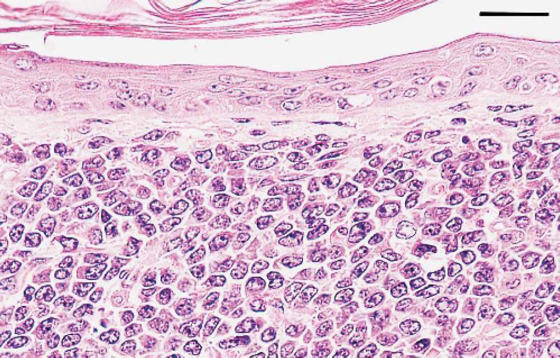
Skin; calf. Neoplastic cells characterized by scant cytoplasm are present in the dermis. HE. Bar = 25 μm.
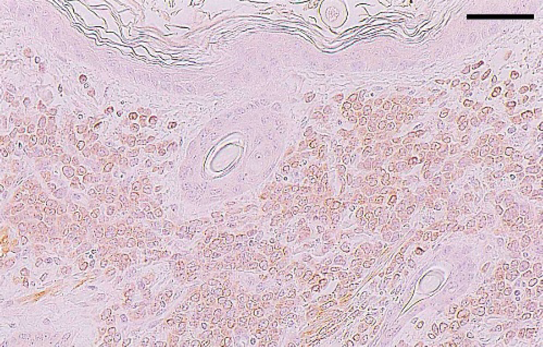
Skin; calf. CD79a-positive neoplastic cells are visible in the dermis, but there are no positive cells in the epidermis and hair follicles. IHC. Bar = 50 μm.
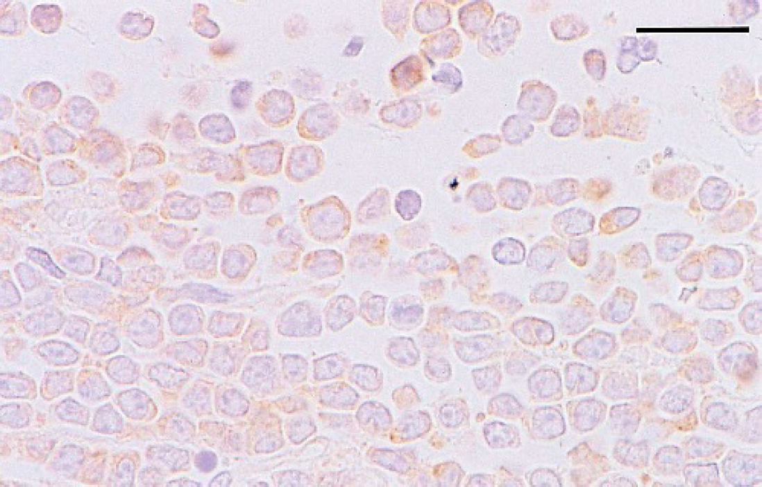
Mesentery; calf. Almost all tumor cells express CD5 in the cytoplasm. IHC. Bar = 20 μm.
A Holstein female calf was euthanized because of an inability to stand, rotation of the head, and multiple cutaneous masses. At necropsy, a large number of tumors, up to 8 × 6 × 6 cm, were present in the subcutis and the dermis or in the subcutis of the neck, trunk, and limbs. They were grayish white and soft, and were well demarcated from surrounding tissue. Both eyeballs protruded because of peribulbar tumor masses. There were tumors in the serosa of the rumen, the reticulum, and the abomasum. Prominent areas of the mesentery and the omentum were replaced by tumor masses, and there were diffuse nodules and plaques on the parietal peritoneum. A few tumors were seen in the mediastinum near the thymus, but the thymus appeared intact. Slightly raised areas were evident on some parts of the pleura.
Tissue samples were fixed in 10% buffered formalin, embedded in paraffin, sectioned at 4 μm, and stained with hematoxylin and eosin (HE). Microscopic examination revealed diffuse neoplastic growths in the mesentery and the omentum, but foci of infiltrating neoplastic cells were rare in the muscularis or the submucosa of the small and large intestines. Near the hepatic hilus, the pancreas was focally invaded by tumor cells. Neoplastic cells invaded the hepatic capsule but not the parenchyma. Sparse neoplastic infiltrates were observed in the muscularis of the rumen, the omasum, the abomasum, and the uterus. In addition, the submucosa was affected in the abomasum. A lesion in the reticulum extended from the serosa to the lamina propria. Gross lesions of mesentery, pleura, mediastinum, skin, and orbit also were composed of sheets of neoplastic cells. In the skin, neoplastic cells in the subcutis extended into the dermis or underlying skeletal muscle, but were less dense (Fig. 1). Although the medulla of some mediastinal lymph nodes were replaced by tumor cells, there were no neoplastic cells in mesenteric, hepatic, and pancreaticoduodenal lymph nodes; spleen; thymus; or bone marrow.
Neoplastic cells were round to oval, and 4–10 μm in diameter, with cleaved or lobulated nuclei. Chromatin was finely clumped, the nucleoli were small or medium sized, and the cytoplasm was scant. Erythrophagia by tumor cells was rare. Mitotic figures were numerous, at a rate of 15 to 30 per 400 × field.
To further characterize the tumor cells, transmission electron microscopy (TEM) and immunohistochemistry (IHC) was performed on formalin-fixed tissues and paraffin sections, respectively. Staining for IHC was performed by an avidin-biotin-peroxidase complex method with avidin-biotin-peroxidase complex kits. a Primary antibodies were rabbit polyclonal antibodies to CD3, b CD5, c and terminal deoxynucleotidyl transferase (TdT), b and a mouse monoclonal antibody to CD79a. b The sections were pretreated by enzymatic digestion with pepsin (CD3) or microwave heating (CD5, TdT, CD79a). The neoplastic cells were positive for CD79a (Fig. 2) and CD5 (Fig. 3), and were negative for CD3; TdT-positive neoplastic cells were frequently seen (Fig. 4) with IHC.
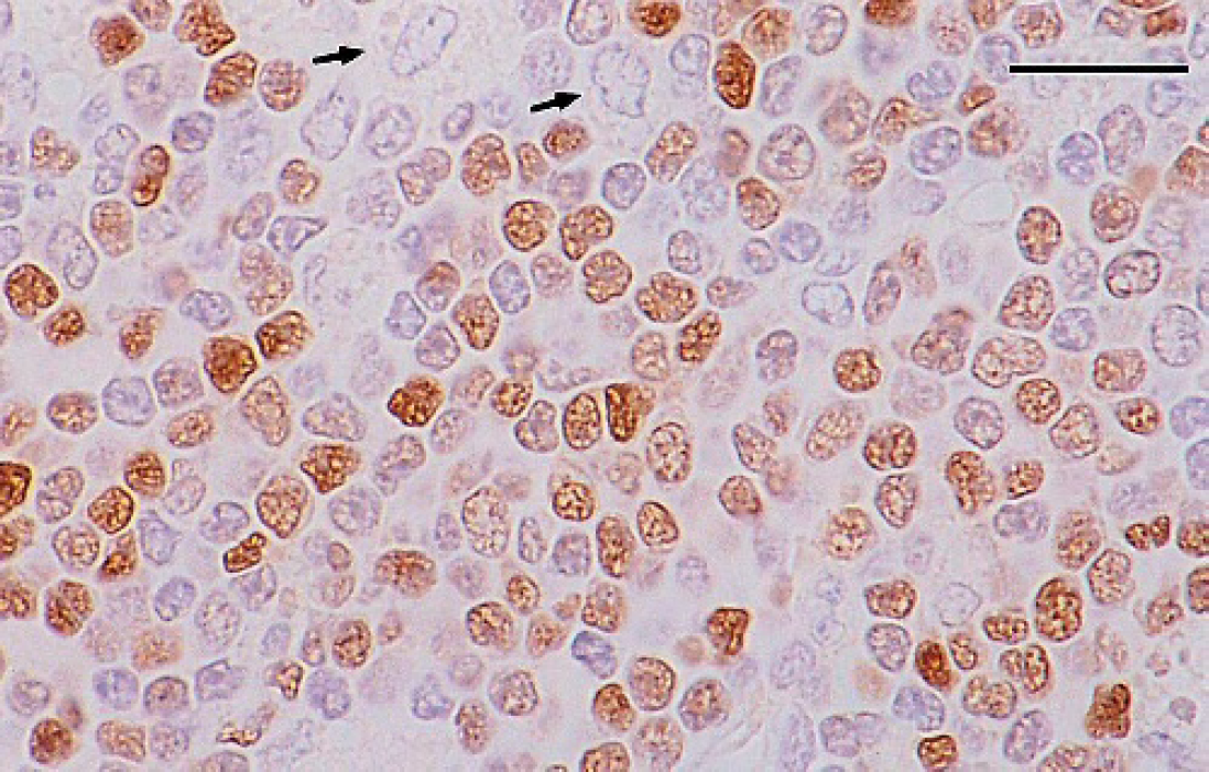
Mesentery; calf. Many neoplastic cells show variable nuclear staining for TdT. Macrophages are negative (arrows). IHC. Bar = 20 μm.
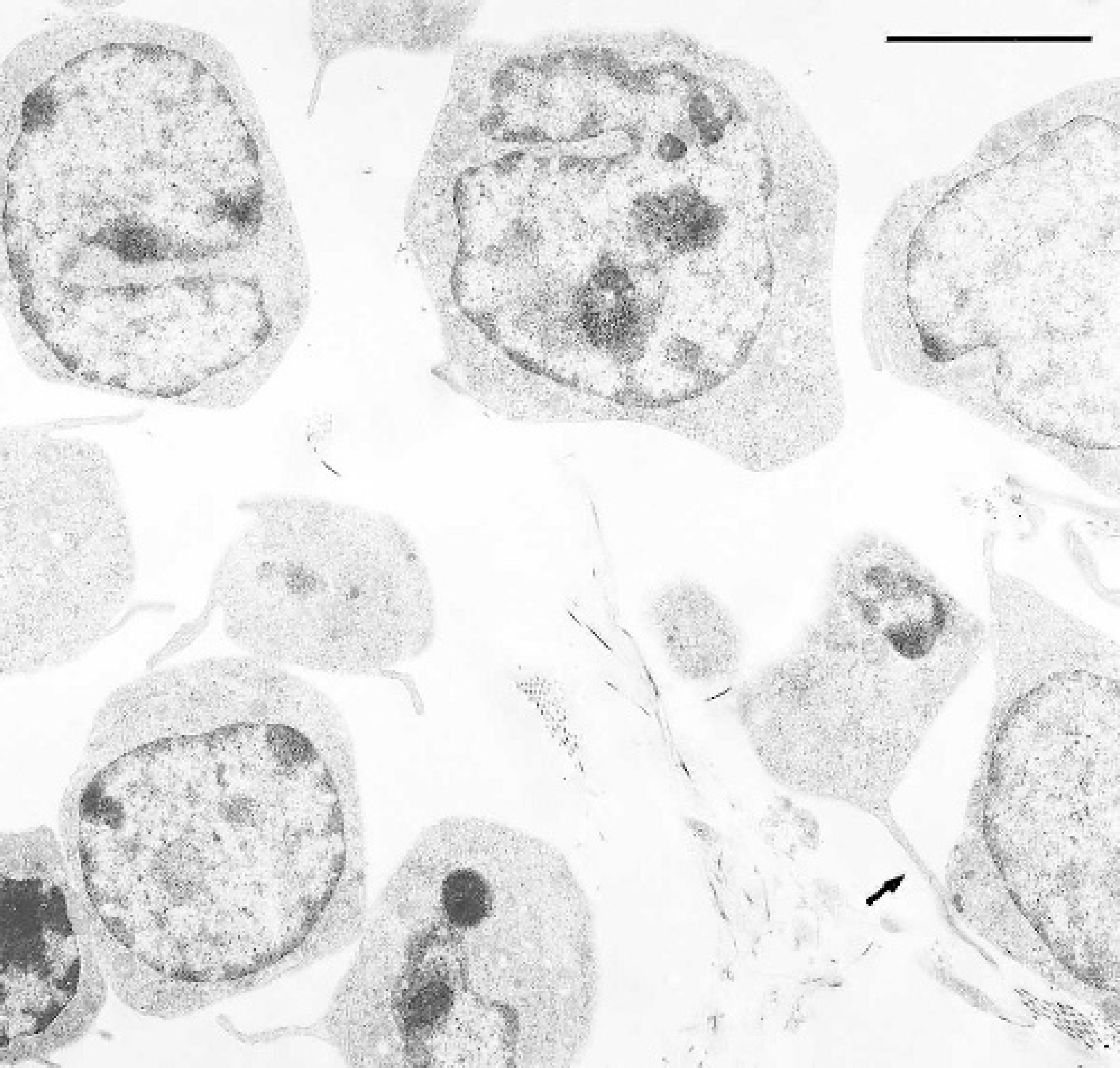
Mesentery; calf. Neoplastic cells reveal cytoplasmic processes like filopodia, one of which is extremely elongated (arrow). TEM. Bar = 4 μm.
Ultrastructurally, the neoplastic cells had irregular nuclei that were mainly euchromatic. Rough endoplasmic reticulum was poorly developed, but occasional cells had a fair number of mitochondria. Small numbers of cytoplasmic projections, which were frequently elongated, occurred in many neoplastic cells (Fig. 5).
B-1 or CD5-positive B cells are a minority subset of B cells that comprise about 5% of all B cells in mice and humans. 1 These cells first appear during fetal development and constitute a large population of the B cells found in the body cavities. 1 In humans, some types of lymphoma, such as CLL and MCL, express CD5 and are considered to be derived from B-1 cells. However, the majority occur in relatively older individuals and commonly involve lymph nodes, spleen, and bone marrow. 12,14 In addition to CD5 expression, the fact that the current lymphoma arose from the abdominal cavity during intrauterine life strongly suggests B-1 cell origin, and a diagnosis of B-1 B cell lymphoma was made. The lymphoma in this calf expressed TdT, which is a marker for precursor lymphoid cells of B, T, or natural killer (NK) cell lineage. 4 In contrast, human CD5-positive B cell lymphomas are negative for TdT and are included in the category of mature B cell neoplasms. 7 Thus, the present lymphoma is a unique form of B cell lymphoma and seems to differ from human CD5-positive cases.
Several lymphocyte subpopulations, such as B-1 cells and epithelial γδ T cells, behave as innate lymphocytes and are located in unusual sites. 9,10 The γδ T cells seem to defend the body surfaces, 9 and their malignant counterpart in cattle may be invasive into the epidermis and mucosal surfaces. 11 In contrast, B-1 cells seem to defend the body cavities. 9 The B-1 cell lymphoma in the calf was present in the body cavities and in the skin. Because B-1 cells are in many ways analogous to epithelial γδ T cells, 9 B-1 cells may play an important role in protecting the superficial zone of the skin.
The calf form of lymphoma is characterized by bone marrow involvement, leukemia, and generalized symmetrical lymphadenopathy; the liver, and the spleen are usually affected. 16 Immunohistochemically, the calf form can be of B cell or T cell lineage. 3,8 The B cell type expresses neither CD5 nor CD11b, and is thought to be of B-2 B cell derivation. 17 In the present CD5-positive lymphoma, the predominant lesions were in the body cavities and the skin, and nearly all lymphatic organs and the liver were unaffected. Thus, the neoplasm was quite distinct from the usual calf form in immunophenotype and tissue distribution. In familial B cell lymphomas with involvement of the thymus in young cattle, the neoplastic cells were positive for TdT but not CD5. 5 Conversely, CD5 was expressed in some cases of adult-form lymphoma, but TdT was absent (K. Kadota).
Electron microscopy revealed cytoplasmic processes, but no other characteristic features. Numerous microvillous projections are observed in human hairy cell leukemia, but broad ruffles, which are a monocytic feature, are also present and may be related to phogocytosis. 6 As in the present case, the neoplastic cells in a previously reported bovine NK-like T cell lymphoma were weakly phagocytic but did not have characteristic surface structures. 13 A few filopodia were observed in human γδ T cells in vitro and were thought to have a relation to active motility. 2 In the B-1 lymphoma reported here, the cytoplasmic processes might be needed for migrating along the peritoneal or pleural surfaces.
In this article, we present an uncommon form of lymphoma in a calf. Other reports of NK-like T cell lymphoma 13 and γδ T cell lymphoma in calves (K. Kadota) may suggest that lymphomas of B-1 cell origin occur more frequently than reported and that may be expected, because their cell of origin is an innate lymphocyte that arises early in ontogeny and is older than conventional lymphocytes in terms of evolutional history. 9
Footnotes
a.
Nichirei, Tokyo, Japan.
b.
Dako, Glostrup Denmark.
c.
Lab Vision, Fremont, CA.