
Abstract
In October 2004, a swine farm in Jinchang, Gansu Province, China, experienced an outbreak of toxoplasmosis. Most of the affected pigs had a rectal temperature greater than 40°C and gradually lost their appetite. Morbidity reached 57%, and mortality was approximately 2%. Analysis of blood samples from affected pigs using hemagglutination inhibition (HI) assay, immunoglobulin G—enzyme-linked immunosorbent assay (IgG-ELISA), and IgM-ELISA tests showed high titers of anti—Toxoplasma gondii antibody. Tachyzoites of T. gondii were found in body fluids of mice inoculated intraperitoneally with ground samples from the heart, liver, spleen, and brain of 2 sick pigs. In addition, the inoculation of 5 pigs with T. gondii tachyzoites caused death in 2 of the pigs. The origin of this outbreak was concluded to be food-borne T. gondii infection.
Toxoplasma gondii (family Sarcocystidae) is an obligate intracellular protozoan that can infect almost all warm-blooded animals including humans. In addition, infection has been documented in some cold-blooded animals, 1,3,4,9 and to date, at least 141 animal species have been confirmed to be pathogenically affected by T. gondii worldwide. 5 Clinical signs of toxoplasmosis in humans are fever, anorexia, lethargy, ocular and nasal discharge, respiratory distress, and spontaneous abortion. 3 Although the parasite is generally harmless to adults, T. gondii infection in humans with compromised immune systems and in pregnant women can be very serious. This disease also causes serious economical losses in livestock due to increased mortality, abortion, and medical costs. 13
In China, T. gondii was first isolated from a rabbit in Fujian Province in 1954, 12 and the first case of human toxoplasmosis was reported in 1964 from Jiangsu Province. 11 Animals are an important source of human toxoplasmosis, and T. gondii infection in pigs is a serious public health concern. A few outbreaks of lethal toxoplasmosis have been reported on pig farms, and most of these cases occurred during hot and humid weather due to the consumption of food contaminated with oocysts from cat feces. 2,6,8,10 It is difficult to eradicate oocysts in food sources. Indeed, it has been reported that porcine toxoplasmosis outbreaks occurred yearly in the same animal house for 5 years on a farm in the Shandong Province of China. 2
Jinchang, located in central Gansu Province, belongs to the arid region in Hehsi Tsoulang (Fig. 1) and is very dry for most of the year. Lethal toxoplasmosis in pigs has never been reported in this area. However, an outbreak of lethal toxoplasmosis on a pig farm in October 2004 in Gansu Province is reported in the current study.
In October 2004, a farm containing 260 sows and 960 fattening pigs in Jinchang, Gansu Province, suffered an outbreak of unknown disease in fattening pigs that resulted in the death of 19 pigs. The morbidity reached 57% (549/960 fattening pigs), and the mortality rate was approximately 2% (19/960 fattening pigs). Most of the affected pigs had a rectal temperature of 40–42.2°C and gradually lost their appetites and became depressed. Therapy with penicillin and streptomycin was without effect. On the third day of the outbreak, the authors were invited to investigate this disease. Since the clinical signs of the affected pigs were similar to T. gondii infection, 10 blood samples were collected from the pigs for anti—T. gondii antibody detection using a hemagglutination inhibition (HI) assay; reagents were prepared by the authors' laboratory. The titers of all the blood samples were >1:256, so it was concluded that T. gondii was the cause of the outbreak. All diseased pigs on this farm were treated with a combination of trimethoprim and sulfamethoxazole, and their food was changed.
Serum was collected from another 144 diseased pigs on the same day and tested for immunoglobulin G (IgG) and IgM antibodies against T. gondii with T. gondii excreted—secreted antigen-based HI assay, 14 IgG—enzyme-linked immunosorbent assay (IgG-ELISA), and IgM-ELISA 7 (the reagents were prepared by the authors' laboratory). For the ELISA, plates were read using a 450-nm filter. Of the 154 sera tested during the outbreak, 138 (89.6%) had IgG antibodies against T. gondii by HI assay, and 122 samples had high titers of antibody (77 samples: 1:256; 39 samples: 1:1,024; 6 samples: 1:4,096; negative control titer: ≤1:4; positive control titer: ≥11,024). Of the 154 tested sera, 142 (92.2%) were positive for IgM antibody, and 147 (95.4%) were positive for IgG antibody. These results indicated that T. gondii was the likely pathogen in this outbreak. Within 3 days after treatment, only 3 pigs died, and mortality was not seen after this time period. Five days later, the diseased pigs gradually regained their appetites, and their body temperatures returned to normal. On the seventh day posttreatment, 11 pigs still had anorexia, but the rest of the pigs had recovered.
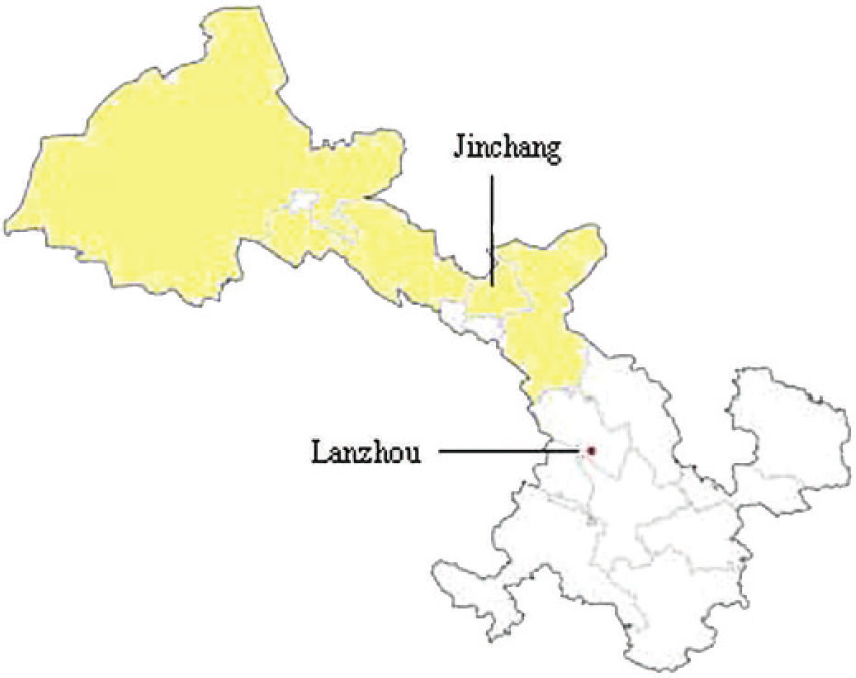
Map of Gansu province (yellow highlighting indicates Hehsi Tsoulang).
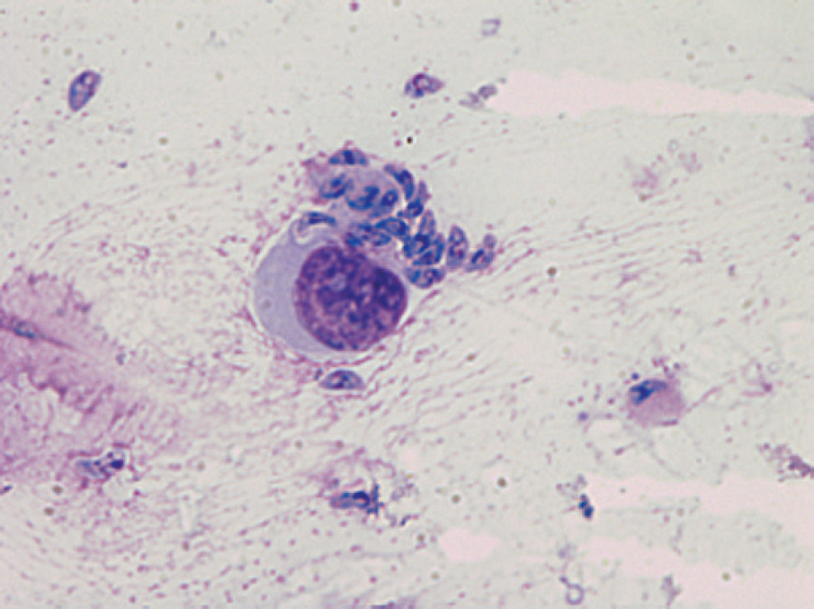
Tachyzoites of Toxoplasma gondii are present in peritoneal fluid. Giemsa stain. 10 × 100 magnification.
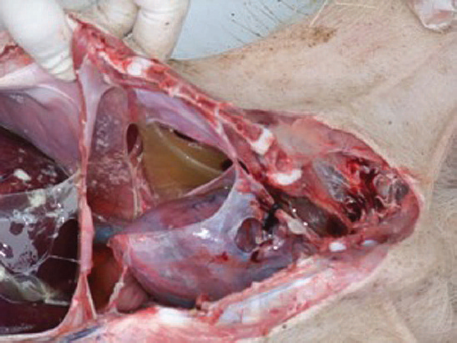
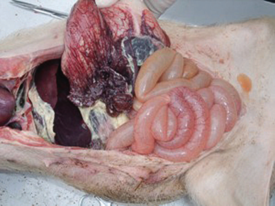
Pleural effusion with a sticky appearance.
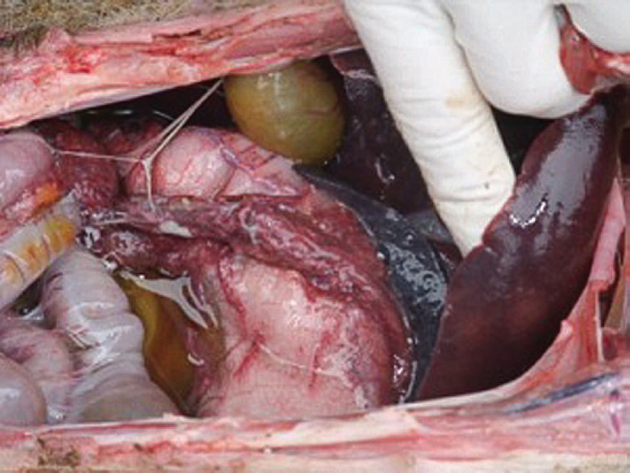
Enlargement of the liver with hyperemia and hemorrhage.
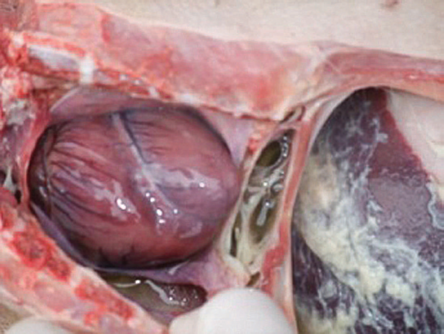
Enlargement of the heart.
Enlargement of the spleen with hemorrhage and necrosis.
Porcine reproductive and respiratory syndrome virus (PRRSV) infection is also prevalent in China. To exclude the possibility of PRRSV infection in the outbreak in the current report, samples from the heart, liver, spleen, and brain of 2 sick pigs (pigs 1 and 2) were collected and analyzed. Five grams of each organ sample from each pig were combined and ground and then diluted 1:10 in phosphate buffered saline (PBS). The PRRSV isolation was performed by 3 blind passages of the mixture in MARC-145 (cloned African green monkey kidney cell line) cultures, and no virus was found. Conventional bacterial isolation was also performed to detect any pathogenic bacteria, but the test results were negative.
To isolate the T. gondii parasite, 15 BALB/C mice without T. gondii infection (anti—T. gondii antibody titers ≤1:4 by HI assay) were allocated into 3 groups with 5 mice per group. The mice were inoculated by intraperitoneal injection with 0.5 ml/mouse of the mixture of pig 1 in group 1 or the mixture of pig 2 in group 2. Mice in group 3 were injected only with PBS and used as a negative control. After 7 days, all of the mice inoculated with mixture samples had signs of toxoplasmosis, but the negative control mice did not exhibit any changes. The mice were euthanized, and the peritoneal cavity was washed with 2 ml PBS per mouse. This suspension from each group was mixed and used to inoculate additional mice (5 mice per group). Five days later, 4 of 5 of the mice in one group and 5 of 5 of mice in the other group developed clinical signs of toxoplasmosis. The mice were euthanized, and the peritoneal cavity was washed as described above. The liquid was collected and examined by light microscopy. T. gondii tachyzoites were found in these samples (Fig. 2). Next, 5 pigs were inoculated by intraperitoneal injection with 1 × 10⁷ T. gondii tachyzoites/pig (preinoculation anti—T. gondii antibody titers were ≤1:4 by HI assay). Seven days later, 2 of the 5 pigs died. Necropsy of these pigs revealed an enlarged heart with superficial hyperemia and hemorrhages. Pleural, peritoneal, and pericardial effusions were sticky. The spleen was fragile, necrotic, and enlarged. The liver was also enlarged, pink to gray, and necrotic. Scattered petechiae were also found on the liver surface (Figs. 3–6). The other pigs developed clinical signs such as fever and depression. The experiments showed that T. gondii infection was the primary cause of death in pigs in this disease outbreak.
The origin of toxoplasmosis was also investigated. All infections occurred in fattening pigs, and cats were found to reside in the feed warehouse. Cat-contaminated feed might be the source of the infection. Furthermore, 100 kg of feed was randomly collected and transported to the laboratory in sealed plastic bags. Five healthy piglets (anti—T. gondii antibody titers ≤1:4 by HI assay) weighing 15 kg were obtained. Approximately 1 kg feed was given to each piglet 4 times a day. The piglets were observed daily. On the seventh day of observation, 3 of the 5 pigs developed fever and depression, but all animals survived. Blood samples were collected from these 5 pigs on observation day 7 to evaluate anti—T. gondii antibody titers using an HI assay detection kit. 14 The antibody titers in the sera from the 3 pigs that exhibited clinical signs were all greater than 1:256, while the 2 remaining pigs had antibody titers ≤1:4. Moreover, T. gondii tachyzoites were also found in the diseased pigs. These findings demonstrated that the T. gondii outbreak in the current report was caused by ingestion of contaminated feed. After October 2004, the animal house was sprayed with 3% sodium hydroxide, and a flame sterilizer was used every 2 weeks. In addition, cats were excluded from the farm. To date, toxoplasmosis has not recurred on this farm.
Acknowledgements This investigation was funded by the director of Lanzhou Veterinary Research Institute.