
Abstract
Epithelioid hemangiosarcoma is a specific variant of hemangiosarcoma that has recently been recognized in domestic animals. These malignant vascular neoplasms histologically resemble, and may be mistaken for, carcinomas. Four epithelioid hemangiosarcomas in the urinary bladders of 4 cows with severe enzootic hematuria are described in the current study. Grossly, the vesicular mucosa of the urinary bladder of each cow contained a single red elevated nodule. Histologically, each neoplasm was composed of short strands, cords, or nests of epithelioid, round, or slightly spindle-shaped endothelial cells that formed small vascular structures. Neoplastic cells were immunohistochemically positive for factor VIII–related antigen and vimentin, and were negative for cytokeratin and desmin. Ultrastructurally, the neoplastic cells often contained cytoplasmic intermediate filaments, a prominent granular endoplasmic reticulum, a Golgi complex, mitochondria, marked pinocytotic activity, and rare Weibel-Palade bodies. These neoplasms were diagnosed as epithelioid hemangiosarcomas based on their histologic, immunohistochemical, and ultrastructural features. The present report widens the spectrum of mesenchymal tumors of the bovine urinary bladder and aids in the characterization of these vascular neoplasms.
Keywords
Vascular neoplasms of the urinary bladder are rare, except in cattle with bovine enzootic hematuria (BEH). Hemangiomas (capillary and cavernous), hemangiosarcomas, 3 and, recently, hemangioendotheliomas are the most common neoplasms associated with BEH. 2 Bladder tumors associated with BEH are seen in adult cattle grazing on bracken fern. 1,11
Epithelioid vascular neoplasms are a specific histologic variant of vasoformative neoplasms that have been described in some veterinary textbooks but have only recently been the subject of detailed examinations in domestic animals. 12 These neoplasms have histologic features that are similar to the epithelioid variants of human vascular tumors. Although the histologic descriptions of brain 4 and urinary bladder 2 vascular neoplasms in cattle do refer to cells with an epithelioid morphology, this pattern was previously only specifically recognized in one epithelioid hemangioma of the skin. 12 Furthermore, the neoplastic cells forming this pattern have not been well-characterized immunohistochemically or ultrastructurally in cattle or in tumors of the urinary bladder from other domestic species.
In the current report, 4 epithelioid hemangiosarcomas occurring in the urinary bladders of 4 Holstein Friesian cows from São Miguel Island, Azores, Portugal were investigated where BEH is endemic. 10 These animals ranged from 6 to 14 years old and presented with hematuria and anemia. The cows were sent to a slaughterhouse where postmortem examination revealed a single, submucosal, red, elevated nodule in the vesicular mucosa of the urinary bladder of each cow. The neoplasms ranged from 3 cm to 10 cm in diameter. Macroscopically, metastases were not observed. Portions of the bladder neoplasms, regional lymph nodes, lung, kidney, and liver were collected for histologic examination from every cow.
Tissue specimens were fixed in 10% neutral buffered formaldehyde and submitted for histologic examination. The tissues were routinely processed, embedded in paraffin wax, sectioned at 3 μm, and stained with hematoxylin and eosin or Gomori's reticulin silver impregnation for light microscopic examination. For immunohistochemical studies, the streptavidin–biotin–peroxidase complex method was used with a commercial detection system a according to the manufacturer's instructions. Primary mouse anti-human monoclonal antibodies were used to detect alpha smooth muscle actin (αSMA), b vimentin, b and desmin. c
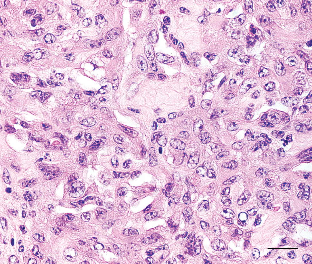
Epithelioid hemangiosarcoma; urinary bladder; cow. Neoplastic endothelial cells have a solid growth pattern. Intracytoplasmic vacuoles displace cellular nuclei. Hematoxylin and eosin. Bar = 20 μm.
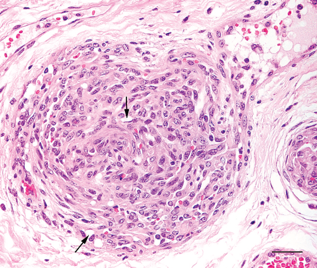
Epithelioid hemangiosarcoma; urinary bladder; cow. Papillary ingrowths of neoplastic endothelial cells are present. Notice the small vascular structures and intracytoplasmic vacuoles (arrows). Hematoxylin and eosin. Bar = 30 μm.
Primary rabbit anti-human polyclonal antibodies were used to detect cytokeratin, d collagen IV, d and factor VIII (FVIII)–related antigen. c All the antibodies specifically cross reacted with bovine tissue. The chromogen was diaminobenzidine tetrahydrochloride, and the counterstain was Mayer's hematoxylin. Negative controls were prepared by omitting the primary antibodies and replacing them with phosphate buffered saline. Internal controls for cytokeratin (urothelium), collagen IV, and FVIII-related antigen (normal blood vessels), vimentin (bladder lamina propria), and αSMA (muscularis mucosa of the bladder) were used as internal positive controls. Additionally, portions of formalin-fixed tissue from the bladder neoplasm of each cow were processed for transmission electron microscopy and scanning electron microscopy.
Histologically, the neoplasms were unencapsulated and infiltrative, consistently involving the vesicular submucosa and muscularis layers of the bladder. These neoplasms were composed of short strands, cords, or nests of epithelioid, round, or slightly spindle-shaped endothelial cells that were adjacent to small vascular structures or exhibited intracytoplasmic lumina that often contained a single erythrocyte (Fig. 1). Occasionally, the lesions had angiocentric growth, which was often occlusive, and papillary ingrowths (Fig. 2). The mitotic rate was greater than 5 mitoses in 10 high-power fields of view and moderate to severe anisokaryosis was present. The stroma was fibrous to myxoid with an intense infiltrate of lymphocytes. None of the other organs examined histologically contained metastases, confirming macroscopic observations.
Numerous neoplastic cells in each tumor were positive for vimentin and FVIII-related antigen, confirming the endothelial origin of these cells. Immunohistochemical staining for FVIII-related antigen was diffuse and strong in well-formed vessels but focal and of moderate intensity in solid areas. All neoplasms were negative for cytokeratin and desmin. Focal periendothelial positivity for αSMA was also seen. After staining the basal lamina with Gomori's silver reticulin and immunohistochemical staining for collagen IV, a tubular network was evident even in solid areas of the neoplasms.
Ultrastructural examination revealed aggregates of large, pleomorphic, endothelial cells with a prominent basal lamina, which was occasionally fragmented. Neoplastic cells contained cytoplasmic intermediate filaments, a prominent granular endoplasmic reticulum, a Golgi complex, mitochondria, marked pinocytotic activity, and rare Weibel-Palade bodies. Neoplastic endothelial cells also contained intracytoplasmic lumina (Fig. 3). Intercellular lumina were also formed by the interdigitation of cell membranes (Fig. 4). Cells consistent with pericytes were observed in all neoplasms.
With scanning electron microscopy, the neoplasms had a spongy appearance because of the presence of numerous irregular vessels, which were poorly defined and had incomplete walls. Neoplastic blood vessels were often oriented perpendicular to and appeared to communicate with the mucosal surface. In all neoplasms, intravascular papillary growths of pleomorphic endothelial cells were observed (Fig. 5). These endothelial cells had numerous microvilli and occasional intercellular junctions. All 4 neoplasms were diagnosed as epithelioid hemangiosarcomas based on similar histologic, immunohistochemical, and ultrastructural features.
In humans, several microscopic variants of hemangiosarcoma are recognized, one of which is the epithelioid subtype. 9 In veterinary medicine, this pattern of hemangiosarcoma has only recently been described (2007) 12 and has yet to be recognized in the World Health Organization Histological Classification. 5,8
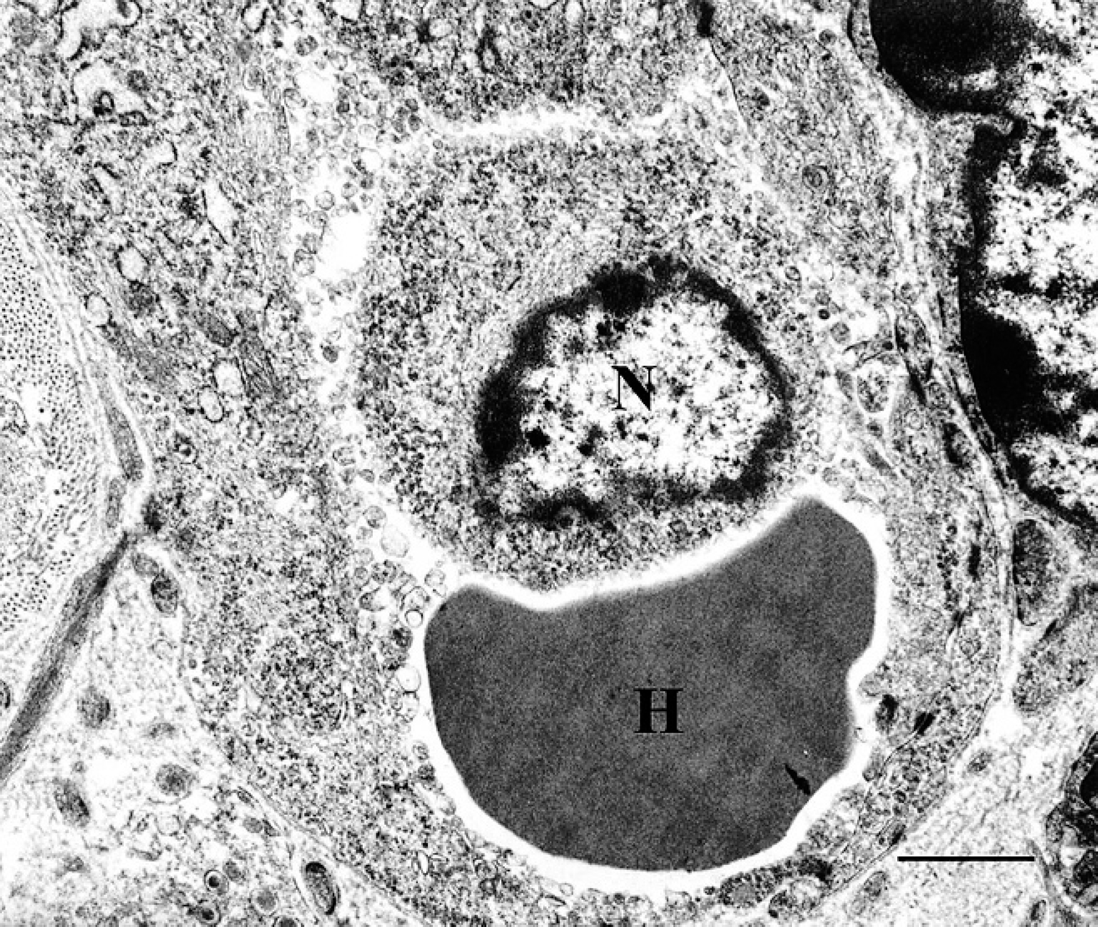
Epithelioid hemangiosarcoma; urinary bladder; cow; transmission electron micrograph. An erythrocyte (H) is present within an intracytoplasmic space adjacent to the nucleus (N) of an endothelial cell. Uranyl acetate and lead citrate. Bar = 2 μm.
Epithelioid vascular neoplasms are characterized by the peculiar epithelioid or histiocytic appearance of the endothelial cells. These tumors are composed predominantly or entirely of plump polyhedral cells that imitate true epithelial cells. Therefore, this pattern may represent a diagnostic challenge because of the possible confusion with carcinoma. 13 Melanomas, clear-cell sarcomas, epithelioid sarcomas, epithelioid leiomyosarcomas, and large cell lymphomas are also included in the differential diagnosis in addition to carcinomas. 9 The definitive diagnosis of malignant vascular tumors often requires immunohisto-chemical evidence of endothelial cell differentiation. Although some authors have suggested the routine use of multiple endothelial cell markers, immunohistochemical staining with antibodies to FVIII-related antigen is sufficient for diagnosis. Other endothelial markers only need to be used if FVIII-related antigen staining is negative, 6 as is possible in some muscle-invasive hemangiosarcomas. 2 Cytokeratin staining alone cannot distinguish vasoformative neoplasms from carcinomas because some human epitheloid vascular tumors are positive for this marker. 7 Additionally, it should be emphasized that the epithelioid variant of hemangiosarcoma must be considered in the differential diagnosis of solid tumors in organs other than the skin and associated soft tissues.
Acknowledgements. The authors thank Mrs. Lígia Bento for expert technical assistance and Dr. Susana Freitas for the revision of the work.
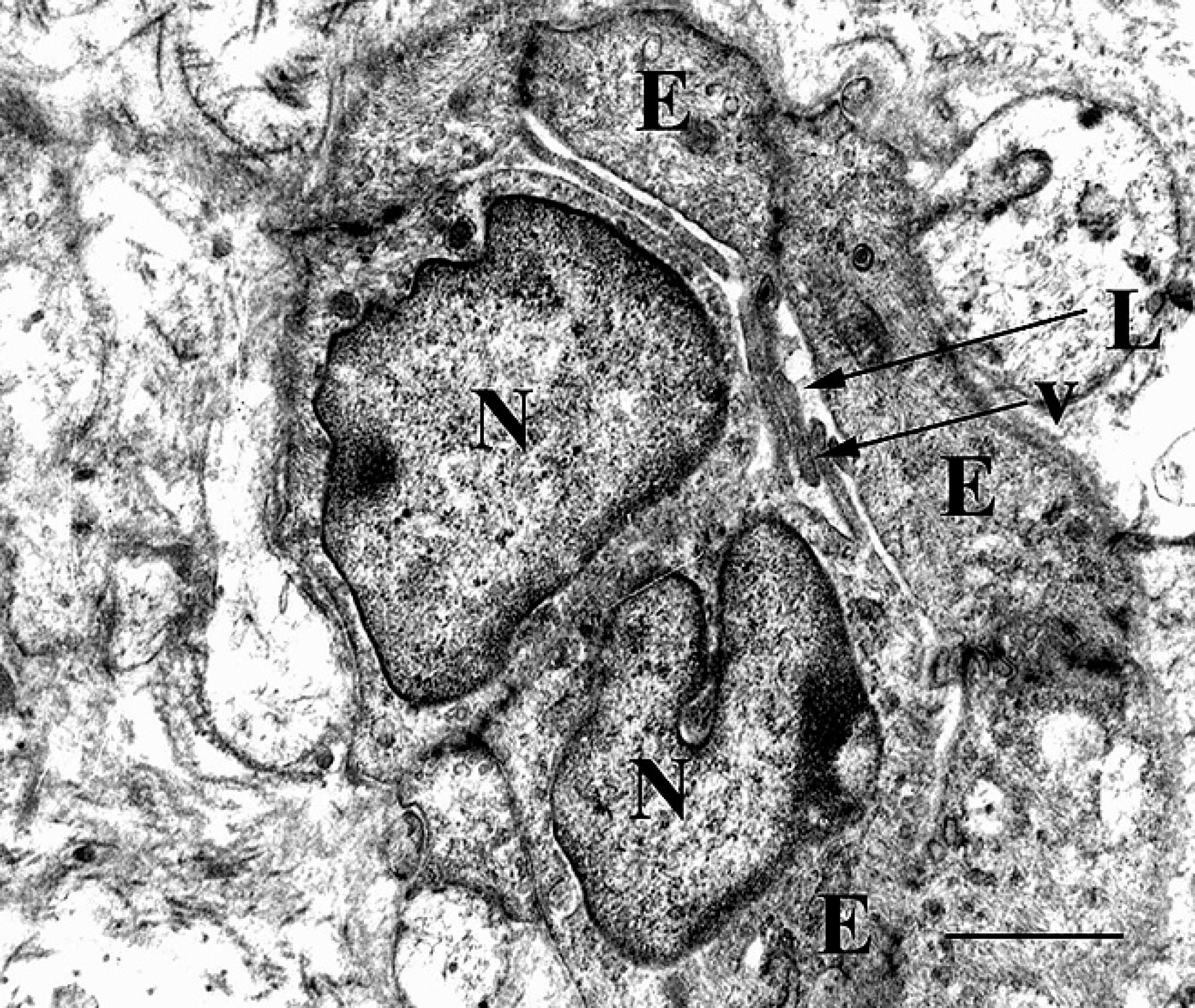
Epithelioid hemangiosarcoma; urinary bladder; cow; transmission electron micrograph. Cytoplasmic processes of the neoplastic cells form an intercellular lumen. Uranyl acetate and lead citrate. Bar = 2 μm. E = endothelial cell; L = lumen; v = interdigitation of cell membranes; N = nuclei.
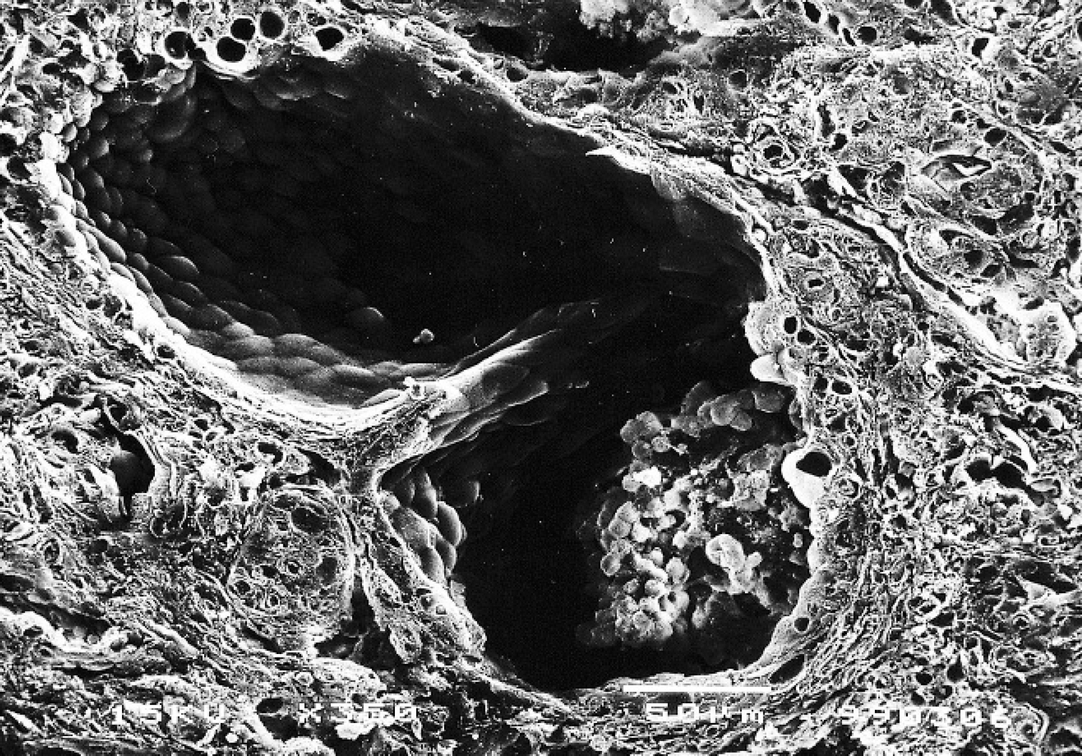
Epithelioid hemangiosarcoma; urinary bladder; cow; scanning electron micrograph. Notice the intravascular papillary growth of neoplastic endothelial cells. Gold-palladium. Bar = 50 μm.
Footnotes
a.
Lab Vision Corp., Fremont, CA.
b.
Novocastra Laboratories Ltd., Newcastle upon Tyne, UK.
c.
Dako North America Inc., Carpinteria, CA.
d.
Chemical Credential, ICN Immunobiologicals, Lisle, IL.