
Abstract
Nocardia spp. are recognized as a cause of bovine mastitis, cutaneous or subcutaneous abscesses, pneumonia, and disseminated disease. Abortion caused by Nocardia spp. is uncommon, and only a few sporadic cases have been reported in horses, pigs, and cattle. In all previous reports, of nocardial abortion, the causative agent was identified as Nocardia asteroides. The current report describes an aborted bovine fetus that was infected with Nocardia farcinica. Placenta, abomasal fluid, lung, liver, and kidney specimens from a lateterm bovine abortion were submitted to the Kansas State Veterinary Diagnostic Laboratory. The gross findings included purulent exudate in the placenta and numerous abscesses in lung. Histologically, there was necrotizing and suppurative placentitis, pyogranulomatous pneumonia, and nephritis with numerous intralesional branching and filamentous, Gram-positive bacteria. Nocardia farcinica was isolated by bacteriology, and the bacteriology result was confirmed by 2 established polymerase chain reaction protocols and by DNA sequencing.
Nocardia are ubiquitous, aerobic, and saprophytic actinomycetes that are present worldwide in soil and decaying organic matter and in fresh and salt water. 9,15 The genus Nocardia currently contains more than 70 species that have been characterized by various phenotypic and molecular methods. Approximately half of these species are recognized human and/or animal pathogens. 1,4,6 Nocardia spp. cause various human infections in immunocompromised hosts, including cutaneous, subcutaneous, pulmonary, cerebral, and lymphocutaneous infections. 1,6 Bovine mastitis is the most common clinical manifestation of nocardiosis among domestic animals and is usually seen in dairy herds with poor hygiene. 13 Other manifestations of nocardiosis in animals are cutaneous or subcutaneous abscesses, pneumonia, and disseminated disease. 13 Abortion caused by Nocardia spp. is uncommon, and only a few sporadic cases have been reported in horses, pigs, and cattle. 2,7,10,11,12,15
In June 2005, 2 late-term, bovine abortions were reported in a herd of beef cattle in Kansas. Placenta, abomasal fluid, lung, liver, and kidney specimens from 1 fetus were submitted to the Kansas State Veterinary Diagnostic Laboratory. Grossly, the placental cotyledons were dark red and covered at multiple foci with white–tan to creamy, purulent exudate (Fig. 1). The entire lung tissue contained miliary abscesses (Fig. 2). No gross lesions were present in the kidney or liver.
Fresh samples of placenta, lung, abomasal fluid, and liver were sent for bacterial isolation. Fluorescent antibody testing of fresh lung tissue for Bovine viral diarrhea virus and Infectious bovine rhinotracheitis virus yielded negative results. Tissues were fixed in 10% buffered formalin, processed, and embedded in paraffin. Deparaffinized sections were stained with hematoxylin and eosin, Gram stain, Grocott methenamine–silver nitrate stain (GMS), and periodic acid–Schiff.
Microscopically, the placental chorionic villi contained multifocal areas of necrosis and neutrophilic infiltration. The intercotyledonary areas were expanded by edema, neutrophilic infiltration, and neutrophilic vasculitis (Fig. 3). The lung parenchyma contained multifocal to coalescing pyogranulomas with multinucleate giant cells (Fig. 4). A few scattered pyogranulomas were also present in the kidney. The liver had no significant histologic lesions.
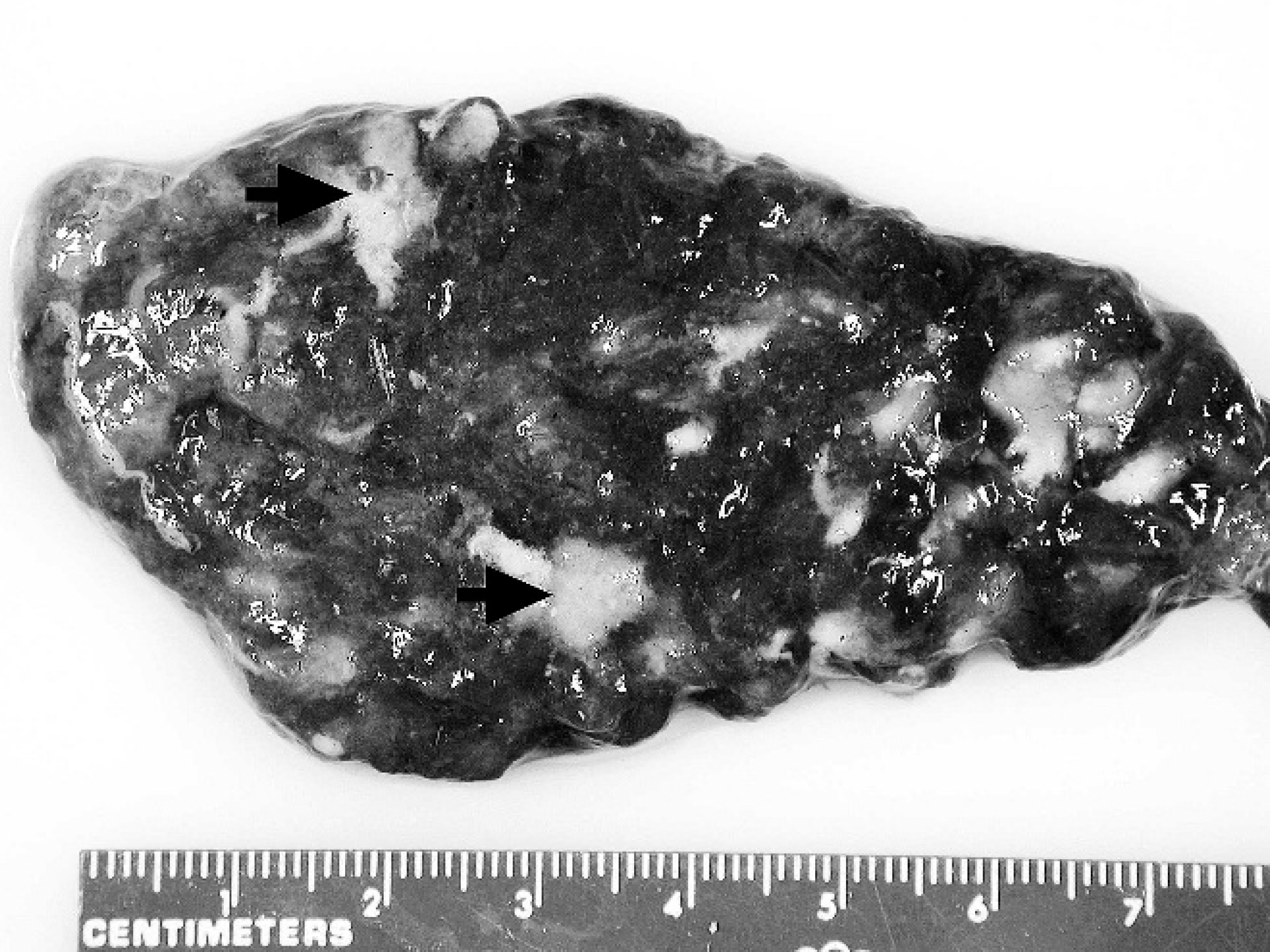
Placenta, bovine. The placenta is covered at multiple foci by white–tan, purulent exudates (arrows).
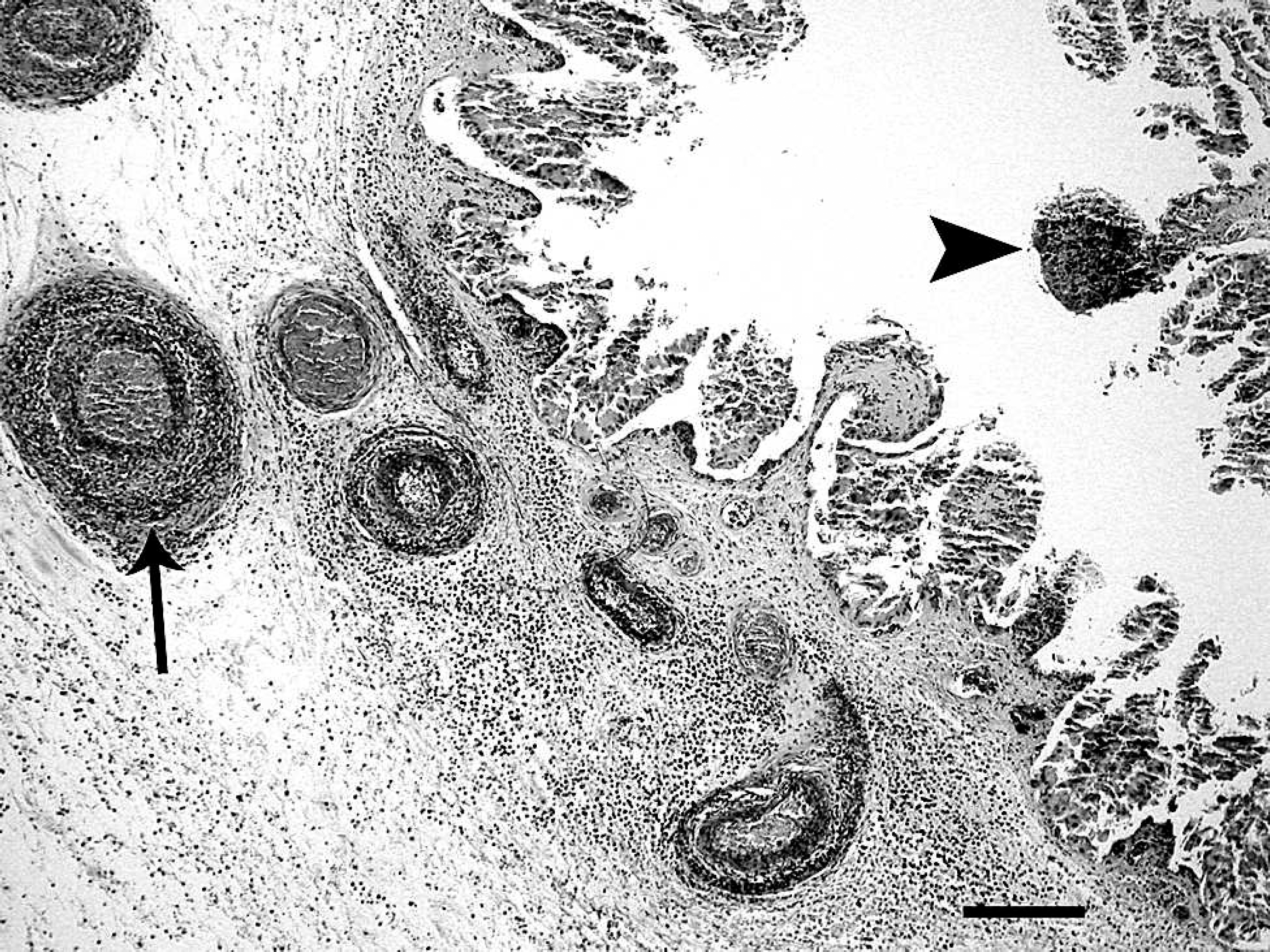
Tissue sections stained by Gram stain and GMS revealed many branching, filamentous Gram-positive bacteria (Fig. 5).
Bacterial culture of lung and abomasal fluid resulted in isolation of colonies that grew after 7 days on tryptic soy agar with 5% sheep blood incubated at 37°C in 5% CO2. Isolated bacteria were Gram-positive bacilli that grew on Sabouraud dextrose agar. The bacteria were nitrate-reduction–positive, urease-positive, and grew at 45°C. Organisms were negative for casein hydrolysis, decomposition of xanthine, and decomposition of tyrosine. Based on the flowchart by Songer and Post, 14 the isolate was identified as Nocardia farcinica. The isolate grew at 37°C and 45°C, and only N. farcinica is reported to consistently grow at both temperatures; however, 50% of N. asteroides isolates can also grow at both temperatures. 8 The isolate was positive for production of nitrate reductase, and in literature, both positive 14 and negative 5 tests are reported for N. farcinica. Hence, the phenotypic identification of Nocardia to the species level using biochemical tests remains problematic, and molecular techniques are the ideal methods that can provide definite identification of Nocardia spp.
Placenta, bovine. Photomicrograph showing mul-tifocal areas of chorionic villi necrosis (arrowhead), diffuse infiltration of chorioallantois with neutrophils, and neutrophilic vasculitis (arrow). Hematoxylin and eosin. Bar = 200 μm.
In humans, the most commonly isolated Nocardia species is N. asteroides. Accurate and prompt identification of N. farcinica, and its differentiation from N. asteroides is important because N. farcinica has a higher degree of resistance to specific antibiotics and has a higher risk of dissemination. 1,5,6 Identification of N. farcinica by poly-merase chain reaction (PCR) assay was recently described by 2 investigators using different sets of primers. Brown et al. 3 designed a pair of primers that amplifies a 314 base pair (bp) fragment, based on a 409-bp randomly amplified polymorphic DNA (RAPD) band that is specific to N. farcinica. The primer pair was tested on 28 N. farcinica isolates, 59 non-farcinica nocardial species, and 41 non-nocardial, but phylogenetically related, species. Their results indicated that the primer pair only amplified the fragment from N. farcinica isolates. A similar approach was used by Hasegawa et al., 8 who identified a 412-bp RAPD band that is specific to N. farcinica and is different from the region described by Brown et al. 3 A pair of PCR primers, amplifying 283 bp, was designed from this 412-bp sequence and tested on 9 N. farcinica and a few nonnocardial species. This PCR amplicon was only produced from N. farcinica isolates.
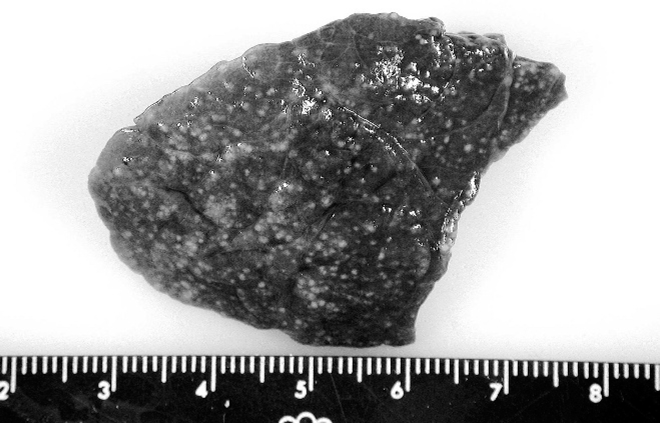
Fetal lung, bovine. Gross photographs of lung showing abscesses in the entire lung tissue.
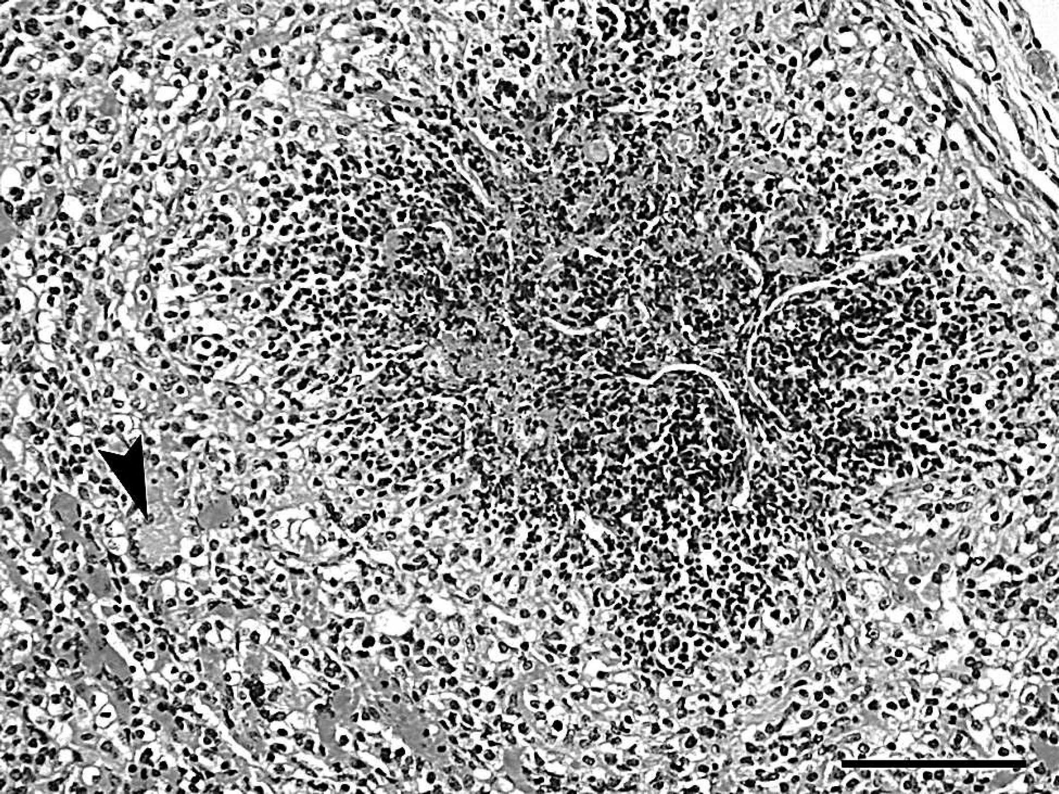
Fetal lung, bovine. Photomicrograph of fetal lung showing pyogranulomatous inflammation with multinucleated giant cells (arrowhead). Hematoxylin and eosin. Bar = 100 μm.
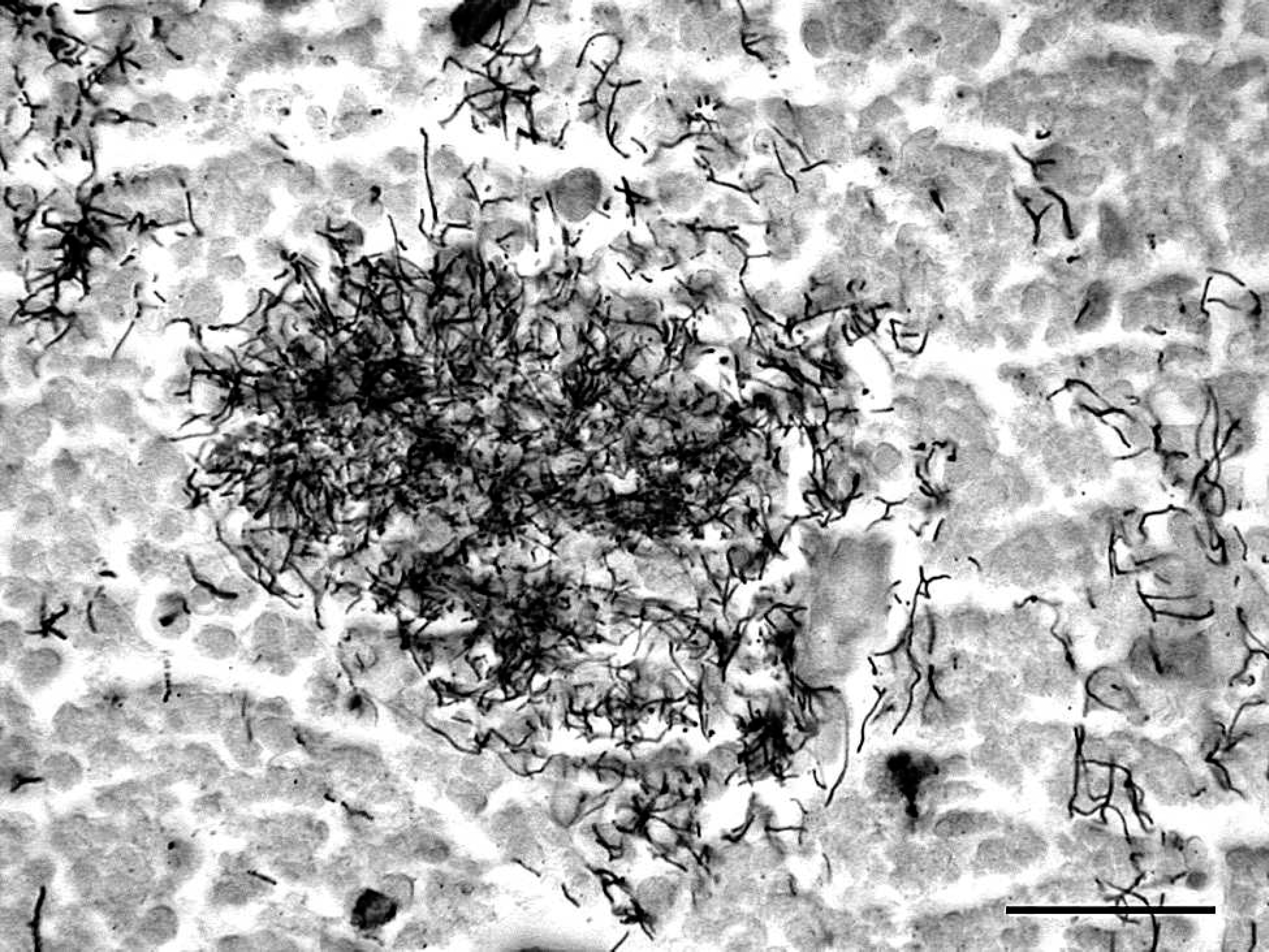
Placenta, bovine. Section of placenta showing filamentous bacteria in the necrotic debris in the chorionic villi. Grocott methenamine–silver nitrate. Bar = 30 mm.
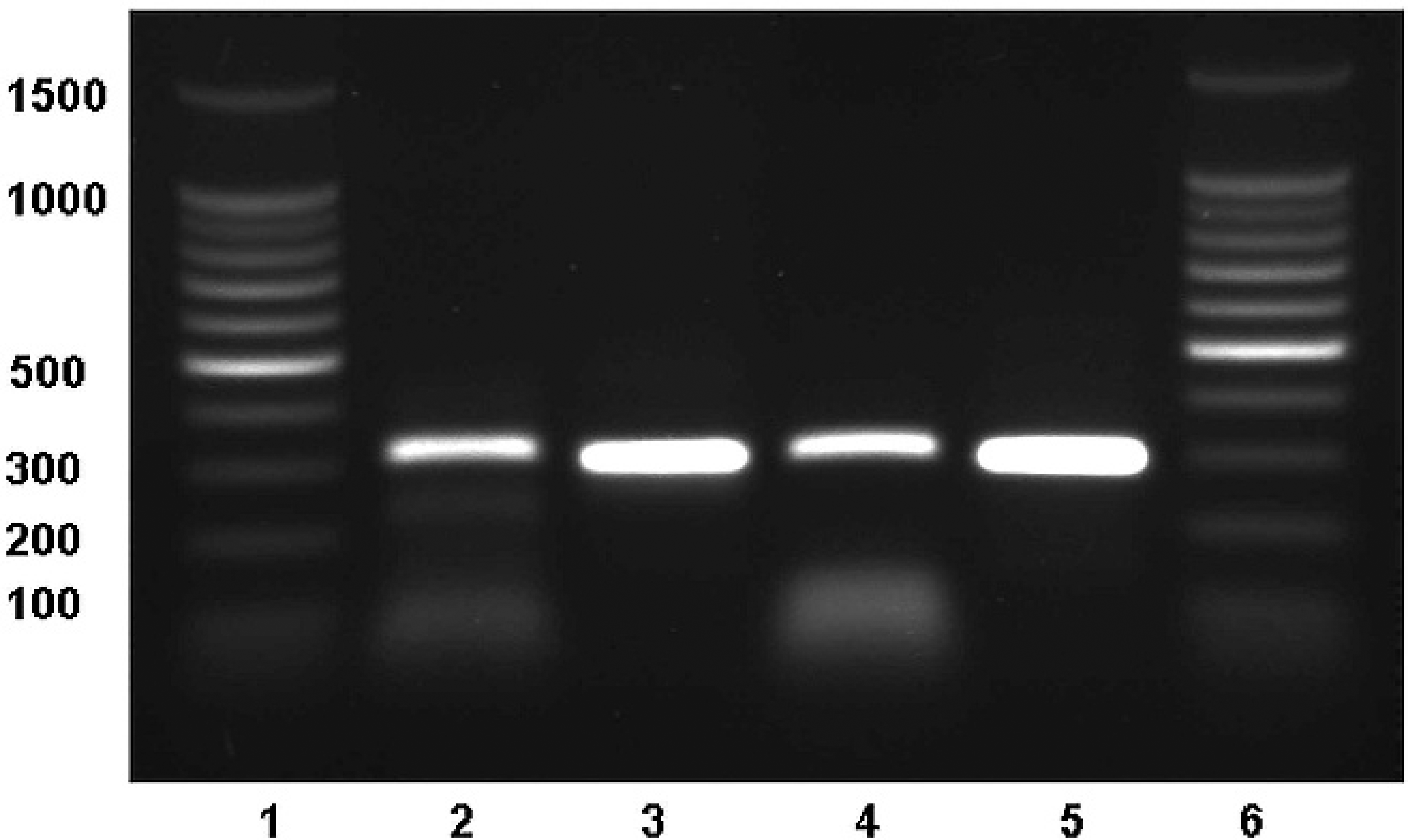
Gel electrophoresis of 4 DNA fragments amplified from Nocardia farcinica. Lanes 1 and 6: DNA size marker; lanes 2 and 3: amplified DNA fragments from placenta using Brown et al. 3 and Hasegawa et al. 6 primers, respectively; lanes 4 and 5: amplified DNA fragments from kidney using Brown et al. 3 and Hasegawa et al. 6 fragments, respectively.
To confirm the bacteriology results, PCR tests for N. farcinica identification were performed. Culture isolates were no longer available, therefore, paraffin-embedded kidney and placental samples were used to isolate genomic DNA with a commercial kit, a following the manufacturer's protocol. Based on the primer sequences in the above-mentioned publications, 2 pairs of primer were synthesized, and PCR was done on both kidney and placenta (total, 4 PCR assays). Primers from Brown et al. 3 amplified a 314-bp fragment from both placenta and kidney. Primers from Hasegawa et al. 6 amplified a 283-bp fragment from both placenta and kidney (Fig. 6). The 4 PCR products were column-purified c and sequenced. d The amplified DNA fragments from placenta and kidney yielded identical DNA sequences. Two individual Basic Local Alignment Search Tool (BLAST) searches in GenBank, using amplified DNA fragments generated by Brown et al. 4 and Hasegawa et al. 6 methods, were performed. Both fragments matched N. farcinica strain IFM 10152 (GenBank accession no. AP006618). The sequences were 96% (Brown et al. 3 method) and 95% (Hasegawa et al. 6 method) identical to IFM 10152. The PCR assay and sequencing data confirmed the presence of N. farcinica in both placenta and kidney.
Bovine abortion caused by Nocardia spp. has been previously reported. 11,12,15 In 2 of the cases, there were no fetal lesions, 11,12 and the third article reported fetal bronchopneumonia and liver necrosis. The specific route of infection is not clearly known. A 1980 study 12 proposed that contamination of the reproductive tract during obstetric manipulation may lead to uterine infection. That, in turn, may progress to placentitis and, subsequently, to fetal systemic infection and abortion. There was no knowledge of pervious obstetric manipulation in this case.
Historically, the nomenclature of the species in genus Nocardia has been confusing. The organism was originally isolated in 1888 by a veterinarian, Edmund Nocard, from a case of bovine farcy. This isolate was later named N. farcinica and was made the type species of the genus Nocardia. However, later in the mid twentieth century, Nocardia asteroides replaced N. farcinica as the type species of genus Nocardia. 4,5 Recently, using different methods, including biochemical characteristics, susceptibility testing, and molecular methods, the species status of N. farcinica was clearly established as separate from that of N. asteroides. 4,13 In the earlier reports 11,12,15 of bovine abortion caused by Nocardia spp., the proposed causative agent had been N. asteroides. This is the first report, to the authors' knowledge, of abortion caused by N. farcinica in animals. Nocardia-associated placentitis and abortion is rarely reported in domestic animals. In the present study, a bovine abortion case presumed to be caused by N. farcinica infection, based on phenotypic and genotypic characterization, is described.
Footnotes
a.
Gentra Puregene Tissue Kit, Qiagen Inc., Valencia, CA.
b.
Integrated DNA Technologies, Coralville, IA.
c.
PCR purification kit, Qiagen Inc., Valencia, CA.
d.
ACGT Inc., Wheeling, IL.