
Abstract
A 20-year-old female cynomolgus monkey (Macaca fascicularis) was presented for an end-of-study necropsy. At necropsy, a 2 cm × 1.5 cm × 1 cm, butterfly-shaped, multilobulated mass extended off the right uterine tube fimbria. Microscopically, the mass was composed of large, plump, finger-like projections lined primarily by simple columnar ciliated epithelium. The interstitium contained a proliferation of smooth muscle stromal cells admixed with varying amounts of collagen. A diagnosis of adenomyofibroma of the fimbria was made. This benign neoplasm should be considered as a differential diagnosis for masses arising from the fallopian tube in old-world macaques.
Neoplasms of the female reproductive tract in old-world monkeys have been reported 3,4 extensively in the uterus and ovaries. However, neoplasms arising from the uterine tube are extremely rare. 14 The current report describes an adenomyofibroma arising from the fimbria of the uterine tube in a cynomolgus monkey (Macaca fascicularis).
A 20-year-old female cynomolgus monkey was housed at the National Institutes of Health, Bethesda, MD. The care of the animal followed the National Institutes of Health's Guide to the Care and Use of Laboratory Animals. The animal was presented to the Division of Veterinary Resources pathology service for routine end-of-study gross necropsy. At necropsy, the animal appeared to be in good nutritional condition and contained adequate adipose stores. Attached to the right fimbria of the uterine tube was an approximately 2 cm × 1.5 cm × 1.0 cm, multilobulated, butterfly-shaped firm mass (Fig. 1). The mass was not attached to the corresponding uterine tube ampulla, ovary, or uterus. No other gross lesions were observed. Select tissues were collected and fixed in 10% formalin, processed routinely, and embedded in paraffin. Five-micron sections were stained with hematoxylin and eosin and Masson trichrome.
Microscopically, the fimbrial mass was composed of large, plump, finger-like projections lined primarily by simple columnar ciliated epithelium with basally oriented, mildly anisokaryotic, oval nuclei with finely stippled chromatin and occasional single small magenta nucleoli (Fig. 2). Focal areas of pseudostratified columnar epithelium and attenuation of epithelium to a simple cuboidal and simple squamous appearance were observed. These latter cell types lacked cilia. Luminal glandular structures lined by similar epithelial cells were observed within the stroma. The stroma was composed of interlacing bundles of uniform cells with fusiform, vesiculated nuclei and streaming eosinophilic cytoplasm (Fig. 3). Linear and patchy areas of collagen deposition, confirmed by Masson trichrome stain, were scattered among the stromal cells and largely constituted the stroma of a few of the finger-like projections (Fig. 4).
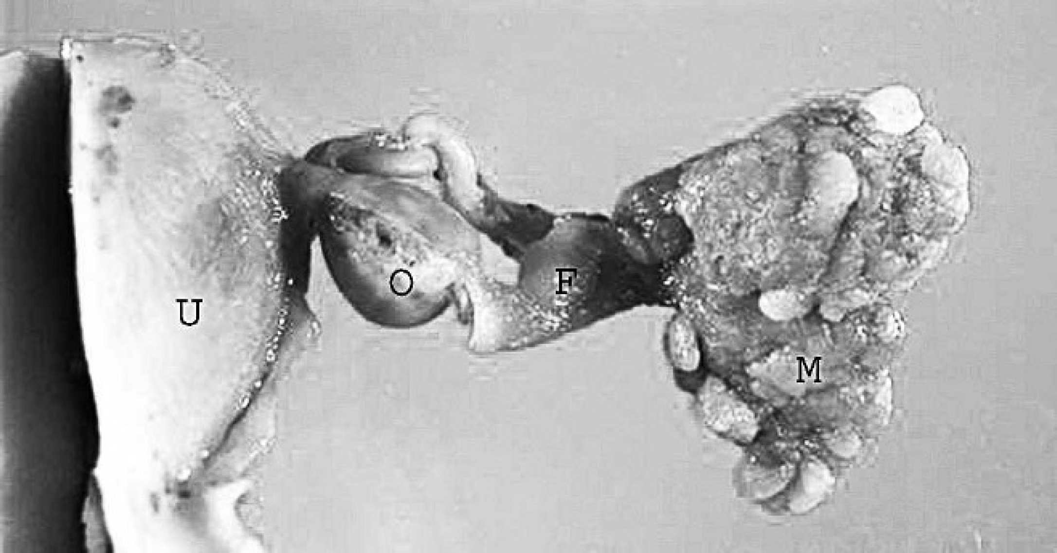
Female reproductive tract; cynomolgus monkey (Macaca fascicularis). A large mass (M) extends off the fimbria (F) of the uterine tube. O = ovary; U = uterus.
To evaluate the stromal cells, immunohistochemistry for desmin and smooth muscle actin was performed. Briefly, slides were deparaffinized, and commercial protein block and hydrogen peroxide were applied to minimize nonspecific staining and to quench endogenous peroxidase activity. Antigen retrieval was performed with heat treatment in citrate buffer, and the tissue sections were incubated with mouse monoclonal anti-smooth muscle actin antibody a and rabbit polyclonal anti-desmin antibody. b Sites of primary antibody binding were localized and visualized with secondary polymerized antibodies c and 3, 3′-diaminobenzidine chromogen. Nonspecific immunoglobulins were used as negative controls. Immunohistochemistry of the stromal cells revealed partial positivity for desmin and diffuse strong staining for smooth muscle actin, indicating the stromal cells to be smooth muscle cells. The gross, histologic, and immunohistochemical findings were consistent with a diagnosis of adenomyofibroma of the fimbria.
Epithelial and mixed (epithelial and mesenchymal) neoplasms of the uterine tube in mammals are rare. Nine neoplasms have been reported in the dog 7,9,10,12 and 1 each in the horse, 10 rat, 8 mouse, 5 Syrian hamster, 11 and cynomolgus monkey. 14 Of these neoplasms, approximately 50% have been described as a mass arising from the fimbria. Ten of the 14 tumors were diagnosed as benign types of adenomas (adenoma, fibroadenoma, adenomyoma, papillary adenoma, adenomatous papilloma, atypical polypoid adenomyoma). The location (fimbria) and diagnosis of adenoma is in concordance with the human literature 1,2 for uterine tube neoplasms. Nonepithelial neoplasms of the mammalian uterine tube are exceedingly rare. One leiomyoma 13 and 1 hamartoma 6 have been described in dogs.
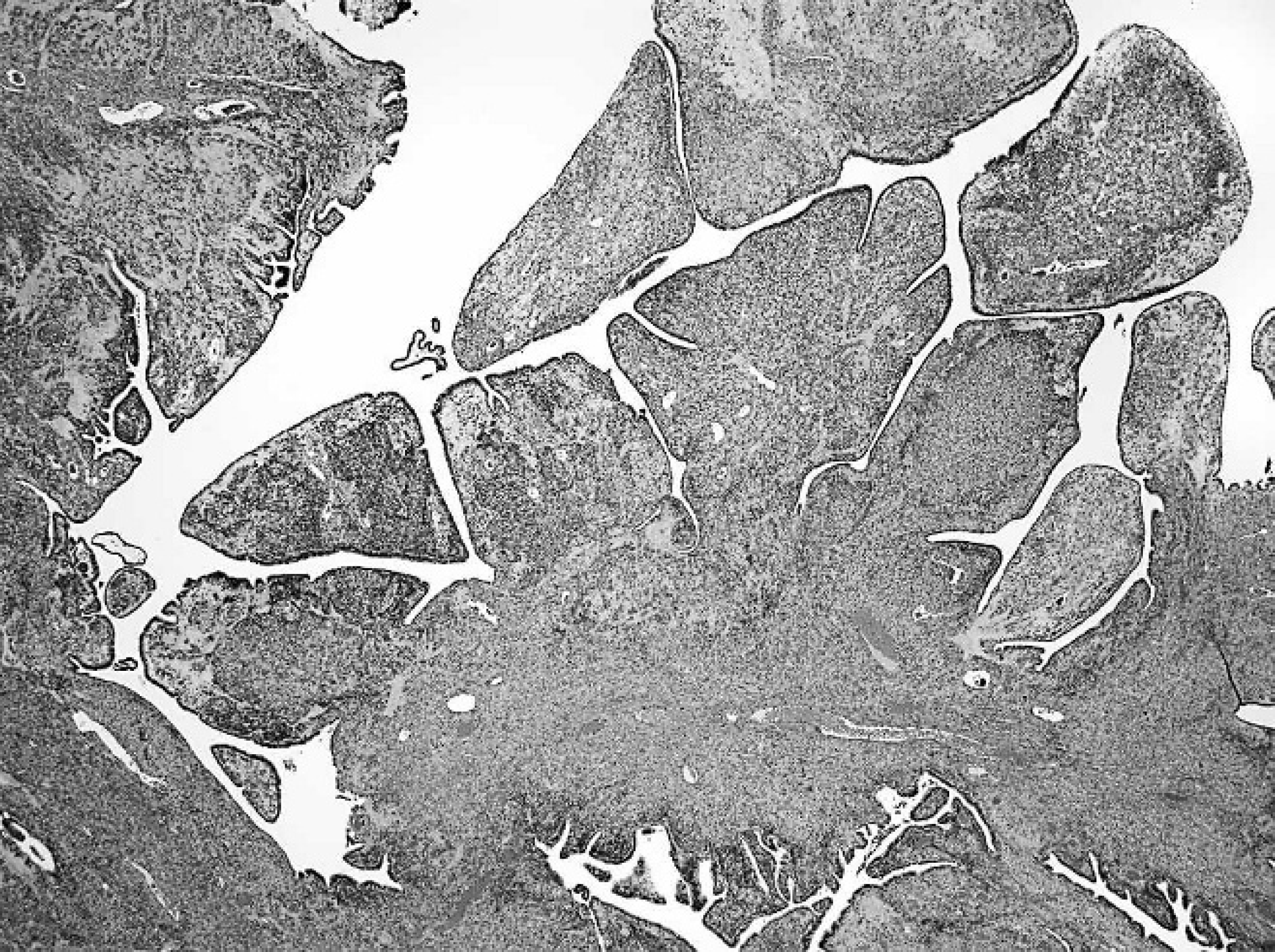
Adenomyofibroma; cynomolgus monkey (Macaca fascicularis). Numerous plump, finger-like projections are lined primarily by simple ciliated columnar epithelium. Hematoxylin and eosin. 50× magnification (5× objective × 10× ocular).
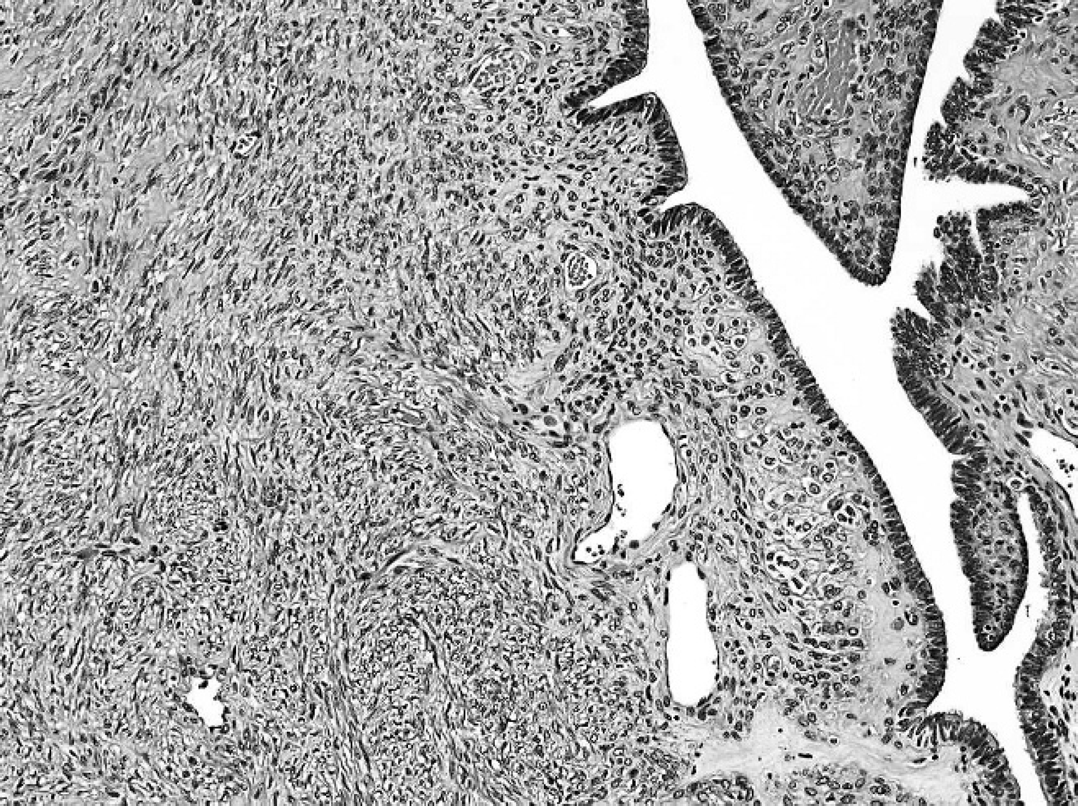
Adenomyofibroma; cynomolgus monkey (Macaca fascicularis). Dense network of interlacing stromal cells that are identified as smooth muscle cells. Hematoxylin and eosin. 100× magnification (10× objective × 10× ocular).
Considering the abundant admixture of smooth muscle stromal cells and collagen within the present neoplasm, a diagnosis of adenomyofibroma was made for the current case. This neoplasm had a few characteristics that were similar to those of an adenomyoma previously observed in another cynomolgus monkey 14 and may possibly represent a variation of it. The presence of abundant smooth muscle proliferation and the epithelial lining cells were similar; however, the present neoplasm had large patchy areas of collagen deposition, which were not observed in the previously reported adenomyoma. In addition, lipid-rich foamy cells were identified in the previous neoplasm, resembling those of ovarian sex-cord tumors of granulosa or Sertoli cells. In the present neoplasm, these cells, or any other ovarian components, were not observed.
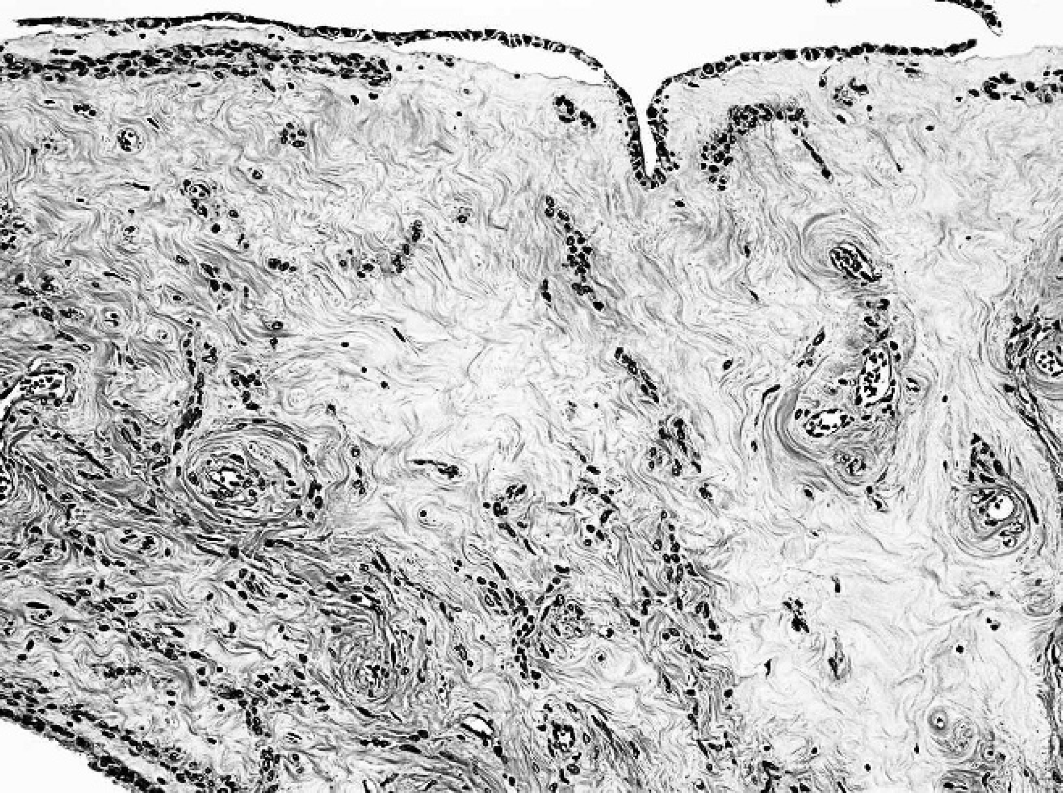
Adenomyofibroma; cynomolgus monkey (Macaca fascicularis). Patchy areas of collagen deposition are present. Hematoxylin and eosin. 100× magnification (10× objective × 10× ocular).
The paucity of neoplasms found in the uterine tube in mammals may be accounted for by their indolent nature (most have been identified as incidental findings at necropsy with no associated clinical signs), the relatively small size of the uterine tube in most mammals, and the young age at which research animals are submitted for necropsy, food animals are submitted for slaughter, and domestic dogs and cats are submitted for elective surgery (ovariohysterectomy).
The pathogenesis of uterine tube neoplasms is not fully known. Tubular hyperplasia has been attributed to reactive, inflammatory, and hormonal changes. Any of these could possibly contribute to the eventual formation of a neoplasm of the uterine tube. Closer evaluation of the uterine tube in animals may reveal higher incidences of neoplasms than have been reported in the veterinary literature and may potentially help explain some reproductive disorders observed in animals.
Acknowledgements. The author would like to thank Jorge Chavez, Annie Merriweather, and Patricia Zerfas for technical assistance. This work was supported by the Intramural Research Programs of the National Institutes of Health, Office of Research Services.
Footnotes
a.
Biocare Medical, Concord, CA.
b.
Lab Vision Corp., Fremont, CA.
c.
Dako North America, Inc., Carpinteria, CA.