
Abstract
To determine if the number of rectal lymphoid follicles decreases with respect to age and sex relative to diagnosis of chronic wasting disease (CWD), rectal biopsies (n = 1,361) were taken from captive Rocky Mountain elk (Cervus elaphus nelsoni) at 4 ranches in the western United States between 2005 and 2008. Rectal tissues were stained with a monoclonal antibody (F99/97.6.1), which selectively stains the abnormal isoform of the prion protein associated with CWD of elk. The number of lymphoid follicles obtained from typical biopsy tissues decreased with the age of the animal. The acceptable number of lymphoid follicles for detection of CWD was not considered to be a problem in elk up to 8.5 years of age, but in elk over 8.5 years of age, the follicle count was considered to be low. Sex of the animal had no effect on the number of lymphoid follicles observed in each age group. Rectal biopsies were an accurate test to diagnose preclinical stages of CWD in elk but may be best suited to elk that are less then 8.5 years of age.
Chronic wasting disease (CWD), a transmissible spongiform encephalopathy, is a devastating disease in captive and free-ranging mule deer (Odocoileus hemionus hemionus), white-tailed deer (Odocoileus virginianus), and Rocky Mountain elk (Cervus elaphus nelsoni). 7,11,12 Currently, there is no effective strategy to control CWD. In captive situations, infected herds are often depopulated when an individual is diagnosed with CWD. Upon depopulation, only 1–3% of an elk herd is typically positive for CWD. 1 Once proven, an accurate diagnostic test to identify infected individuals during early preclinical or nonclinical stages of CWD would enable managers to remove these individuals and spare noninfected members of the herd. Further, by removing infected individuals as early as possible, the spread of CWD could be minimized.
Preclinical tests for scrapie in domestic sheep, using biopsy samples of lymphoid tissues from the palatine tonsil, 5 third eyelid, 4 and rectal mucosa, 2,3 have been described. Preclinical diagnostic tests for CWD have been described for mule deer using biopsy tissues of palatine tonsil 10 and rectal mucosa. 13
A rectal mucosal biopsy technique has recently been described 6,9 for detecting CWD in Rocky Mountain elk.
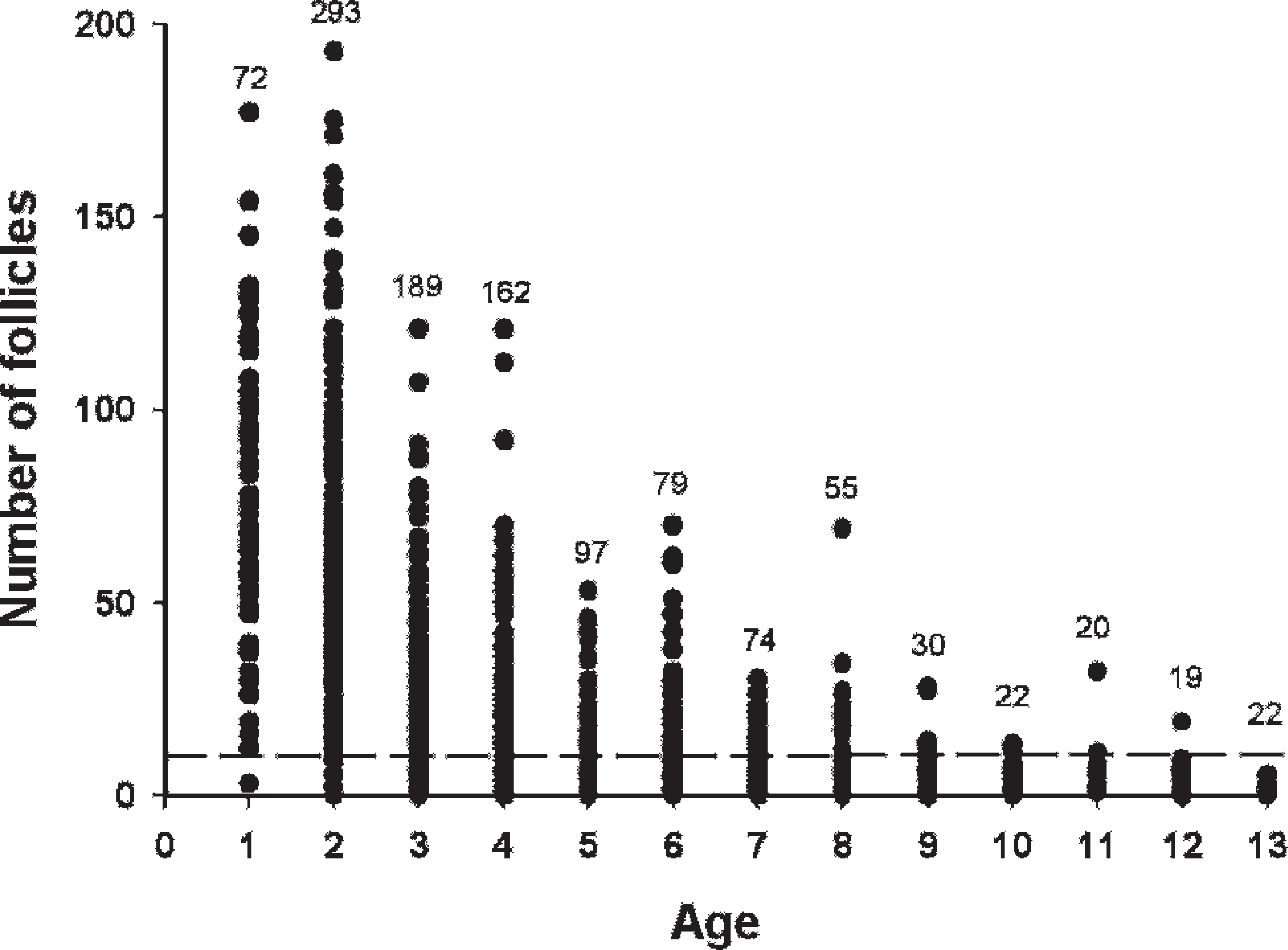
Number of follicles from 1,134 rectal biopsies for Rocky Mountain elk (Cervus elaphus nelsoni) of known age in captive facilities in Colorado, 2005–2008. Number above each age class represents sample size. Dashed line represents the 10 follicles that are believed to be needed to detect prion protein associated with chronic wasting disease (PrPCWD), if it is present. 9
Examination of postmortem rectal mucosal tissue sections for evidence of the prion protein associated with CWD (PrPCWD) has been reported 8 in elk for diagnosis of CWD, and data indicate that the technique may be useful in the diagnosis of preclinical cases of CWD. The use of rectal mucosal biopsies has also been reported 9 in a herd of 40 ranch-raised elk, and 5 nonclinical cases of CWD were found with PrPCWD in lymphoid follicles of rectal mucosal. Both of these studies 8,9 indicate that rectal biopsies may be a useful technique for diagnosing preclinical CWD in elk, but they also indicate potential limitations. The lymphoid tissues may not be affected in all elk with PrPCWD, and PrPCWD may not be present in lymphoid follicles of rectal mucosae in extremely early cases. In addition, lymphoid follicle counts may decrease with age 6,9 and vary by sex. The objective of the current study was to address the possible decrease in rectal lymphoid follicles with respect to age and sex of individual elk.
Mean (±standard deviation [SD]) number of follicles by age class from rectal biopsies of ranch-raised Rocky Mountain elk (n = 368 individual elk) sampled for ≥ 2 consecutive years in Colorado, 2005–2008.
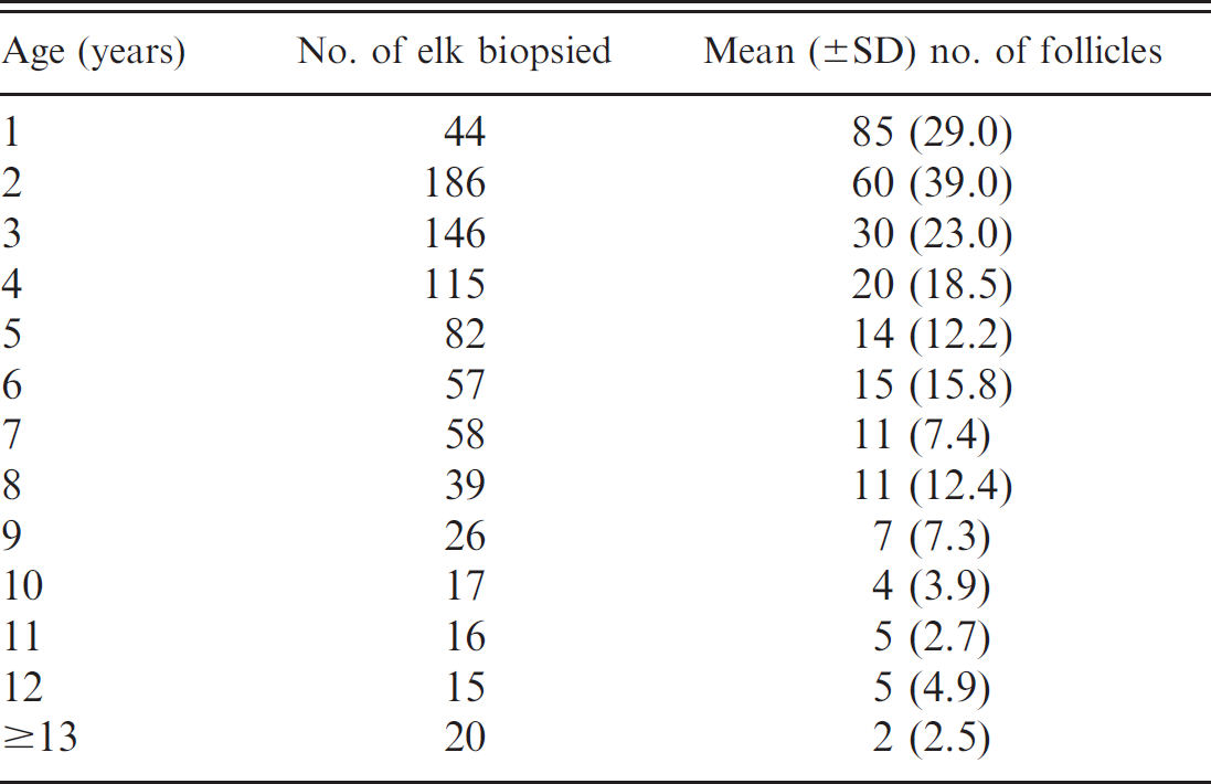

Mean (±standard deviation) number of follicles from 821 rectal biopsies of Rocky Mountain elk (Cervus elaphus nelsoni) of known age sampled for ≥l2 years (n = 368 individual elk) in captive facilities in Colorado, 2005–2008. Means with similar letters for each age class indicate no difference in mean number of follicles at P = 0.05. Dashed line represents the 10 follicles that are believed to be needed to detect prion protein associated with chronic wasting disease (PrPCWD), if it is present. 9
Rectal biopsies of elk from 4 relatively large captive herds were performed during 4 consecutive years (2005: n = 297; 2006: n = 268; 2007: n = 353; and 2008: n = 443). The age and sex of each elk was recorded at the time of biopsy. The technique used for taking the biopsies has been previously described. 9 Biopsies were taken by 4 of the authors (RDM, TLG, KCV, TRS) and 3 veterinarians that were trained on the technique by the authors. Rectal biopsies were fixed in formalin solution, processed routinely, and embedded in paraffin. Tissue sections were stained for PrPCWD in lymphoid follicles using an immunohistochemical technique, as previously described. 7,8 Using light microscopy, slides of rectal mucosal tissues were examined, and the total number of follicles in stained sections from each biopsy was recorded.
Follicle counts were compared between age and sex using a 2-way analysis of variance (ANOVA) with post-hoc differences determined by Tukey's multiple comparison tests. Differences between the numbers of follicles collected from the same elk for ≥2 consecutive years were analyzed by ANOVA for repeated measures, with post-hoc differences determined by the Tukey's multiple comparison test. Elk that were ≥13 years of age were combined into a single age class because of a lack of sufficient sample size.
Of 1,361 rectal biopsies collected from 871 individual elk from 2005 to 2008, 227 had no age data, which resulted in 1,134 rectal biopsies for age-related analysis. The number of follicles from rectal biopsies decreased in the older age classes (Fig. 1). There was no interaction of sex or age for number of follicles (F 9, 1, 078 = 0.71, P = 0.704). There were significant differences in the number of follicles by age (F 12, 1, 078 = 82.36, P < 0.001), with younger elk having more follicles than older elk. There was no difference in the number of follicles between male and female elk (F 1, 1, 078 = 0.36, P = 0.547).
There were 821 rectal biopsies collected for ≥2 consecutive years from 368 individually identified elk (Table 1). The mean number of follicles differed among several age classes (P < 0.001), with the greatest number of follicles occurring in the 1-4-year-old age classes (Fig. 2). Lymphoid tissues are currently used in preclinical testing to diagnose scrapie in sheep and CWD in mule deer and elk. A limitation of using lymphoid tissue from the rectal mucosal biopsy technique in elk appears to be a decrease in the number of follicles as elk age. 6,9 If the number of follicles decreases too much, then the test would decrease in sensitivity and be of limited use in older animals.
As previously acknowledged, the minimum number of lymphoid follicles needed for an accurate diagnosis of CWD using rectal mucosal biopsies in elk is unknown but is estimated to be 10 follicles. 9 Although this question was not addressed in the current study, a minimum of 10 follicles indicates that it may be more difficult to acquire enough follicles from elk that are >8.5 years of age. However, the follicle numbers obtained in the present study were adequate to detect PrPCWD in 8 nonclinical elk that were <8 years of age. As the typical age of ranch-raised elk with CWD is 3–6 years of age (TR Spraker, unpublished data), the rectal biopsy technique provides enough follicles to have confidence in the test results much of the time. Further, some rectal mucosal biopsies in elk >8.5 years of age produced adequate numbers of lymphoid follicles and could be used for diagnosis of CWD in elk. There were animals in every age group in which follicles were not found. It is believed that this result was partially due to the inexperience of the person taking the biopsy, the movement and position of the animal being biopsied, or the restraint of the animal at the time of the biopsy and probably had little to do with the actual number of and location of follicles within the rectal mucosa.
The current study documents that although follicle counts decrease with age, enough follicles are present to have confidence in test results for all but the oldest individuals in a herd. For individual elk that are >8 years of age, it may be advisable to take larger or multiple biopsies. The rectal biopsy technique demonstrates promise for detecting nonclinical cases of CWD in infected herds. This technique may prove useful in preventing movement of infected animals from herds of unknown CWD status if they are tested prior to movement. Though progress has been made in the current study, it is imperative to remember that a negative CWD test in an individual elk should not be interpreted to mean that the elk is not incubating CWD. Further research must be done using this technique to delineate its usefulness and limitations as a screening test to identify infected herds as part of an integrated management strategy to reduce the incidence of CWD in captive and free-ranging elk.
Acknowledgements. The authors are especially grateful to Dennis and Stephanie White (owners of Velvet Ridge Elk Ranch) and the 3 other anonymous elk ranch owners who allowed their elk to be biopsied. The authors wish to thank Richard Brewster and Jack Ellithorpe for taking some of the rectal biopsies; Jennifer Dartez, Tara Boss, Tracy Nichols, and Crystal Meyerett for recording data; and Justin Fischer, Michael Lavelle, Tara Ruby-Camenische, and the many ranch hands who handled elk in chutes. The authors thank Leah Powers for cutting and placing the tissues into cassettes and Robert Zink and Bruce Cummings for embedding, cutting, and immunohistochemical staining of the tissues. Also, special thanks to Katherine O'Rourke for providing guidance and reviewing the manuscript. This project was partially funded by Specific Cooperative Agreement 58-5348-2-0678 with ARS/USDA (Pullman, WA); the Colorado State University Diagnostic Laboratory, College of Veterinary Medicine and Biomedical Sciences, Colorado State University (Fort Collins, CO); and the USDA/APHIS/WS/National Wildlife Research Center.