
Abstract
This report describes a pituitary acidophil macroadenoma in a goat. Antemortem clinical findings included hypothermia and rumen stasis. Clinicopathologic findings included refractory hypoglycemia, low total thyroxin and insulin concentrations, elevated bile acid concentration, and hyposthenuria. In addition to the pituitary macroadenoma, bilateral atrophy of the zona reticularis of the adrenal glands was observed histologically.
Keywords
A 22.7-kg, 8-year-old, female pygmy goat (Capra hircus hircus) with a body condition score of 3 out of 5 and mild to moderate dehydration presented to the Louisiana State University Farm Animal Medicine Service (Baton Rouge, LA) with a 2-month history of abdominal distension (with the left side more distended than the right). Initial abdominal radiography was consistent with a markedly distended rumen. An initial fecal examination, with an egg count, complete blood cell (CBC) count, and biochemical profile, was performed. Fecal analysis identified trichostrongyliasis with a count of 1,200 eggs/g. The CBC revealed mild leukopenia (6,500 white blood cells [WBCs]/μl, reference [ref.] interval: 9,600–18,400 WBCs/μl). 4 Abnormalities in the biochemical profile included mild hypoglycemia (49 g/dl, ref. interval: 62.6–87.5 g/dl 11 ), a increased aspartate aminotransferase activity (AST; 110 U/l, ref. interval: 17.3–52.9 U/l 11 ), a and hyperproteinemia (10.3 g/dl, ref. interval: 5–8.5 g/dl 11 ). a , 5 Trichostrongyliasis with an egg counts >500 eggs/gwas considered an important finding. Differential diagnoses for the mild fasting hypoglycemia included insulinoma or tumors producing insulin-like growth factors, growth hormone deficiency, cortisol deficiency, glucagon deficiency, hepatic insufficiency, and sepsis. However, the mild hypoglycemia was not considered an important contributing factor to distention of the rumen. Laboratory error and prolonged storage were excluded as causes of the hypoglycemia because all blood samples during the hospitalization of this patient were run immediately on a benchtop wet chemistry analyzer. a Neonatal diseases were excluded based on signalment. Furthermore, severe malnutrition, uremia, and severe polycythemia as causes of hypoglycemia were excluded by additional laboratory testing. The mildly increased AST activity was attributed to muscle damage during phlebotomy and initial therapeutic interventions, although liver involvement could not be conclusively eliminated. The hyperproteinemia was thought to reflect mild to moderate dehydration based on physical examination. Although the total absolute leukocyte count was slightly decreased, the absolute counts of each leukocyte subtype were still within published ref intervals for this species of goat (ref. intervals for pygmy goats had not been established for the laboratory performing the testing). 4,8
The goat was treated with laxatives, oral surfactants, an anthelminthic, intravenous fluids, and glucose supplementation. Neither clinical improvement nor defecation were observed. Two days later, additional laboratory testing revealed continued mild hypoglycemia (59 g/dl) a and increased AST activity (632 U/l). a However, the total protein concentration returned to the ref interval, presumably because of adequate rehydration. The increased AST activity was attributed to additional muscle damage related to treatment and prolonged recumbency.
Because of the poor clinical response to treatment, the goat was anesthetized, and a routine flank rumenotomy was performed to evacuate the rumen. Postoperatively, the goat continued to receive maintenance fluids. In addition, analgesics (flunixin meglumine, 22.5 mg intravenously [IV], 3 times a day [TID]), antibiotics (procaine penicillin G, 1.5 ml subcutaneously [SQ], twice a day [BID]), thiamin hydrochloride (150 ml IV TID), and prokinetics (metoclopramide, 11 mg SQ TID) were administered along with transfaunation from a healthy adult Boer goat. Three days later the goat appeared clinically normal and was discharged from the hospital.
Nine days later, the goat presented with depression, recurrence of left-sided abdominal distension, lack of gut sounds, and severe hypoglycemia (blood glucose = 14 mg/dl). c The blood glucose value was assumed to be artificially low because of the nonlinearity of the point-of-care instrument, but it did correlate with depressed mentation and clinical signs of illness. Dextrose supplementation was provided intravenously. Several doses of detergent were administered orogastrically, and transfaunation was performed. Abdominal distension decreased, and gastrointestinal sounds returned, but any time the goat was weaned off of IV fluid administration, the blood glucose concentration would stabilize between 20 and 35 mg/dl. a,c Additional laboratory testing revealed a normocytic normochromic anemia (hematocrit = 18.4%, ref. interval: 30–38% 3 ), hypoalbuminemia (2.0 g/dl), a increased preprandial and postprandial bile acid concentrations (71 and 44 μmol/l, respectively), decreased total thyroxin concentration (3.33 μg/dl, ref. interval: 6.1–8.3 μg/dl 3 ), and decreased insulin levels (1.7 μIU/ml and 1.8 μIU/ml, ref. interval: 2–20 μIU/ml). The insulin levels were determined on frozen serum samples obtained 1 day apart and analyzed at a local ref laboratory. Both the hypoalbuminemia and anemia were attributed to the previously heavy Trichostrongyle spp. infestation. Increased bile acid concentrations were consistent with cholestasis, the cause of which was undetermined. Decreased thyroxin concentration was consistent with hypothyroidism or, potentially, a euthyroid, but sick, animal. None of the drugs this goat received had been reported to depress thyroid hormone levels, but numerous illnesses have been associated with decreased hormone levels in other species (“sick euthyroid”). 10 Decreased insulin concentrations were appropriate for the low blood glucose levels and inconsistent with an insulinoma. Urinalysis revealed hyposthenuria (specific gravity = 1.005 g/dl), which indicated decreased renal tubular conservation of water, suggesting either overzealous fluid therapy or diabetes insipidus.
Because of the inability to maintain glucose homeostasis, euthanasia was elected, and a necropsy was performed. Gross necropsy findings included a markedly enlarged pituitary gland, measuring approximately 2 cm long and 1 cm in diameter, and erosion of the sphenoid bone with enlargement of the sella turcica to accommodate the enlarged pituitary gland (Figs. 1, 2). The thyroid glands were enlarged bilaterally and measured approximately 3 cm long by 1.5 cm in width. Multiple 1–2-mm-diameter cystic follicles were present within the parenchyma. Gross inspection of the incised adrenal glands revealed a decreased amount of cortical tissue, but both glands were expanded by 5-mm-diameter corticomedullary cysts that were filled with serosanguineous fluid.
Histologically, the pituitary gland was expanded by a nonencapsulated, well-demarcated mass that appeared to arise from the pars intermedius and was compressed against the adjacent parenchyma. The mass was composed of round to polygonal cells that were individualized or formed islands, cords, pseudorosettes, and follicles. These cells had round, centrally placed nuclei, and single or double prominent nucleoli within euchromatic chromatin (Fig. 3). A minimal to moderate amount of cytoplasm was present, and cellular borders were sharply defined. Many of the cells had eosinophilic cytoplasmic granules consistent with pituitary acidophils. Other cells had amphophilic to slightly basophilic cytoplasm, suggesting degranulated neuroendocrine cells. Anisocytosis and anisokaryosis were moderate to marked, and mitoses were rare.
The meninges at the base of the thalamus had several tubular to follicular structures lined by basophilic cuboidal cells consistent with neuroendocrine cells. These cells stained positively for chromogranin A and B on immuno-histochemistry (IHC), further supporting the cells' neuroendocrine origin. One area of follicle formation had basophilic colloid in the center (Fig. 4), but cells and colloid were negative for thyroglobulin on IHC.
Adrenal gland architecture was largely effaced by large expansile cortical cysts that were lined by flattened epithelial cells and compressed the adjacent parenchyma. In both adrenal glands, the zona reticularis was markedly attenuated to nonexistent, and the corticomedullary junction was thickened with fibrous connective tissue (Fig. 5). In many areas, the zona fasiculata was also attenuated. Mild multifocal nodular cortical hyperplasia was present in the superficial cortex and extending into the capsule. In the thyroid glands, follicles were often markedly distended with colloid and lined by flattened epithelial cells.
A previous report 11 of pituitary tumors in goats described prolactin-secreting acidophil adenomas in 2 goats. Differentiation of pituitary adenoma from pituitary carcinoma can be difficult if histopathology is the sole criterion used for diagnosis. In fact, human pathologists consider nonfunctional pituitary tumors to be particularly problematic from a diagnostic standpoint unless extensive clinicopathologic data are available. Even with such data, there are few prospective clinicopathologic studies that have been used to develop a clinically useful classification system. 9 Furthermore, cellular morphology of adenomas and adenocarcinomas may be similar, and the mitotic rate is typically low for both neoplasms. However, some adenocarcinomas may have more frequent mitoses, occasional giant cells, and a greater degree of nuclear pleomorphism. 2 Because of erosion and invasion of the sphenoid bone and meninges and presumed loss of function of somatotropes and corticotropes, an acidophil adenocarcinoma cannot be excluded with certainty. Macroadenoma was diagnosed by default because definitive evidence of malignancy, vascular invasion, and distant metastases were not seen.
The clinical history could be consistent with partial hypopituitarism. In humans, pituitary neoplasia is the most common cause of hypopituitarism. 1,14 Pituitary tumors can cause hypopituitarism by mechanical compression of normal pituitary tissue, impaired blood flow to normal tissue, and interference with delivery of hypothalamic-regulating hormones through the hypothalamic-hypophyseal portal system. 14
Persistent hypoglycemia was the primary clinical problem in the goat in the current case. Deficiencies of growth hormone and/or adrenocorticotropic hormone (ACTH) are thought to contribute to the development of fasting hypoglycemia in individuals with hypopituitarism. In hypoadrenocorticism, there is insufficient secretion of glucocorticoids to stimulate adequate hepatic mobilization and production of glucose. Hypoglycemia occurs despite an appropriate decrease in insulin levels because reduced insulin levels must be accompanied by an increase in hepatic gluconeogenesis to correct hypoglycemia. Growth hormone is also a key counterregulatory hormone involved in hepatic glucose synthesis and secretion. It is also necessary for glucose production during states of hypoglycemia and hypoinsulinemia. 1,7,12,14
Although the brain has historically been thought to have an absolute requirement for glucose as an energy source in many species, volatile fatty acids can be converted to glucose for use in the central nervous system of ruminants, making the consequences of severe hypoglycemia less severe than in nonruminants. Rumen fermentation provides large amounts of acetate, propionate, and butyrate, which are rapidly metabolized to acetate and other volatile fatty acids. Thus, volatile fatty acids are used to supply the majority of energy requirements in ruminants. Consequently, ref intervals for blood glucose in ruminants tend to be approximately 70% of those in nonruminants. Because of this species-related difference in energy supply, there were no seizures or severe neurologic consequences in the goat in the current case, despite the markedly low blood glucose concentrations.
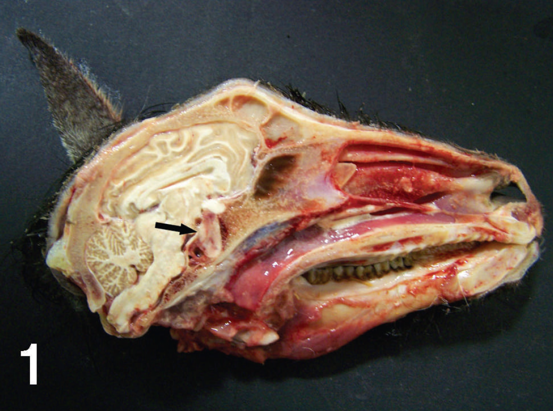
Gross photograph of the skull and pituitary gland (arrow) of a pygmy goat demonstrating enlargement of the gland and erosion of the surrounding sphenoid bone.

Higher magnification photograph of the pituitary mass (arrow). Notice the erosion and hemorrhage in the sphenoid bone surrounding the sella turcica. Bar = 1 cm.

Photomicrograph of a macroadenoma of the pituitary gland of a pygmy goat. Hematoxylin and eosin stain. Bar = 500 μm.

Photomicrograph of neoplastic pituicytes invading the meninges of the overlying brain. Hematoxylin and eosin stain. Bar = 100 um.

Photomicrograph of the adrenal gland depicting loss of the zona reticularis and large band of corticomedullary fibrosis. Hematoxylin and eosin stain. Bar = 500 μm.
Normal total thyroxin concentrations in goats has previously been determined. 3 Based on that determination, the goat in the current report was considered to be hypothyroid. Ideally, a species-specific ref interval should be available from the laboratory performing the testing or an age-matched control pygmy goat should have been used for comparison, but neither was available. It is common to have increased thyroid mass with primary hypothyroidism because decreased concentrations of T3 and T4 stimulate the release of increased concentrations of thyroid-stimulating hormone (TSH), resulting in thyroid hyperplasia and increased iodine uptake (goiter). The fact that there was bilateral thyroid enlargement in the goat suggests that thyrotropes in the pituitary gland retained their ability to secrete TSH. If hypothyroidism occurred from decreased pituitary function, the thyroid glands should have been atrophied. Therefore, the data obtained indicated minimal thyroid function and did not support a diagnosis of pituitary-dependent hypothyroidism (the hypothalamicpituitary-thyroid axis was not evaluated with stimulating hormone testing or dynamic hormone tests). Natural hypothyroid states other than goiter have not been reported in goats. In the goat in the current report, the cause of goiter was not determined; the clinical history did not indicate exposure to goitrogens; glandular inflammation was not seen histologically, and the iodine supplementation status was not known. Despite the lack of a dietary history, iodine-deficiency goiter is not uncommon in goats, and iodine supplementation was initiated shortly before necropsy. Other defects in thyroglobulin synthesis and secretion can also cause goiter, but this possibility was considered unlikely given the goat's mature age.
The histologic changes in adrenal morphology are consistent with hypopituitarism and decreased ACTH production. Both the zona fasiculata and zona reticularis were decreased to nonexistent histologically. Both adrenal glands had an overall loss of cortical parenchyma, but the large corticomedullary cysts gave a false impression that the adrenal glands were of normal size or slightly enlarged. Although hypoadrenocorticism was not confirmed during the clinical evaluation of the goat, the histologic changes in the adrenal glands are supportive of this possibility.
Cholestasis has been described along with hypoglycemia in congenital hypopituitarism. 13 Although histopathologic evidence of cholestasis was not observed in liver sections from the goat in the current report, the increased bile acid concentrations were supportive of cholestasis. The mechanism responsible for cholestasis in hypopituitarism remains controversial but may involve a combination of growth hormone, ACTH, and/or thyroid hormone deficiencies. 6 Several hepatic enzymes have been shown to be growth hormone and ACTH dependent, suggesting biochemical impairment. 13 Hypothyroidism is a well-recognized cause of cholestasis. Hypothyroid cholestasis also has an uncertain pathogenesis but may be related to reduced synthesis of primary bile acids by delayed maturation of normal enzymatic pathways and abnormalities in the structure and/or function of bile canaliculi or the pericanalicular cytoskeleton (suggested in congenital forms). 13
Hyposthenuria in the face of mild clinical dehydration could be compatible with deficient antidiuretic hormone (ADH) production by the neurohypophysis or diabetes insipidus. Repeat urinalysis, water deprivation testing, or ADH response testing would have been necessary to determine whether there was posterior pituitary compromise in addition to evidence of partial anterior hypopituitarism.
The normocytic normochromic anemia seen in the goat could have been from a variety of causes, including trichostrongyliasis or a deficiency of somatotropes. Growth hormone causes hepatocytes to up-regulate transcription of transferrin. Therefore, growth hormone deficiency leads to decreased concentrations oftransferrin, which decreases iron transport, resulting in iron deficiency or iron-lack anemia.
The goat in the current report also had rumen stasis and abdominal distension. Human patients with hypopituitarism often present with acute abdominal pain and distension of uncertain etiology. Conversely, in the face of dehydration and hypoglycemia, rumen stasis in this goat was not totally unexpected.
Descriptions of the histomorphologic changes in endocrine target organs of animals with acquired hypopituitarism appear to be rare or nonexistent. Descriptions of target organ changes in human patients are also difficult to locate; most reports of acquired hypopituitarism focus only on clinicopathologic changes. It is suspected that the alacrity of marked definitive target organ lesions attributable to hypopituitarism in this goat reflect the short clinical course of illness. Longer-standing disease may have resulted in more profound adrenocortical atrophy.
In summary, the goat in the present report had a pituitary macroadenoma and suspected endocrinopathies, suggesting pituitary dysfunction. Although pituitary dysfunction could not be proven, circumstantial evidence suggests that this entity was responsible for the failure to correct the goat's persistent hypoglycemia. When an index of suspicion for pituitary neoplasia exists, baseline serum hormone concentrations and dynamic hormone testing should be considered to identify pituitary dysfunction in a timely manner.
Footnotes
a.
As determined on nonstored fresh serum on an Olympus AU 640E serum chemistry analyzer, Olympus, Center Valley, Pennsylvania.
b.
As determined on fresh ethylenediamine tetra-acetic acid (EDTA)-preserved whole blood on an ADVIA 120 blood counter, Siemens, Munich, Germany.
c.
As determined immediately on an unspecified point-of-care blood glucose analyzer.