
Abstract
Malignant catarrhal fever (MCF) is a fatal, systemic disease of cattle and other domestic and wild ruminants that, in Europe, is caused by Ovine herpesvirus 2 (OvHV-2). American bison (Bison bison) are highly susceptible to the disease. An adult American bison, housed in a zoo in southern Italy in close cohabitation with a group of domestic sheep (Ovis aries aries) displayed clinical signs that resembled the acute form of MCF. By real-time polymerase chain reaction, OvHV-2 DNA was detected intravitam in blood, in nasal and ocular swabs, and postmortem in tissue samples of the bison. By indirect fluorescent antibody test, high MCF antibody titers were found in the bison serum. Ovine herpesvirus 2 DNA and antibodies were also found in blood samples from the domestic sheep, thus suggesting a potential role of these animals as a source of the infection. To the authors' knowledge, this is the first MCF case in captive ruminants in Italy and the second confirmed case in captive bison of European zoos.
Malignant catarrhal fever (MCF) is a fatal disease syndrome responsible for mortality in several ruminant species, such as cattle, deer, and bison. Two major epidemiologic forms of MCF are known, both caused by rhadinoviruses that belong to the Gammaherpesvirinae subfamily. Most cases of MCF in domestic cattle in the United States and Europe are caused by Ovine herpesvirus 2 (OvHV-2), which is responsible for the sheep-associated MCF (SA-MCF), mainly carried by domestic sheep (Ovis aries aries). 10 In the last few years, MCF caused significant economic losses in commercial American bison industries. 9,11 In the current report, the first reported case of SA-MCF in captive American bison in Italy is described. In September 2007, a 3-year-old, male American bison (Bison bison) housed in the safari park of Fasano (Brindisi, Italy) was found lethargic and depressed. The zoo is home to about 2,000 animals housed in free safari or exhibits, 200 of which belong to species susceptible to MCF. A group of 9 bison (4 males and 5 females) is exhibited to the public in an enclosure of about 400 m 2 allocated within the safari and with free access to various animals, including 4 Cameroon sheep (3 rams and 1 ewe). Cameroon sheep are a small, hardy domestic breed of domestic sheep (Ovis aries aries) from West Africa that requires little management and is mainly used for meat production. Although no direct contact with other potential carrier animals was reported, other ruminants known to be involved in the transmission of MCF were housed in separate areas of the zoological garden, including domestic goats (Capra aegagrus hircus) and mouflon sheep (Ovis orientalis musimon). After the onset of the clinical signs, the ill bison displayed a loss of appetite and nasal discharge, and nasal and ocular swabs were collected from the animal. In the subsequent days, the general condition of the animal appeared to worsen, with marked depression, corneal opaqueness, mucopurulent ocular and nasal discharge (Fig. 1A), watery and hemorrhagic diarrhea, and erosions of the oronasal mucosa. Ethylenediamine tetra-acetic acid treated blood and serum samples, and nasal, oral, and ocular swabs were collected from the sick bison and from the 4 sheep for virologic and serologic investigations. Because of the severity of the disease, at day 7 after the onset of the clinical signs, the bison was euthanized and necropsied. Tissue samples, urine, and pericardial fluid were collected and stored at −80°C until virologic investigations were carried out. Fragments of tissue samples were also withdrawn for histopathology. To avoid animal stress, no samples were taken from the other bison that did not show any clinical sign during the outbreak. However, these animals remained healthy in the subsequent 6 months.
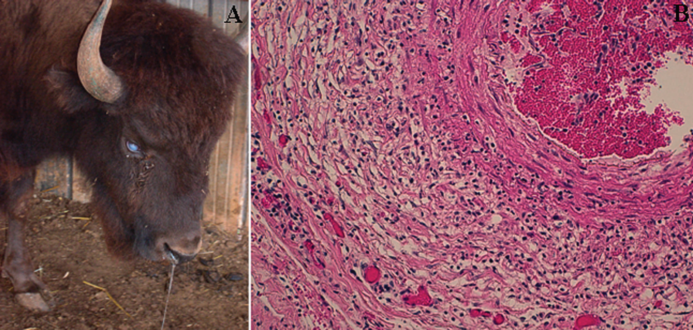
Malignant catarrhal fever-affected bison.
At postmortem examination, brain and major organs (nasal mucosa, lung, heart, liver, spleen, kidney, and urinary bladder) were collected from the sampled animal, fixed in 10% neutral-buffered formalin, and routinely processed for histopathologic examination. Brain was cut into 5-μm-thick coronal sections, whereas the other organs were sectioned at 3–5 μm. Sections for microscopic examination were deparaffinized, rehydrated, and stained with hematoxylin and eosin for evaluation of the pathologic changes. Nasal, oral, and ocular swabs taken from all animals were homogenized in Dulbecco's minimal essential medium, the suspensions were centrifuged at 4,000 × g for 15 min, and the pellets were collected. Peripheral blood leukocytes (PBL) were isolated from whole blood samples of the affected bison and healthy Cameroon sheep by means of density gradient centrifugation. a DNA was extracted from all samples (swabs, PBLs, tissues, and urine and pericardial fluid) by using a commercial kit, b according to the manufacturer's instructions. Samples were stored at −80°C until used as templates for real-time polymerase chain reaction (real-time PCR). Nucleic acids for reverse transcription PCR (RT-PCR) and real-time RT-PCR assays were extracted by using a commercial kit. c The DNA extracts were tested by a TaqMan assay able to recognize OvHV-2. 5 Samples collected from the affected bison were also analyzed for detection of the main viral pathogens of the Bovidae family, such as Bovine viral diarrhea virus, Bovine herpesvirus 1 and 4, Bovine coronavirus, Bovine torovirus, rotaviruses, caliciviruses, and Bovine respiratory syncytial virus by using established molecular protocols. 2 Bacteriologic investigations were also carried out by using standardized methods. Detection of MCF antibodies in the serum samples of the bison and the 4 Cameroon sheep was performed by an indirect fluorescent antibody test (IFAT) according to a previously described method 7 with minor modifications. 3
The clinical form displayed by the bison was highly suggestive of MCF. The main gross lesions noted at necropsy were mucosal ulceration of the oral and nasal cavities, pneumonia, and hemorrhages in several organs, including segmental areas of the intestinal mucosa, heart, urinary bladder, and kidney. Pericarditis with abundant pericardial effusion, lymphadenopathy with scattered hemorrhages, and enlargement of the gallbladder were also observed. Histologic examination of the brain performed at the level of the cortex, pons, and cerebellum revealed a rarefaction of the granular layer and a faint loss of Purkinje's cells. Major lesions were localized in medium-sized vessels of all the organs examined and were represented by accumulation of mononuclear cells (lymphocytes) in the adventitia and fibrinoid necrotizing arteritis and phlebitis. Generalized lymphoid hyperplasia was present. Infiltration of mononuclear cells in the epithelium and associated glands was present in the nasal mucosa, whereas the lungs showed diffuse interstitial chronic pneumonia with alveolar hemorrhages. Furthermore, periportal accumulation of mononuclear cells in liver and diffuse hemorrhagic cystitis in the urinary bladder were evident (Fig. 1B).
Ovine herpesvirus 2 (OvHV-2) DNA titers detected by real-time polymerase chain reaction in samples from malignant catarrhal fever-affected bison and carrier Cameroon sheep.*
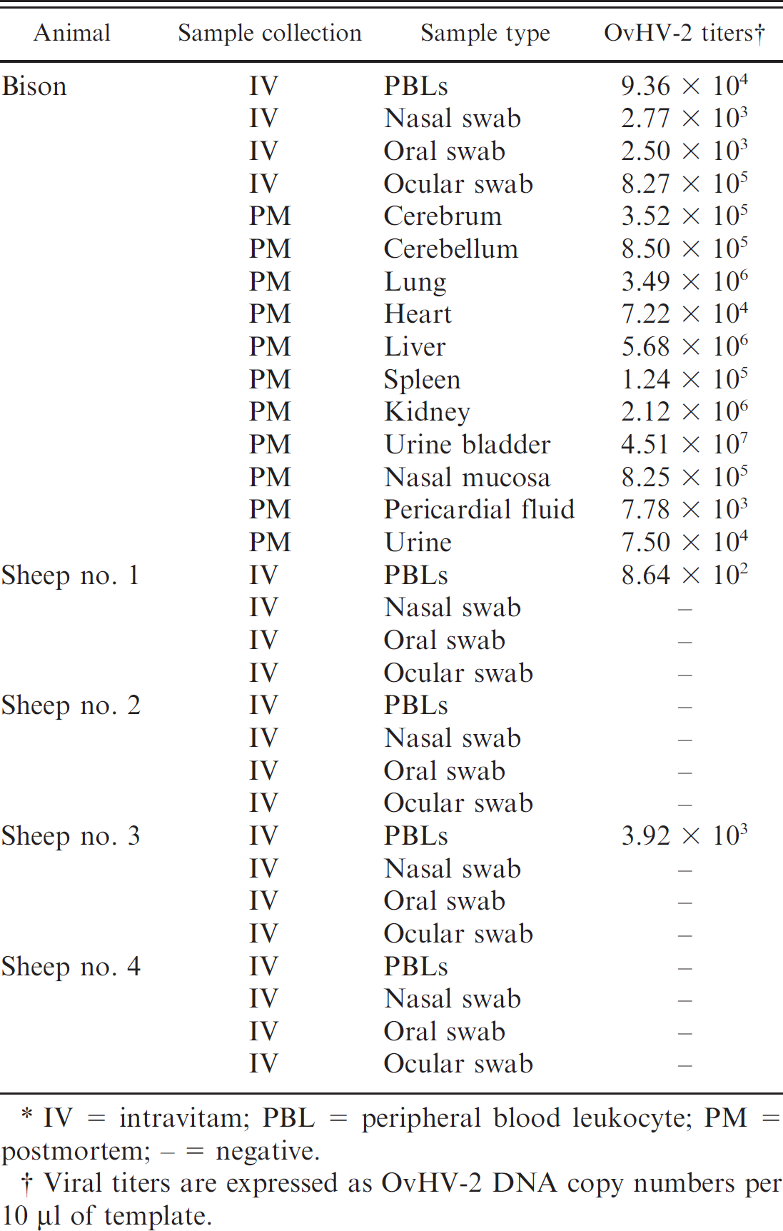
IV = intravitam; PBL = peripheral blood leukocyte; PM = postmortem; − = negative.
Viral titers are expressed as OvHV-2 DNA copy numbers per 10 μl of template.
Ovine herpesvirus 2 DNA was detected in PBLs (9.36 × 10 4 per 10 μl of template) and in all swabs collected intravitam from the bison, with the highest viral load of 8.27 × 10 5 DNA copies per 10 μl of template in the ocular swab (Table 1). The viral DNA was demonstrated in all tissue samples collected at postmortem, which showed a very wide distribution of the virus in the organism and reached the maximal values in the bladder mucosa (4.51 × 10 7 DNA copies per 10 μl of template). Huge amounts of viral DNA were also detected in the nervous tissues, with titers of 3.52 × 10 5 and 8.50 × 10 5 DNA copies per 10 μl of template in the cerebrum and cerebellum, respectively. The molecular assays gave negative results for other viruses, and bacteriologic investigations failed to detect common pathogenic bacteria. Sheep nos. 1 and 3 showed low OvHV-2 DNA titers in PBLs, whereas the blood of the remaining sheep tested negative (Table 1). All swabs collected from the sheep tested negative to OvHV-2 DNA. Malignant catarrhal fever virus-specific antibodies were detected in the serum samples of the infected bison, with a titer of 1:800, and in all 4 sheep with lower titers (Table 2).
Malignant catarrhal fever antibody titers in the affected bison and carrier sheep sera calculated by indirect fluorescent antibody test.
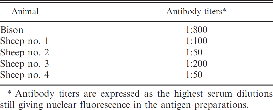
Antibody titers are expressed as the highest serum dilutions still giving nuclear fluorescence in the antigen preparations.
The clinical signs observed in the bison were characteristic of MCF. 10 The severity of symptoms reflects what was previously described in bison outbreaks. 1,6 The lack of clinical signs in the other bison confirms that these ruminants are susceptible to clinical MCF, but they are not able to transmit the OvHV-2 infection to herd mates (dead-end hosts). 1 Malignant catarrhal fever was corroborated in the ill bison by histopathology, whereas the etiologic agent was identified as OvHV-2 by specific realtime PCR. All 4 Cameroon sheep that had free access to the enclosure housing the MCF-affected bison were found to have MCF antibodies, and 2 of them tested OvHV-2 positive in their blood. Accordingly, a close contact with the Cameroon sheep may be hypothesized as a source of infection, when considering that adult sheep can sporadically shed OvHV-2 in their nasal and ocular secretions. 8 Nevertheless, when taking into account that long-distance spread of OvHV-2 was recently reported in a bison outbreak, 6 occasional transmission by other potential MCF-carrier ruminants cannot be definitively ruled out, although those animals were housed in separate and distant enclosures of the zoological garden. The healthy conditions in which the other 8 bison remained in the subsequent 6 months support an occasional transmission of the virus and confirm previous reports that suggest that MCF-affected ruminants are unable to transmit the virus to other susceptible animals. 10
American bison have been shown to be highly susceptible to SA-MCF, which is now causing severe outbreaks in bison herds, mainly in North America. 1,6 Malignant catarrhal fever outbreaks in Europe were reported in free-ranging and captive ruminants. 4,12 To date, there is a single report of MCF in captive bison in Europe, 4 whereas there are no data available about the presence of MCF in Italian zoos, thus, this is the first report on MCF in captive ruminants in Italy and the second report on MCF in captive bison in Europe.
Acknowledgements. The authors thank the director and keepers of the Zoosafari of Fasano (BR), Italy, for their kind cooperation and assistance. The authors also thank Dr. Monika Engels (Institute of Virology, Vetsuisse Faculty, University of Zurich, Switzerland) for providing OvHV-2 plasmid and MCF-negative sheep serum, and Dr. Georgina Ibata (Department of Viral Diseases, Veterinary Laboratories Agency, Weybridge, UK) for providing A1HV-1 strain WC 11.
Footnotes
a.
Lympholyte®-H, Cedarlane, Hornby, Ontario, Canada.
b.
DNeasy™ Tissue Kit, Qiagen SpA, Milan, Italy.
c.
QIAamp® Viral RNA Mini Kit, Qiagen SpA, Milan, Italy.