
Abstract
An occipitoatlantoaxial malformation was diagnosed in a 1-year-old Murciano-Granadina goat. At clinical examination, the head and cranial part of the neck were deviated to the right. Clinical signs of spinal cord or brain disease were not observed. At necropsy, morphological abnormalities were seen in the craniovertebral junction and cervical vertebrae, characterized by a firm attachment and incomplete articulation between the occipital bone and the atlas, and scoliosis in the cervical regions. The definitive diagnosis was bilateral asymmetrical occipitoatlantoaxial fusion with rotation of the atlas and atlantoaxial subluxation. To the authors' knowledge, this case report is the second occipitoatlantoaxial malformation described in a goat and the first description in an adult goat.
In domestic mammals, anomalies of the craniovertebral joint are referred to as occipitoatlantoaxial malformations (OAAMs). Such malformations involve different degrees of symmetrical or asymmetrical, unilateral or bilateral fusion of the atlas to the skull. Moreover, both fusion and subluxation between the atlas and the axis may exist. 6,13 Although rare, it has been recorded mainly in horses, 3,13 and less frequently in cattle, 1,6 sheep, 10 a dromedary camel, 8 a pig, 4 a dog, 11 and a cat. 12 In goats, only 1 case of unilateral atlantooccipital fusion in a kid, which survived 18 hr, has been reported. 7 Occipitoatlantoaxial malformation is assumed to be congenital and represents a disturbance in the normal development of the caudal occipital and cranial cervical sclerotomes with occipitalization of the atlas, 2 although an etiology of postnatal fracture, trauma, or inflammation has been reported in some cases. 6,10 To the authors' knowledge, this is the first report of congenital bilateral asymmetrical occipitoatlantoaxial fusion in an adult goat.
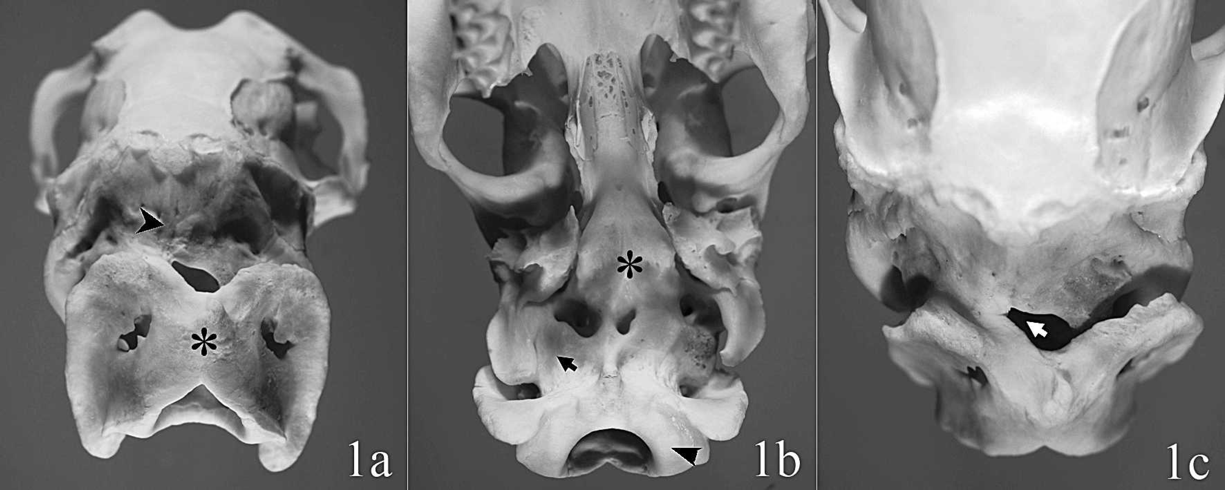
A 1-year-old female Murciano-Granadina goat was discarded for breeding because of a deviated head and was referred to the Murcia University Veterinary Teaching Hospital for necropsy. Clinical examination revealed a neck “click,” extended neck posture, stiff neck, restricted atlantooccipital movement, atlantoaxial luxation, and a palpable “bump” at the location of the atlas and axis. 5,13 The head was deviated to the right. Clinical signs of spinal cord or brain disease were not observed. Necropsy disclosed a mildly atrophic muscle mass, but other significant soft tissue macroscopic lesions were not observed. The macroscopic skeletal findings, following maceration, revealed severe morphological abnormalities in the craniovertebral joint and cervical vertebrae, characterized by fusion between the occipital bone and the atlas, and scoliosis in the cervical region (Fig. 1). The malformation comprised fusion of the basioccipital bone and the atlas (Fig. 1). Right rotation and lateral deviation of the atlas on the sagittal plane with atlantoaxial subluxation and narrowing of the vertebral canal also were observed (Fig. 1A). Hypoplasia of the occipital condyles and asymmetry of paracondylar processes were evident. The basioccipital bone was fused with the left wing of the atlas and the adjacent cranial articular fovea. This fusion was incomplete dorsally and ventrally (Fig. 1). The right paracondylar process appeared hypoplastic, fused to the atlas wing down to the allantoidea fossa, and partially fused to the adjacent cranial articular fovea of the atlas (Fig. 1B, 1C). A proliferation of fibrous tissue around the spinal cord without macroscopic alteration was noted in the vertebral canal in the fusion zone (Fig. 2). The caudal articular facets of the atlas appeared convex (Figs. 1B, 2), and mild hypoplasia in the axis dens was observed.
For histological evaluation, samples of the central nervous system were placed in 10% neutral buffered formalin. Formalin-fixed tissues were routinely processed, embedded in paraffin, sectioned at 5 μm, and stained with hematoxylin and eosin. Microscopic examination showed no lesions within these tissues. The diagnosis was made by evaluation of clinical and physical signs, palpation of the occipitoatlantoaxial region, and subsequently confirmed by postmortem examination. The definitive diagnosis was bilateral asymmetrical occipitoatlantoaxial fusion with rotation of the atlas and atlantoaxial subluxation.
The most valuable clinical aids for the diagnosis and differentiation of OAAM are neck posture, palpation, and radiographic interpretation of the occipitoatlantoaxial region. Neurological signs such as ataxia and paralysis may or may not be observed. The present case was diagnosed by clinical and physical findings and palpation because neurological signs were not observed. 5 Most OAAMs are associated with neurological signs related with spinal cord compression and injury induced by narrowing of the foramen magnum and vertebral canal, and by an incomplete joint resulting from atlantoaxial subluxation. Clinical signs, if present, are a result of a progressive and focal compressive myelopathy of the cervical spinal cord. 3 In the present case, the degree of narrowing was insufficient to compress and injure the spinal cord. However, the proliferation of connective tissue around the spinal cord could have encroached upon the vertebral canal, causing a gradual and progressive compression of the spinal cord that might have led to the development of neurological signs at 2–3 years of age, as has been described in horses. 14
In goats, congenital malformations related to genetic and environmental factors constitute a substantial proportion of cases of bone pathology, but OAAM has only been reported previously in 1 kid. 7 In domestic mammals, atlantooccipital fusion usually is asymmetrical and bilateral, 2,11 although it can be symmetrical in some individuals. 5 In the aforementioned goat, 7 the atlantooccipital fusion was reported to be asymmetrical and unilateral. The present case represents the first OAAM report in an adult goat and coincides with an asymmetrical occipitoatlantoaxial fusion with congenital cervical scoliosis and a deviated head 5 with the atlas rotated to the right. This OAAM in an adult goat, as in other species, may have been congenitally induced, representing a disturbance in the normal development of the caudal occipital and first cervical sclerotomes with occipitalization of the atlas. In Arabian horses, the origin of this condition is assumed to be hereditary. 11
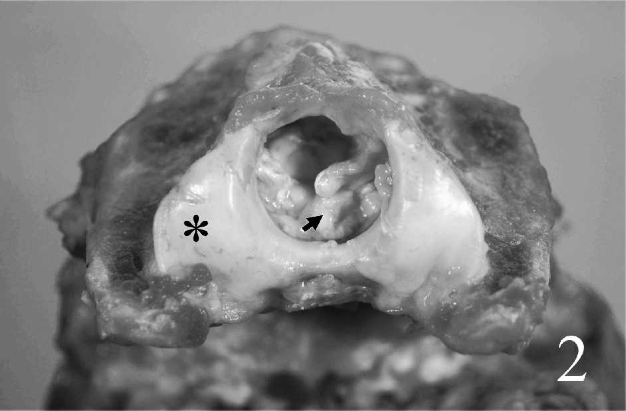
Caudal view of the vertebral canal in the occipitoatlantoaxial malformation; proliferation of connective tissue (arrow) occupies space reserved for the medulla. Note the occipitalization and the convex caudal articular facets of the atlas (asterisk).
The clinical and physical findings in the present suggest a differential diagnosis that includes torticollis, inflammation, postnatal fracture, or trauma, 2,6 but examination of skeletal changes at the craniovertebral joint and cervical vertebrae provided no evidence to suggest such alterations. Also, the differential diagnosis of congenital malformation includes fetal infection with a teratogenic virus, ingestion of certain toxic plants, and vitamin D imbalance. 9 In the present case, there was no clinical evidence to suggest that viral infection, plant toxicosis, vitamin D imbalance, or other idiopathic teratogenic factors were capable of inducing a congenital malformation in a single individual from a large flock. The fusion of the bones involved in this malformation, together with the morphological characteristics of the malformation, led to the possibility that the caudal occipital and the first cervical sclerotomes had not separated correctly. 6 This defect would have occurred sometime between the formation of primordial cartilage and the differentiation of the ossification centers in the axial skeleton.
Although the etiology of this congenital malformation could not be determined, the rarity of the lesion in the absence of a teratogenic epidemiological pattern of flock disease suggests a congenital skeletal defect. 9 Farming practices, with numerous animals and a variety of production conditions, could explain why the alteration malformation was not detected until the animal reached its reproductive age.