
Abstract
A case of malignant melanoma, originating at the base of the left horn, was diagnosed in a white 11-year-old Pygora doe. At necropsy, the goat had an ulcerated, black-pigmented, 2.5-cm mass at the base of the left horn. Firm masses diffusely black on the cut surface were present at the left horn base, subcutaneously over the frontal bones, and infiltrating into the frontal sinuses, the submandibular lymph node, and left parotid salivary gland. The left maxillary premolars and molars were loosened from the periodontia. Multiple black foci of metastasis were observed in the liver. Histologically, the masses consisted predominantly of moderately pleomorphic, polyhedral to spindle cells that contained variable amounts of dark brown intracytoplasmic pigment granules. The tumor cells were positive for Melan A by immunohistochemical staining.
An 11-year-old white pregnant Pygora doe goat weighing 43 kg was examined for a mass at the base of the left horn that had been present for at least 1 month. There was no known history of trauma to the base of the horn. No prior treatment had been performed, and the mass was apparently becoming larger. Physical examination revealed a black mass at the base of the left horn that was exuding a foul-smelling brown-black discharge. The horn was firmly attached, and there was no associated regional lymphadenopathy. No other abnormalities were found on physical examination.
Hair around the mass was clipped, and the mass was cleaned with Chlorhexidine a scrub to allow better visualization. The mass was firm, apparently not painful, spherical, and approximately 2.5 cm in diameter. The goat was anesthetized with isoflurane b via nasotracheal intubation. The mass was surgically excised. The site was heat cauterized using an electric dehorning iron. A section of the mass was submitted for histopathologic evaluation. Immediately postsurgery the goat was treated with single doses of procaine penicillin G c (30,000 IU/kg) and flunixin meglumine d (2.2 mg/kg). The goat was sent home the same day with instructions to the owner to administer 2 doses of long-acting oxytetracyline (20 mg/kg) 3 days apart starting the following day.
Histologically the mass was entirely composed of round-shaped and spindle-shaped neoplastic cells of variable sizes. The cells contained variable amounts of cytoplasmic melanin pigment, and sections from the biopsy specimen were consistent with malignant melanoma.
Approximately 3.5 months later the goat was examined due to recurrence of the mass at the base of the left horn, which was again producing a foul-smelling black exudate. The goat now weighed 28 kg. She had recently given birth to 2 kids that died within a few hours of birth. The owner reported that the goat had been anorexic for the last several days and had become progressively lethargic. The left eye was ruptured. While restraining the goat for physical examination the left horn broke off at the base with minimal force. Palpation of the head revealed unilateral enlargement in the region of the left parotid salivary gland. Thoracic radiographs performed to check for pulmonary metastases revealed no abnormal findings. Due to the poor condition of the goat, the owner elected to have the goat euthanized. The goat was submitted for postmortem examination.
On gross postmortem examination, the tissue at the site of the horn fracture was ulcerated and extensively discolored black. There was a perforating corneal ulcer approximately 1 cm in diameter on the left eye. A firm black mass, approximately 3 × 3 × 2 cm, extended from the base of the horn subcutaneously over the frontal bones, invading the bones and infiltrating into the frontal sinuses as multiple nodules ranging from 0.5 to 1.0 cm in diameter. The submandibular lymph node and the left parotid salivary gland were markedly enlarged, firm, and diffusely black on cut surface. The left maxillary premolars and molars were loosened from the periodontia. Multiple black foci (0.5 to 1.0 cm) were observed in the liver. Histologically, the masses consisted predominantly of moderately pleomorphic, polyhedral-shaped to spindle-shaped cells containing variable amounts of dark brown intracytoplasmic pigment granules. Mitotic figures were numerous. The tumor cells were positive for Melan A by immunohistochemical staining. Deparaffmized sections were labeled for Melan A using the indirect immunoperoxidase method and using the envision polymer (Dako) as the second step. The primary antibody was used at a 1:100 dilution. Negative controls included nonspecific immunoglobulin as the primary antibody. As a positive control, melanocytes from the skin of an adult caprine were labeled appropriately using the same technique. No oral masses or other epithelial masses were present. The mass was diagnosed as a malignant melanoma arising from the horn base with local spread to the frontal sinuses and metastases to the parotid salivary gland, regional lymph nodes, and liver (Figs. 1–3).
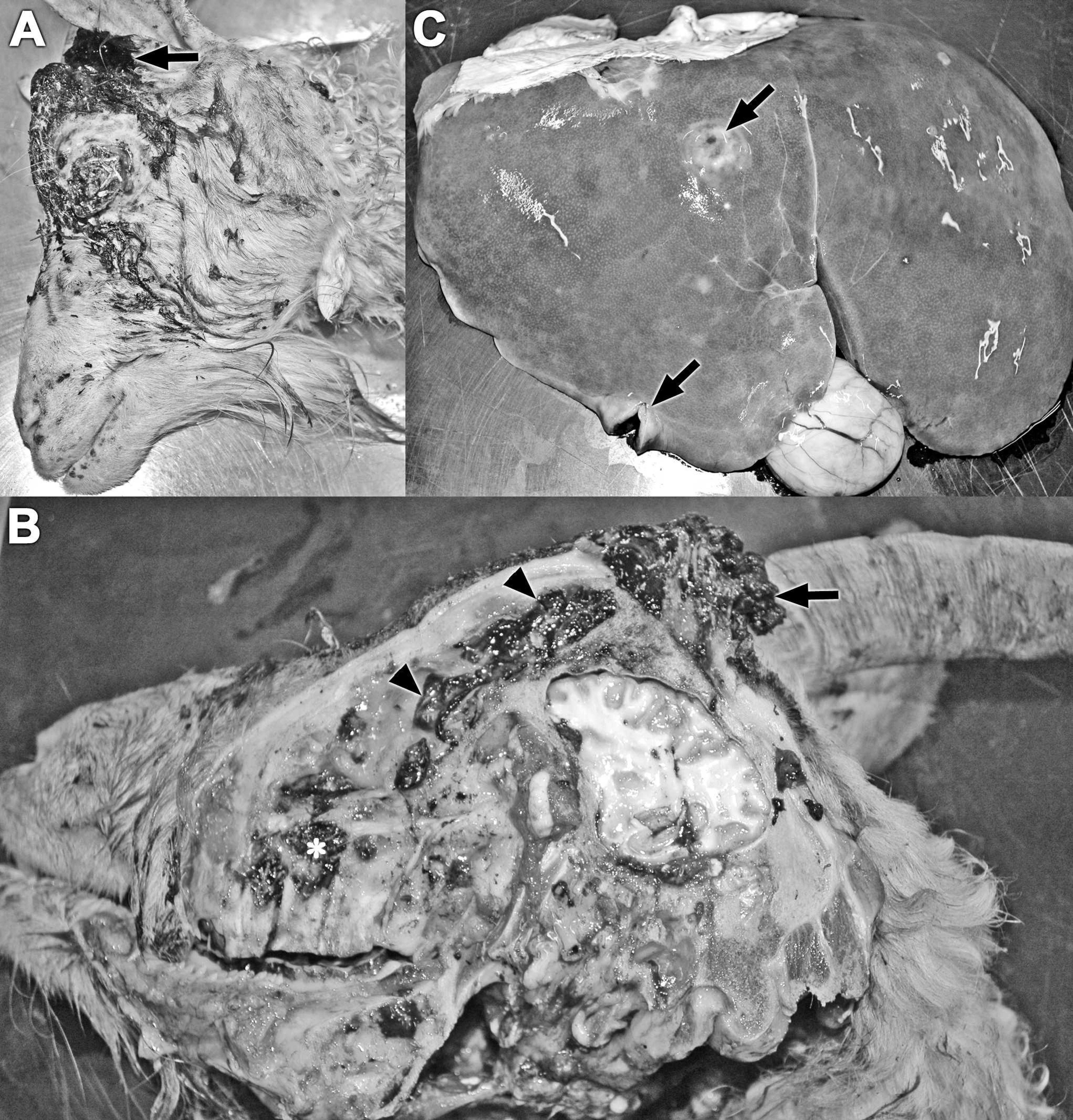
Gross presentation of the primary mass at the left horn base (
Melanomas in goats have reported occurrences ranging from rare 1,6,11 to as high as 40%. 3 Based on 1 report, melanomas are most often seen in female goats. 7 The highest age range reported for the occurrence of melanomas in goats is 4 to 5 years. 7 In the present study, the animal was 11 years old.
Melanomas may occur as solitary or multiple lesions and may be dermoepidermal or subcutaneously located. Reportedly, melanomas of the goat are highly malignant, locally aggressive, and commonly metastasize to other organs via lymphatics and blood stream. 1,7–9 Reported primary sites for malignant melanomas in goats include the skin, coronary band of the hoof 9,11 and the horn buds. 10 Initially the epidermis overlying the tumor may be intact, but becomes ulcerated with rapidly growing tumors. 12 Common sites for metastasis include regional lymph nodes, lungs, and liver, but other sites of distant metastasis have been reported. 7,9 Surgical excision is the treatment of choice; however, prognosis of malignant melanomas in goats is guarded to poor. 7,10 Recurrence following surgical excision has not been evaluated. 7
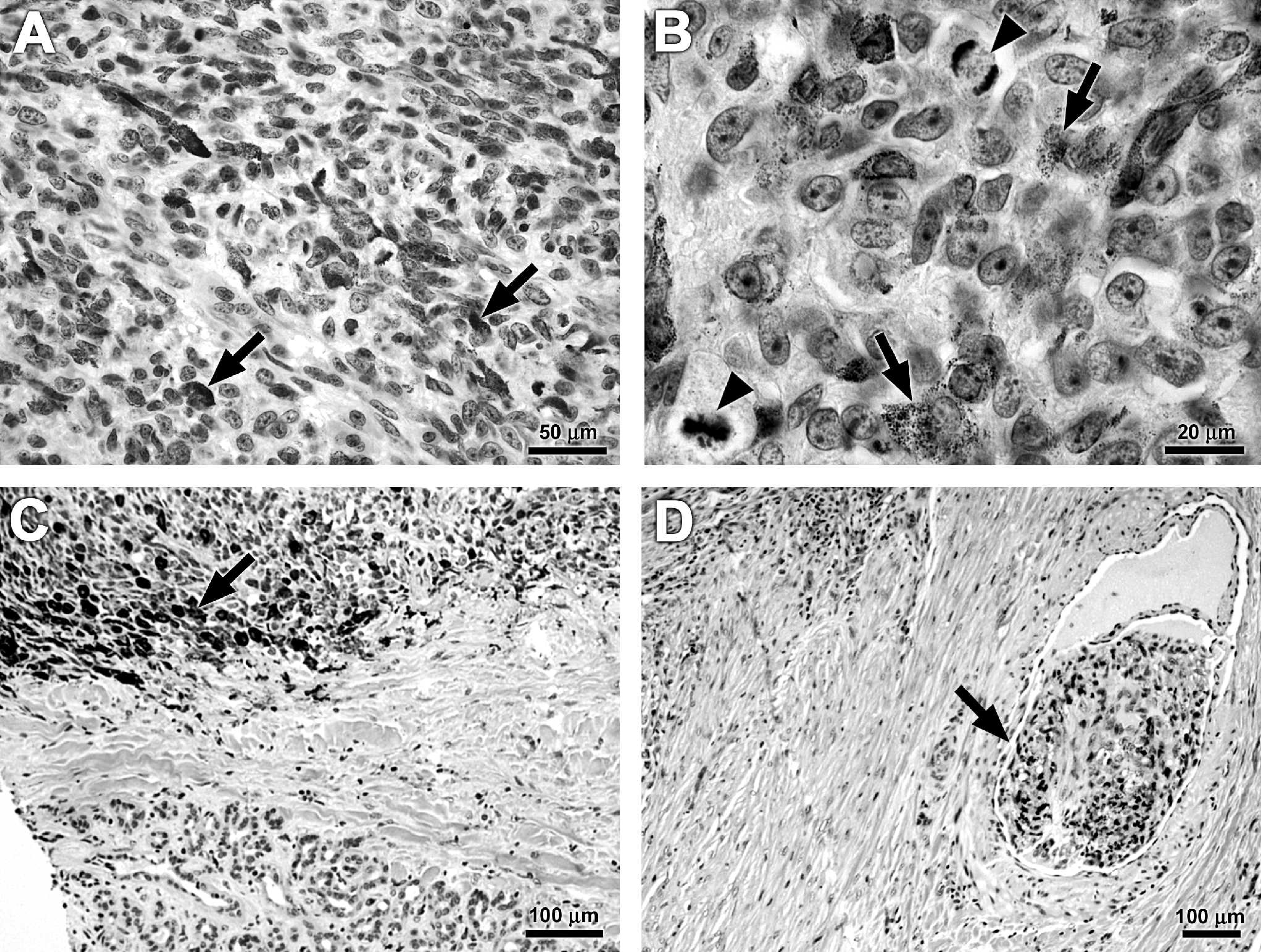
Hematoxylin and eosin stained sections of the horn base (
The malignant melanoma in the present case was locally invasive with metastasis to the regional lymph nodes, the ipsilateral parotid salivary gland, and the liver with no evidence of pulmonary metastasis. Previously reported cases associated with horn buds have occurred after trauma to the horn bud. 10 The case reported herein had no history of trauma to the horn. The horn was fully developed and was firmly attached on initial examination. One similar case has been reported in an Angora goat. 5
The causes of melanomas are uncertain. In humans, risk factors such as race, lack of skin pigmentation, excessive exposure to sunlight, and presence of preexisting nevi have been described. 4 Risk factors for melanomas in most domestic animals have not been fully described. In horses, the loss of melanin pigment with age and a tendency to develop on animals with gray or white hair coats have been identified as risk factors. 4 Some reports suggest that dark-skinned and hairy goats are more often affected. 6,11 Other domestic animals in which melanomas are associated with dark skin include cattle, sheep, pigs, and dogs. 4,6 Conversely, several cases of cutaneous malignant melanomas have been reported in Angora goats that were predominantly white. 5,10 Additionally, sites of occurrence tend to be sparsely covered with hair such as the ear, face, anus, vulva, tail, and udder. 2,5,11 Recent reviews suggest that the occurrence of malignant melanomas in Angora goats is secondary to mutations induced by ultraviolet solar radiation. 2,8 Angora goats frequently have variably sized areas of pigmentation called lentigines, which are more prevalent on the skin sites exposed to solar radiation. Lentigines are considered possible, but not obligate, melanoma precursors similar to melanocytic nevi in human beings. 2
The reported high incidence of malignant melanoma in Angora goats in South Africa may be a reflection of the Angora goat being a very common breed in that country, or may represent a true breed predilection. In 1929, a study suggested that selection for high quality mohair in Angora goats by inbreeding might have introduced a hereditary predisposition to the development of malignant melanomas in this breed. 10 The goat in the present report was a Pygora, which is derived from crossbreeding Pygmy and Angora goats. The parents of this particular goat were both Pygoras. To the authors' knowledge, this is the first report of a horn base malignant melanoma in a Pygora goat. Given the apparent predilection of Angora goats to malignant melanoma, the fact that Pygoras have Angora ancestry may have predisposed the goat reported here to malignant melanoma. Additional risk factors included the age and sex of the goat and exposure of the horn base to sunlight.

Immunohistochemical labeling of Melan A in cytoplasm of cells in neoplasm at horn base. Bar = 100 μm. Inset, Higher magnification of labeled cells. Bar = 25 μm.
Acknowledgement. The authors thank Howard Wilson for technical assistance.
Footnotes
a.
Chlorhexidine Gluconate, Purdue Products L.P., Stamford, CT.
b.
Isoflurane, Hospira, Inc., Lake Forest, IL.
c.
Procaine Penicillin G, IVX Animal Health, Inc., Fort Dodge, IA.
d.
Banamine, Schering-Plough Animal Health Corp., Kenilworth, NJ.