
Abstract
Three Francois' langurs (Trachypithecus francoisi) were found dead, without previous clinical signs, over a 48-hour period at a zoological institution after transfer to a new exhibit. A hybrid yew shrub (Taxus baccata × T. cuspidata) was found in close proximity to the exhibit perimeter fence. Despite clinical suspicion of yew intoxication, thin-layer chromatography performed on gastric contents was negative. However, microscopic examination of gastric contents revealed multiple yew fragments, and taxine alkaloids were detected by gas chromatography and mass spectrometry of the gastric contents to confirm yew intoxication. Acute death of the animals prevented treatment. The fourth langur in the collection survived, most likely because of its low rank in the troop's hierarchy, with a suspected small amount or none of the plant ingested. To the authors' knowledge, this case report is the first yew intoxication documented in a nonhuman primate species. Taxus spp. intoxication is an often fatal condition reported in domestic animals and humans. In comparison with these species, mortality appeared delayed in the Francois' langurs, most likely because of their unique gastrointestinal anatomy, with both foregut and colonic fermentation. Plant intoxication should be a differential diagnosis when multiple acute deaths are observed after recent introduction to a new enclosure.
Francois' langurs (Trachypithecus francoisi) are Asian monkeys, roughly 8 kg (17.6 lb), with a slender body and a very long, thin, nonprehensile tail. They are solid black, except for the white band that extends between the angle of the mouth across the cheeks to the pinna. The head has a pointed coronal crest. Newborns have orange pelage that changes slowly to the adult coloration by 1 year of age. Langurs are folivorous (leaf-eating) monkeys with a very large and complex gastric chamber that resembles a fermenting rumen. 3 However, results of digesta marker passage studies indicate that prolonged retention of digesta for fermentation occurs in both the stomach and the haustrated colon. The digestive strategy of these monkeys, therefore, is defined as gastrocolic fermentation, unlike that of other forestomach fermenters in which the hindgut fermentation is of secondary importance. 3 This digestive strategy is unique among folivorous mammals in general and among primate species in particular.
In late afternoon (day 1), an 8-year-old female langur, 8 kg (17.6 lb), was found on the ground and was unresponsive. The monkey was pronounced dead on arrival of the veterinarian minutes later. The caretaker reported that the entire troop (1 male and 3 females) had been visualized 1 hour earlier as alert and active. The abdominal area of the deceased langur was substantially distended on palpation, suggestive of frothy bloat, which was supported by postmortem radiographs. In addition to frothy bloat noted grossly, cerebral edema and moderate visceral congestion, suggestive of cardiovascular collapse, were detected histologically.
The langurs were moved into their indoor holding for the night. The next morning, the 12-year-old male langur, 9 kg (19.8 lb), was found dead on the floor. This individual was under treatment for known hypertrophic cardiomyopathy. The 2 remaining animals were free of clinical signs but were placed under close observation for the next 24 hours, when suddenly a third animal, an 11-year-old female, 7.4 kg (16.3 lb), collapsed and then died, despite emergency resuscitative efforts (day 3). Once again, no specific gross or histopathologic lesions were observed on necropsy for these 2 cases, except for moderate visceral congestion in both animals, mild frothy bloat in the female, and myocardial hypertrophy with right heart dilatation in the male, attributed to the previously diagnosed cardiomyopathy. The second deceased female was found pregnant with a mid-second trimester fetus.
The 4 langurs had been introduced to this exhibit 48 hours before the death of the first animal. The exhibit had previously been inhabited by white-cheeked gibbons (Hylobates leucogenys) that are mostly frugivorous (fruit eating) rather than folivorous primates. In light of the peracute disease course, the high mortality rate, and the lack of specific evidence of underlying disease, intoxication was suspected, and the zoo horticulture department was contacted to search the exhibit and the peripheral area for toxic plants. Although no toxic plants were detected directly in the exhibit, an ornamental hybrid yew (Taxus baccata × T. cuspidata) was found just outside the exhibit perimeter fence, roughly 0.5 m away, with some evidence of leaf consumption (Fig. 1).
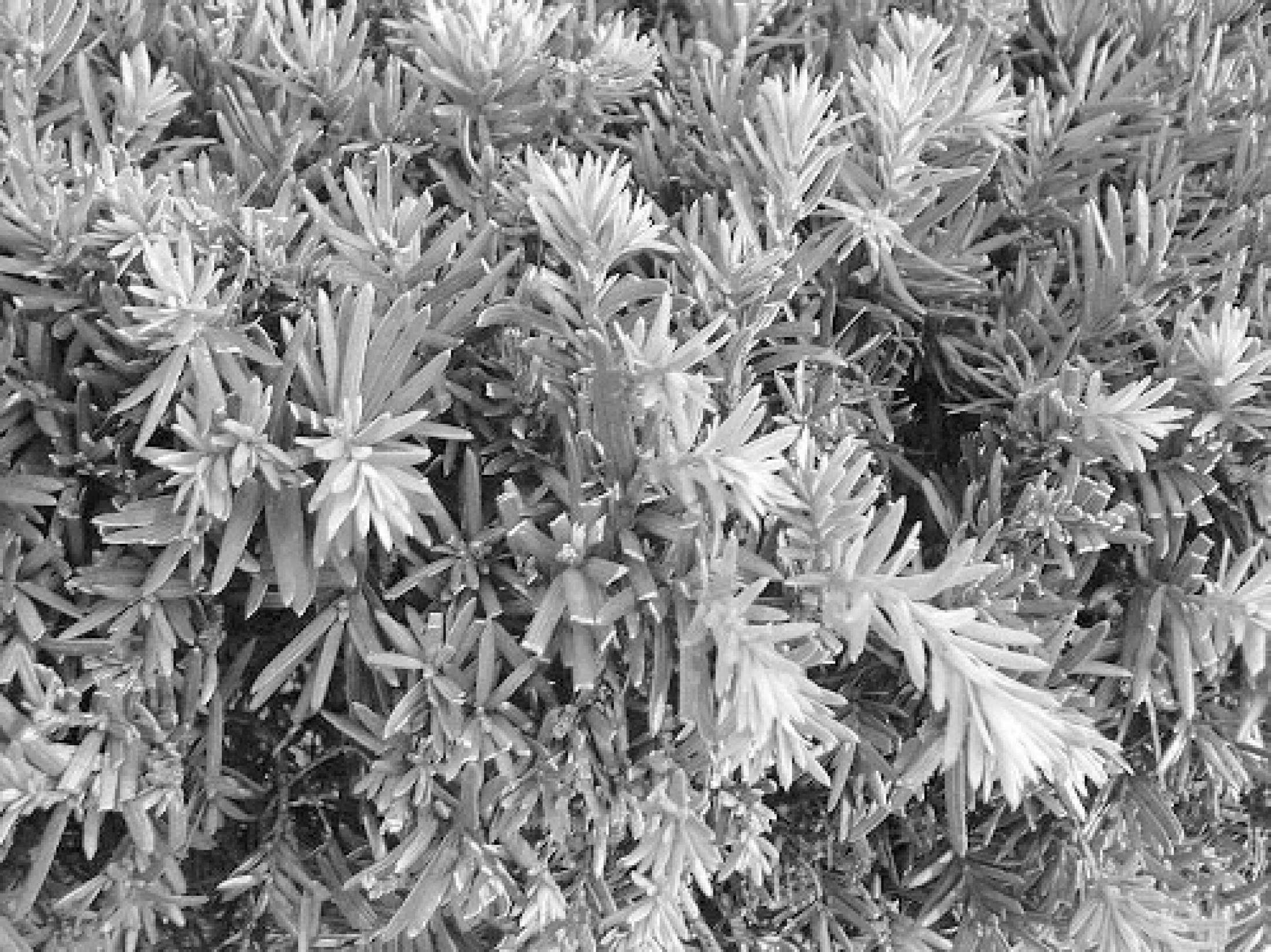
Yew plant (Taxus baccata × T. cuspidata) ingested by 3 Francois' langurs that died of Taxus intoxication.
Toxicologic testing of postmortem samples was performed to confirm yew intoxication. Initially, thin-layer chromatography (TLC) did not detect the alkaloid toxin of the plant (taxine) within the gastric contents of the langurs. However, gastric contents from the first case were submitted for microscopic examination, and a large amount of highly processed yew leaf fragments were observed (Fig. 2). The leaf epidermis had numerous stomata with sunken guard cells, morphological characteristics of Taxus spp. A unique feature of this plant is the arrangement of the stomata in rows on the lower leaf surface. These findings, and the almost cuboidal rows of cells where few or no stomata were present, aided in the conclusive identification of the plant fragments. 4
Gastric contents from the 3 animals were subsequently submitted to another laboratory for analysis and were found positive for Taxus alkaloids by using a previously reported extraction procedure and a modified gas chromatography/mass spectrometry (GC/MS) method. 5,16 Briefly, 5 g of the contents were extracted with 100 ml of 5% ethanol in ethyl acetate a (v/v) after the addition of 1 ml, 10 N sodium hydroxide, a and 50 g sodium sulfate. a A 40-ml aliquot was extracted with a total volume of 15 ml 0.5 N hydrochloric acid, after the addition of 100 ml hexane. a The aqueous extract was sparged with a stream of nitrogen, and the pH increased to greater than 10 when using 10 N sodium hydroxide. The extract was then adsorbed on a polymeric C18 SPE column. b The Taxus alkaloids were eluted with 2 ml ethyl acetate. The extract was evaporated to dryness and derivatized with bis (trimethylsilyl) trifluoroacetamide. c This extract was qualitatively analyzed by using GC/MS and a 12 m × 0.2 mm × 0.33 μm HP −1 capillary column. d Three major chromatographic peaks with retention times of 17.3, 17.4, and 17.9 minutes were present in the extract of the T. baccata reference plant. The largest peak eluted at 17.4 minutes and generated a mass spectrum with m/z 492 (relative intensity, 100), 420 (1), and 265 (9). Similar peaks with identical retention times and spectra were present in the stomach contents from all 3 langurs. The concentration of Taxus alkaloids in the gastric contents for the first, second, and third langurs were estimated to be 4%, 5.3%, and 0.9%, respectively, based on comparison of the peak area of the alkaloid eluting at 17.4 minutes to the peak area of the control matrix spiked with 1% T. baccata at the same retention time. The chromatographic peaks at 17.3 and 17.9 minutes, with mass spectra of m/z 420 (100), 212 (27), and m/z 490 (23), 400 (88), 310 (39), and 282 (100), respectively, were also used to confirm the presence of Taxus alkaloids in the gastric contents. The match qualities of these spectra were 94% and 99%, respectively, when compared with the spectra identified in the extract of T. baccata.
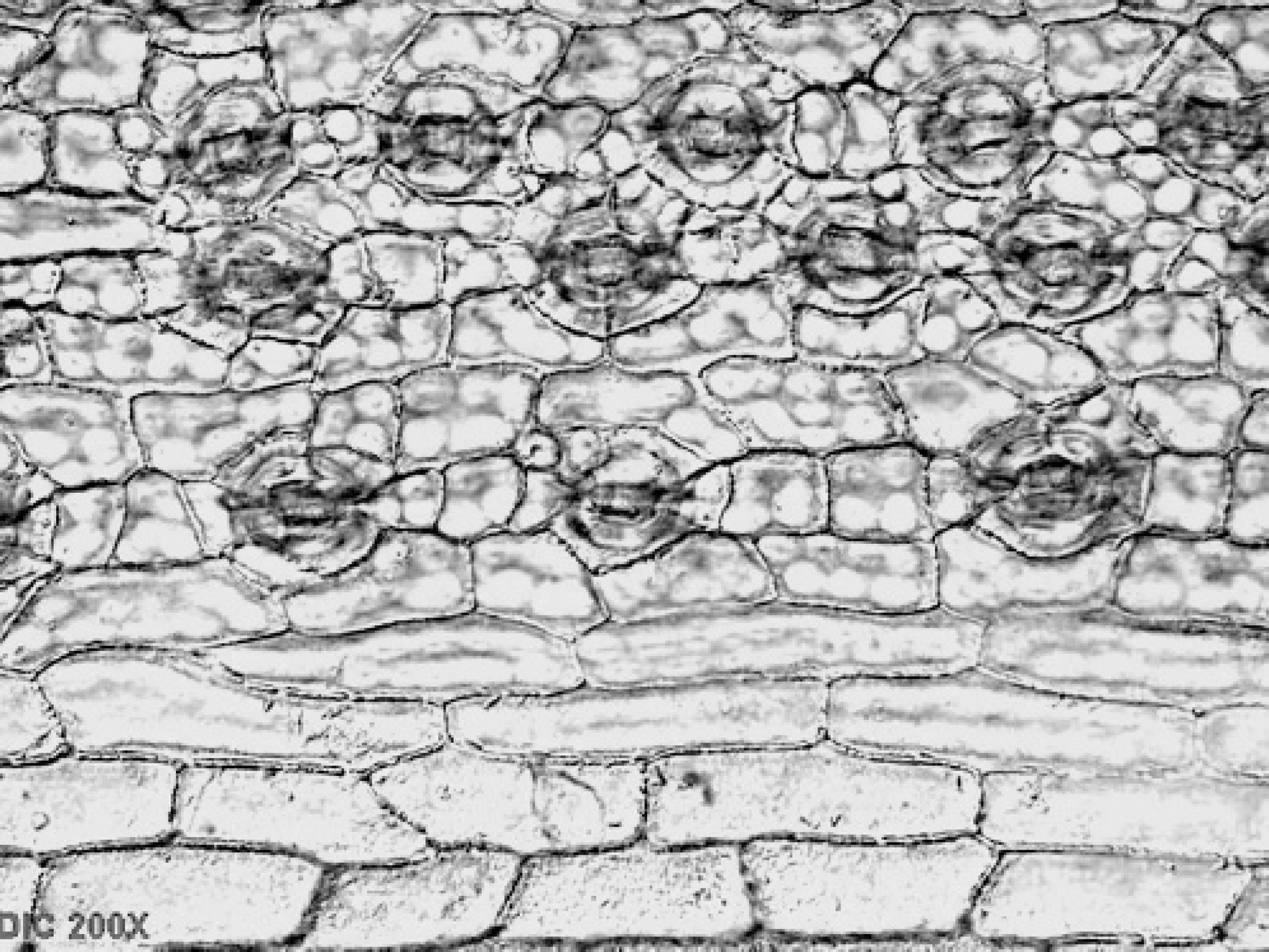
Yew fragments observed microscopically (200X) in the gastric contents of a langur that died from Taxus intoxication. The leaf epidermis had numerous stomata with sunken guard cells arranged in rows, morphological characteristics of Taxus species.
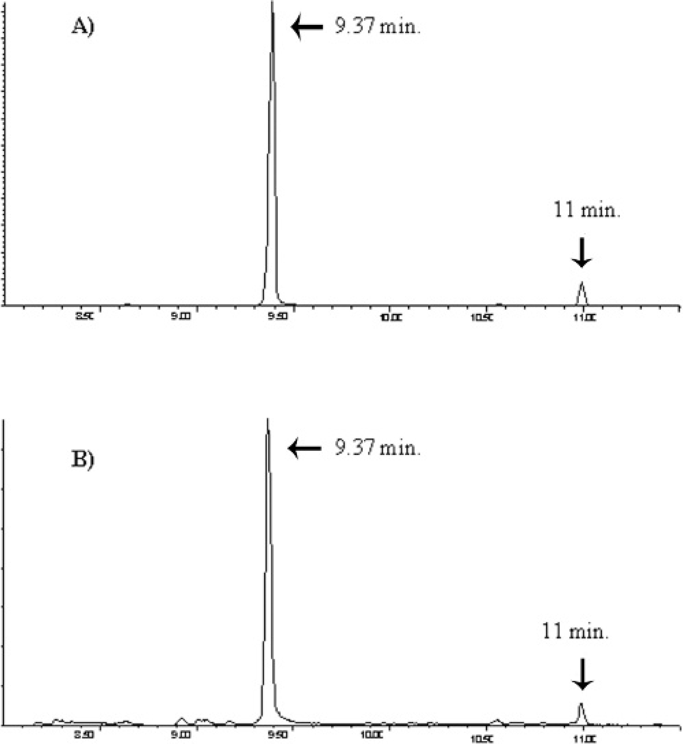
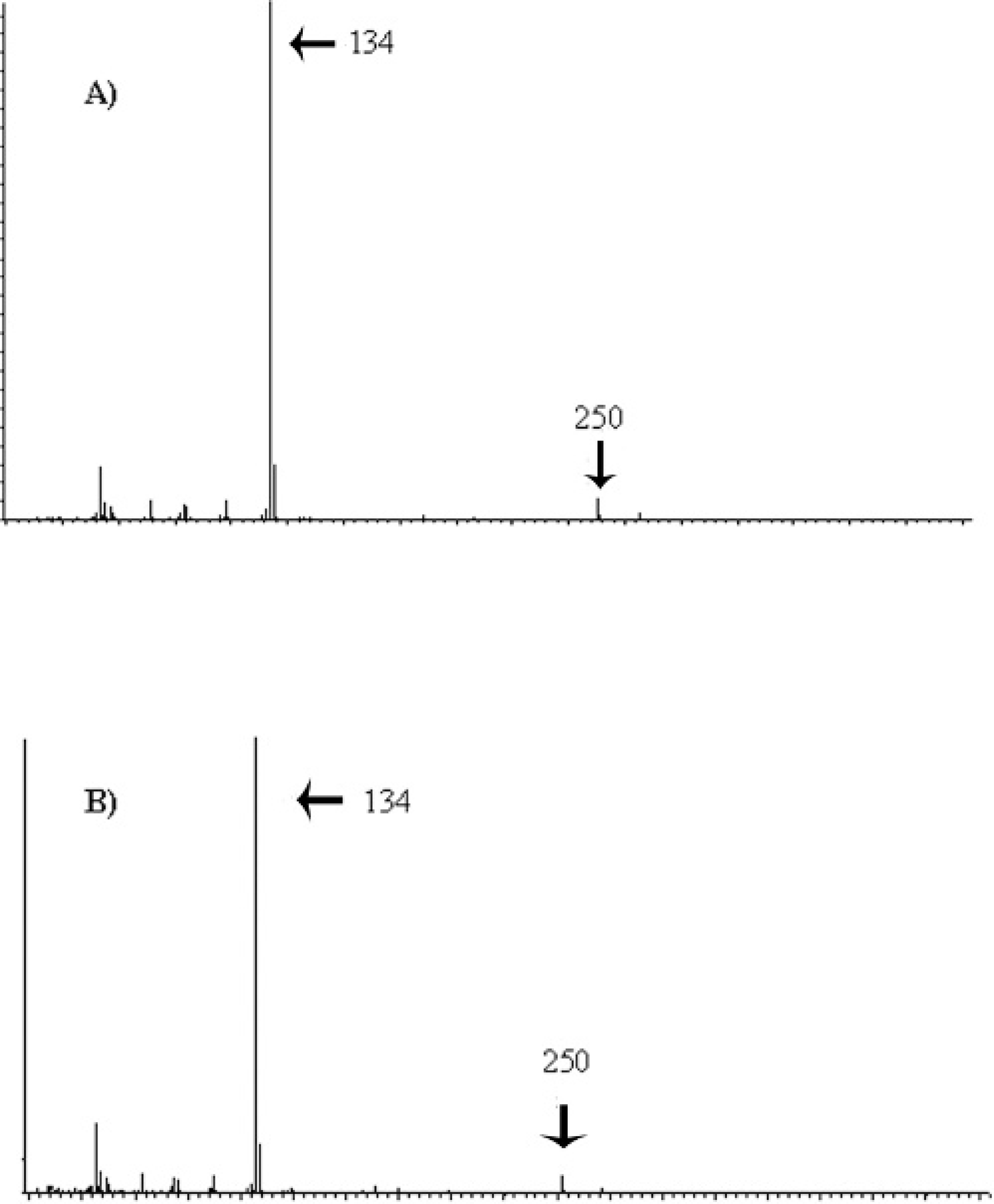
For further evidence of Taxus exposure, the ion chromatogram for m/z 134 was evaluated in the reference plant and gastric contents. This ion was found as characteristic of a chromatographic breakdown product of both taxine A and B in a previous investigation and was postulated to be the phenyldimethylamine moiety common to taxines. 8 A large peak was detected at approximately 9.37 minutes in both the reference plant and gastric contents from 2 affected langurs (Fig. 3). The ion chromatogram of the reference plant was identical to that in the gastric content samples (Fig. 4). A pattern of other peaks in the m/z 134 ion channel with similar spectra were detected in the reference plant eluting between 20 and 25 minutes (not shown). Similar patterns of chromatographic peaks were detected in the 2 gastric content samples. Other diagnostic samples analyzed with the langur gastric content samples did not contain any of the reported peaks from the reference plant. Although these results do not allow for the conclusive identification of taxines, the similarities between the plant and the ingesta in the chromatographic peaks and their associated spectra, particularly those in the m/z 134 ion channel, provided strong evidence of exposure to Taxus sp. in these animals.
Spectra of peak from 9.37 minutes in an extract of T. baccata (
Similar peaks were detected in a urine sample from the male langur but were not detected in liver and heart blood samples from the 2 females when using similar methodology. The negative results may have been because of the rapid metabolism of absorbed plant components or because of a relative insensitive test method. The toxicology results, combined with evidence of cardiovascular collapse and the absence of any additional significant underlying disease, were consistent with acute cardiac failure because of Taxus sp. ingestion and intoxication.
The yew family includes 3 genera and approximately 15 species that are found in both hemispheres. The most common of the Taxus species include T. baccata (English yew), Taxus cuspidata (Japanese yew), Taxus canadensis (American yew), and Taxus brevifolia (Western yew). 19 Yew plants are extremely toxic, with reported fatalities in many domestic mammals, including swine and rodents, as well as in birds and humans. 2,12,13,14,17,20 Most intoxication cases have been reported in horses and cattle. 1,9,10,15,16 Leaves are waxy, linear, dark green, and pointed. The toxic alkaloids are found in all parts of the plant, with the exception of the fleshy aril surrounding the seed. 6 Taxine is present in greatest concentrations in the plant in the winter. 15 The plant is often present in the animal's oral cavity or gastric contents on postmortem examination, because it is essentially immediately lethal. Taxine produces an immediate cardiotoxic effect by inducing conduction abnormalities and cardiac arrest, with few premonitory signs. The toxic component, taxine B, inhibits both calcium and sodium transport across the cell membrane of the myocardial cells. 19 In monogastrics, if clinical signs are observed before death, they are usually nervous in character, with ataxia and clonic convulsions preceding death. 11,14 In subacute poisonings, mainly in rodents and humans, gastroenteritis may be evident. However, this inflammation is most likely because of irritant oils present in the plant and not taxine. 11,19
Selected ion chromatograms for m/z 134 in an extract of T. baccata (
A time interval of 2 days before death has been reported in some ruminants. In such subacute intoxications, animals presented with clinical signs of nervousness, bradycardia, trembling, dyspnea, ataxia, gastroenteritis, and diarrhea. Spontaneous recovery has occurred. 6 The stress of moving or handling cattle often precipitates acute collapse and death. 11 Rumen degradation of taxine alkaloids has been shown to help protect white-tailed deer against intoxication. 18 The timeline of death in these 3 langurs suggested a delay in toxicity in this species, most likely because of their unique gastrocolic fermentation process, but the fatal effects of the taxine alkaloids were not prevented. Only 1 animal of the troop survived and was believed to be the lowest in the hierarchy, therefore, consumption of the yew plant may have been prevented by the other more dominant animals of the troop. During the case investigation, trimmings of the plant were obtained that approximated the size of the branch presumed accessed by the monkeys. The weight of the entire stem would account for 0.01% of body weight for all 3 animals exposed. The minimal toxic dose (LDmin) of taxine varies between animal species. Comparatively, horses are very sensitive (LDmin of 1–2 mg/kg) and chickens are least sensitive (LDmin of 82.4 mg/kg). 19 Taxus baccata has been found to be toxic to monogastric animals at 0.05%-0.1% of the animal's weight and for ruminants at 0.5% of the animal's weight. 1 This information suggests that langurs are either exquisitely sensitive to taxine alkaloids or that they accessed additional portions of the plant that were not apparent. Because of the protracted timeline and heavy concentration of Taxus found within the gastric contents, the latter conclusion is more likely. The relative sensitivity of the Francois' langur to taxine is unknown.
Taxus poisoning in cattle is usually confirmed by examination of the rumen contents for intact leaf material; but, in horses and other nonruminants, anatomic confirmation is more difficult, because the plant can be degraded in the stomach. Gas chromatography-mass spectrometry and liquid chromatography and mass spectrometry (LC/ MS) are reported as sensitive techniques for detecting taxine alkaloids in equid gastric contents in suspected Taxus poisoning cases. 7,8 In general, TLC is a less sensitive method than either GC/MS or LC/MS; this is the most likely reason that initial analysis of ingesta by TLC was negative. The major peaks identified by GC/MS in this case were in good agreement with those found in the gastric contents of a horse intoxicated by Taxus. 16 Unfortunately, standards are not commercially available for taxine A or B, which precludes their unambiguous identification in diagnostic samples. The laboratory results from these 3 cases demonstrate the need for multiple testing protocols to confirm diagnosis of yew intoxication. In this particular case, although the initial TLC results were negative, the history and clinical signs indicated that Taxus sp. intoxication should still be included in the differential diagnoses. Subsequent microscopic analysis of the stomach contents revealed the presence of yew leaves and chemical analysis with more sensitive methods indicated evidence of yew ingestion.
It has been demonstrated in feeding trials with a cow, a goat, a horse, and a sheep, that Taxus spp., which contain an irritant oil, are not highly palatable. 1 In most intoxication cases in domestic animals, the plant was either diluted in a palatable vehicle or animals were malnourished or did not have access to good quality hay. Young animals, however, tend to ingest the plant more often because of their curious nature when plant material may be inadvertently available to them. 10 The langurs were receiving an appropriate diet, and it is unknown why they chose to consume this plant, which was inconveniently placed. However, the species may have been motivated by curiosity within a new enclosure and increased interest in leaves over the prior frugivorous inhabitants.
After these cases, all yew plants were removed on zoo grounds, but these plants remain popular ornamental shrubs and are encountered frequently in the surrounding neighborhood. This plant is common in urban settings, and Taxus intoxication should be a differential diagnosis when multiple acute deaths are observed after recent introduction to a new enclosure.
Acknowledgements. The authors thank Drs. John C. Reagor (Texas Veterinary Medical Diagnostic Laboratory System) and Steve Hooser (Purdue University) for diagnostic assistance, as well as Marcia Boothe (California Animal Health and Food Safety Toxicology Laboratory, School of Veterinary Medicine, University of California) for test analyses.
Footnotes
a.
Fisher Scientific, Pittsburgh, PA.
b.
Transgenomic, Omaha, NE.
c.
Pierce, Rockford, IL.
d.
Agilent, Palo Alto, CA.