
Abstract
Objective
The feasibility of placing longer, larger diameter double-threaded screws into the pedicle for good fixation in osteoporotic patients with lumbar spondylolisthesis was investigated via robot-assisted optimal access planning.
Method
A total of 80 patients with degenerative lumbar spondylolisthesis needed posterior incision decompression and bone grafting combined with pedicle screw fixation due to spondylolisthesis. The patients were equally and randomly assigned to a robot-assisted group and a bone cement-strengthened group. The operative time, intraoperative blood loss, and intraoperative radiation dose were recorded. X-ray and CT scans were routinely reviewed after the surgery. The ratio of screw diameter to pedicle width (SD/PW) was calculated. The pedicle position was graded. The Bub score assessed proximal facet joint invasion. Visual analogue pain scale (VAS) was recorded before surgery and 3 days after surgery. The Oswestry Disability Index (ODI) and health Survey Summary Form (SF-36 to assess patients’ quality of life) were performed before surgery and 6 months after surgery. The rate of screw loosening, removal, complications and revision were evaluated by X-ray and CT 12 months after operation.
Results
VAS score on day 3 after surgery was significantly better in the robot-assisted group than in the bone cement-strengthened group. (p = 0.027). The operative time and intraoperative radiation dose of the robot-assisted group were lower than those of the bone cement-strengthened group (p < 0.001). The ratios of screw length, screw diameter, and SD/PW in both groups were significantly better in the robot-assisted group than in the bone cement-strengthened group (p < 0.001). The incidence of screw small joint invasion was 10.2% in the robot-assisted group and 19.1% in the bone cement-strengthened group, with a statistically significant difference between the two (p = 0.020). The Oswestry Disability Index (ODI) and Health Survey Summary Form (SF-36) at 6 months after surgery were significantly improved in both groups.
Conclusion
Patients with osteoporotic lumbar spondylolisthesis who use robot assistance to implant longer, thicker-diameter double-threaded screws achieved a similar fixation effect as those of bone cement-reinforced screws. Meanwhile, the operation time was shorter, the radiation damage was less, and the difficulty of later revision surgery was reduced. Thus, the proposed surgical protocol can be applied as a new option for patients with osteoporotic lumbar spondylolisthesis.
Lumbar spondylolisthesis is a partial or total slippage of the upper and lower vertebrae induced by the abnormal connection between the adjacent vertebrae of the lumbar spine. It is a relatively common disease occurring after spinal surgery, 1 which often progresses to waist and leg pain, numbness, and other conditions, and, in serious cases, it can lead to incontinence and even paralysis. 2 Although most patients with spondylolisthesis (I° and II° type) can achieve remission after strict conservative treatment, most are prone to relapse and aggravation in a short time. Some patients also require surgical treatment as the conservative treatment is completely ineffective. 3
The most common disease of lumbar spondylolisthesis is degenerative, which occurs in middle-aged and elderly people. 4 In elderlies, it is often accompanied by osteoporosis because of the imbalance of bone metabolism. With the acceleration of the aging process of the social population, the number of patients with osteoporotic lumbar spondylolisthesis is also increasing. Due to the osteoporosis of the vertebral body, the holding force of the pedicle screws on the vertebral body is decreased, and issues such as screw loosening and pulling out occur easily. 5 To resolve this concern, the researchers used polymethyl methacrylate (PMMA) to strengthen the nail path so as to increase the local bonding strength for meeting the fixed strength requirements. 6 However, it also increases the risk of bone cement leakage and significantly increases the difficulty of secondary revision surgery. To this end, researchers have attempted to increase the screw length, thread, and diameter to enhance the stability between the screw and the cone as well as the pullout resistance. However, the pedicle channel is narrow, and the peripheral nerve and blood vessels are densely packed, which is easy to damage and even lead to the rupture of the pedicle. 7 A finite element analysis in humans reported that when compared with the use of conventional pedicle screws, double-threaded pedicle screws significantly increase the stability of the pedicle screws in the cone, and this stability is closely related to bone density; lower the bone mass, more obvious is the advantage. 8 In recent years, the spinal robot has been widely used in a clinical setting owing to its accuracy of nail placement.9–11 This approach facilitates the clinical placement of longer, larger-diameter pedicle screws. As such, we attempted to explore the feasibility of placing longer, larger-diameter double-threaded screws into the pedicle to achieve a good fixation in osteoporotic patients with lumbar spondylolisthesis through robot-assisted planning of optimal access.
Methods
General information
A total of 80 patients requiring posterior incision decompression and fusion due to degenerative lumbar spondylosis were selected from the Mianyang Orthopedic Hospital, Sichuan Province between August 2022 and June 2024. Dual-energy bone mineral density (BMD) and QCT examination of all patients indicated osteoporosis. The patients were then randomly assigned to two groups: the robot-assisted group and the bone cement-reinforced group. The robot-assisted group was implanted with double-threaded pedicle screws. In the bone cement-strengthened group, the pedicle screws were implanted with the bone cement-strengthening technique under fluoroscopic guidance.
The following were the patient inclusion criteria: ① patients without surgical contraindications; ② imaging examination and clinical symptoms consistent with the diagnosis of degenerative lumbar spondylolisthesis; ③ BMD suggestive of osteoporosis; (4) surgical evidence of slip reduction.
The following were the exclusion criteria: ① Patients with other spinal diseases, such as congenital spinal deformities, ankylosing spondylitis, etc.; ② Patients with severe comorbidities who cannot tolerate surgery; ③ Patients lost to follow-up during the follow-up process.
This study was formulated in accordance with the Declaration of Helsinki and has been approved by the Ethics Committee of Mianyang Orthopedic Hospital, Sichuan Province. According to the literature review and the pretest results, the bilateral test level α = 0.05 was set, and the certainty was 90%. According to the sample size calculation formula, n = 33 cases could be obtained. Considering the 1:1 randomized grouping, that is, the placement of 33 subjects in each group and the exclusion of 20% due to follow-up data loss, the final sample size of each group was 40 patients.
Robot-assistance group
The PSIS-A type robot and the DaBo double-thread pedicle screw rod system were used for internal fixation. All patients underwent routine X-ray, CT, and MRI examinations before the operation. The preoperative CT data of the patients were imported into the robot computer for preoperative planning. After general anesthesia, the patients were placed in the prone position, and the sliding vertebrae were located under C-arm fluoroscopy. The skin and fascia of the lumbar region were incised, and the muscle tissue along the spinous processes and intervertebral ligaments was dissected to expose the bilateral laminae and articular processes. The intraoperative images were matched with the preoperative planning through feature point matching, alignment of the screw holes and the thickness, length of the screws. After the matching was completed, the mechanical arm carried the guide device and moved itself along the planned axis of pedicle screw implantation. Again, C-arm fluoroscopy was performed, and based on the intuitive image, it was determined that the positioning was correct. Then, manually percutaneously puncture the corresponding vertebrae pedicle at the direction of the guide device, enter the anterior 1/3 of the vertebrae under anteroposterior and lateral fluoroscopy, confirm the puncture was correct, and then insert the guide wire, drill and expand the channel, use the robot-selected length and size of the screw to complete the screw implantation. The C-arm fluoroscopy was satisfactory, and intervertebral disc decompression, intervertebral bone graft fusion were performed, followed by slippage reduction, and the surgery was completed. The images during the operation and before and after the surgery are shown in Figure 1. Robot-assisted surgery process and images before and after surgery. (a) Preoperative lateral X-ray of the lumbar spine; (b) Preoperative sagittal CT images of the lumbar spine; (c) Before operation, the robot planned the path of the fixation cone; (d) Lumbar lateral radiographs were reviewed after surgery; (e) Postoperative sagittal CT images of the lumbar spine. The patient, a 78-year-old female, presented with lumbar four pyramidal I° spondylolisthesis.
Bone cement-strengthened group
Internal fixation was performed using the Dabao bone cement pedicle screw-rod system. During the operation, the displaced vertebra was positioned under C-arm fluoroscopy. The skin and fascia of the lumbar region were incised, and the muscle tissue along the spinous processes and intervertebral ligaments was dissected to expose the bilateral laminae and articular processes. Under fluoroscopic guidance, bone cement screws of the pre-measured size were selected and implanted. Each screw was inserted with approximately 2-3 ml of bone cement. The process of injecting the bone cement needed to be carried out slowly under fluoroscopy to avoid leakage of the bone cement into the spinal canal and outside the vertebrae. After all the screws were implanted, intervertebral disc decompression, intervertebral bone graft fusion, and dislocation reduction were performed, and the surgery was concluded. See Figure 2. Images before and after surgery in the bone cement group. (a) Preoperative X-ray films; (b) Preoperative CT data; (c) Postoperative X-ray films; (d) Postoperative CT data. The patient, a 67-year-old female, presented with lumbar five pyramidal II° spondylolisthesis.
Postoperative treatment
All patients were administered intravenous antibiotics 24 h after the surgery to prevent infection. Lumbar and back muscle exercise was initiated on the first day after surgery, and all activities were conducted under brace protection after day 3 of the surgery.
Observation indicators
The operative time, intraoperative blood loss, and intraoperative radiation dose were recorded. X-ray and CT scans were routinely reviewed in all patients. The screw length and diameter of all patients were calculated. The ratio of the screw diameter to the pedicle width (SD/PW) was calculated based on the CT images. Based on the CT images, the dimension with the maximum screw deviation was selected to evaluate the accuracy of the implant and facet joint invasion. The pedicle position was graded by the Gertzbein and Robbins scales 12 : in grade A, the screws were located entirely within the pedicle. Grade B, pedicle cortical invasion <2 mm; Grade C, 2 mm < pedicle cortex invasion <4 mm; Grade D, 4 mm < pedicle cortex invasion <6 mm; Grade E, pedicle cortex invasion >6 mm. The excellent (Grade A) and clinically acceptable (grade A+ B) rates were compared between the two groups. Bub score 13 (proximal facet joint invasion) was classified as grade 0 (the screw is not in the facet), Grade 1 (the screw is in the lateral facet, but not in the facet), grade 2 (the facet joint penetrates <1 mm), and grade 3 (the facet joint penetrates >1 mm or moves in the facet plane).
Visual analogue pain scale (VAS) was recorded before surgery and 3 days after surgery. The Oswestry Disability Index (ODI) and health Survey Summary Form (SF-36 to assess patients’ quality of life) were performed before surgery and 6 months after surgery. X-ray and CT examinations were performed at the last follow-up (i.e., 12 months after the surgery) to evaluate the rate of screw loosening, removal, complications, and revision.
Quality control
① Quality control standards should be established before the test, and relevant personnel should be trained; ② Patients were collected in strict accordance with the criteria of inclusion, exclusion and shedding and signed informed consent; ③ Reduce the occurrence of adverse events; ④ The quality control of the study is supervised by the Science and Technology Department of the hospital and the Drug Clinical Trial Management (GCP) department.
Statistical methods
SPSS 27.0 statistical software was used to analyze the data. Measurement data conforming to normal distribution were represented by mean ± standard deviation, and inter-group comparison was performed by independent sample t test. Measurements of non-normal distribution were expressed by median (lower quartile, upper quartile), and Wilcoxon rank sum test was compared between groups. Statistical data were represented by the number of cases and percentage, and Pearson χ2 test was used for comparison between groups. The mean difference was used for the effect size of the normal distribution measurement data, and the median difference was used for the skew distribution measurement data, and 95% confidence interval (CI) was calculated for the mean difference and the median difference. Bilateral test level α = 0.05.
Results
General situation
A total of 80 patients were included, of which 40 were assigned to the robot-assisted group and 40 to the bone cement-strengthened group. The ratio of male to female [22:18 versus 19:21; χ2 = 0.200, p = 0.654], weight [(61.38 ± 7.15) versus (63.45 ± 8.57) kg; t = −1.176, p = 0.243], age [(68.23 ± 5.84) versus (67.78 ± 6.12); t = 0.339, p = 0.735], with no statistical significance. Preoperative QCT values [(65.90 ± 6.19) versus (66.00 ± 6.66); t = −0.700, p = 0.945] showed no significant difference.
Comparison of VAS scores before and after operation between the two groups
Comparison of the VAS scores before and after operation between the two groups [M (QL, QU), score].
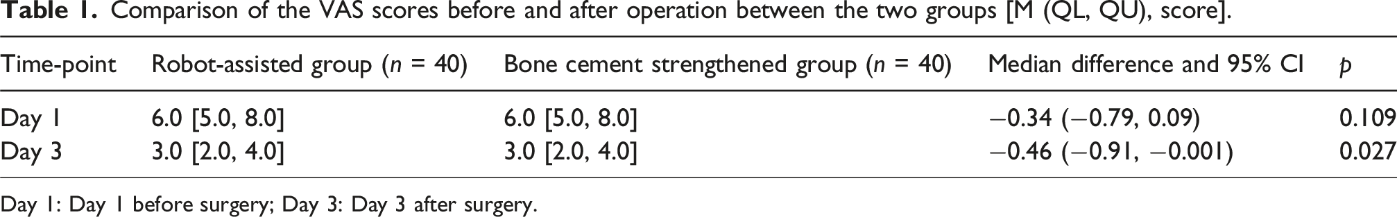
Day 1: Day 1 before surgery; Day 3: Day 3 after surgery.
Intraoperative observation indicators
Comparison of intraoperative observation indexes between the two groups.

A: Operation time[M(QL,QU),min]; B: Intraoperative blood loss
The screw length, diameter, and ratio of screw diameter to pedicle width (SD/PW) used during the operation
Comparison of the ratios of screw length, screw diameter, and screw diameter to pedicle width (SD/PW) in both groups revealed significantly better results in the robot-assisted group than in the bone cement-strengthened group (p < 0.001).
Comparison of implant accuracy and small joint invasion rate between the two groups
Screw length, diameter, and the ratio of screw diameter to pedicle width (SD/PW) in the two study groups.

S L: Screw length(
Comparison of implant accuracy and small joint invasion rate between the two groups [%].

Dysfunction score ODI
ODI scores.

Quality of life assessment
Quality of life assessment (SF-36).

PF (Physical function); RP (Role limitations due to physical health); BP (Somatic pain); GH(General sense of Health); VT (Vital Vitality); SF (Social function); RE (role limitations due to emotional problems); MH (Mental Health).
Effect of slip reduction and postoperative complications
All patients showed satisfactory reduction of slip by DR and CT after surgery. X-ray and CT examinations were performed at the follow-up visit at the end of the 12 months of surgery. No loosening or breakage of screws was detected in any of the patients, and there were no revision cases.
Discussions
Lumbar spondylolisthesis is a common orthopedic disease, which is one of the important causes of lumbar and leg pain in adults, seriously affecting the quality of life of patients, with the incidence in adults being 5.4%. 14 Wiltse 15 classified lumbar spondylolisthesis into degenerative, isthmic, traumatic, congenital, pathological, and iatrogenic types. For patients with mild lumbar spondylolisthesis, conservative treatment is generally available, 16 which includes activity restriction, the use of anti-inflammatory drugs and epidural steroid injections, and physical rehabilitation exercises such as ultrasound and electrical stimulation. However, due to the lack of in-depth research, no mature conservative treatment system has yet been reported. 17 Despite the prevalence of the disease, the optimal treatment strategy for patients with degenerative lumbar spondylolisthesis remains controversial, with surgical treatment appearing to have better clinical outcomes compared with nonsurgical treatment, 18 including decompression only and decompression fusion. 19
However, in recent years, some scholars have suggested that simple posterior laminectomy without fusion will destroy the stability of the spine and aggravate further spondylolisthesis of the lumbar spine 20 The following four types of decompression fusion treatment are mainly used 21 : posterior lumbar interbody fusion, transforaminal lumbar interbody fusion, anterior lumbar interbody fusion, and oblique lateral lumbar interbody fusion. Posterior lumbar interbody fusion is the most widely used of them. The nail rod system is the key to the return and fix the slip. With the further aggravation of aging in our country, the number of elderly patients with osteoporosis lumbar spondylolisthesis is increasing every year. The conventional pedicle screws used in surgery for internal pedicle fixation in patients with osteoporosis do not achieve the expected therapeutic effect, mainly because of the insufficient bonding force between the screws and bone trabeculae, as well as the possibility of the occurrence of postoperative complications such as unstable pedicle screws, protrusion, and even vertebral dissection. 22 Past studies have demonstrated that the incidence of pedicle screw loosening and surgical failure in patients with osteoporosis ranges from 0.6% to 25.0%. 23 Therefore, in recent years, the surgical treatment of patients with osteoporosis has mainly been attempted through strengthening the pedicle nail canal or changing the design of pedicle nails so as to enhance the stability of pedicle nails and the adhesion of bone trabeculae. 24 Presently, the technique of bone cement strengthening is considered to be reliable and feasible, as it can increase the holding force of screws by reinforcing the nail path, and it is widely practiced in clinical settings. However, the defects of this procedure are also quite obvious, with a risk of leakage of bone cement into the spinal canal, foramen, or vertebral venous plexus during the operation. 25 In addition, the bone cement releases excessive heat during the polymerization reaction. When bone cement is close to the spinal cord or nerve roots, there is a risk of nerve damage, 26 and more serious complications include pulmonary embolism, 27 paraplegia, 28 or death. 29 Meanwhile, the second surgical revision is undoubtedly difficult for patients with a reinforced bone cement nail path. The results showed that the biomechanical stability of the screw implantation is closely related to the screw diameter and thread depth. In addition, only when the screw insertion depth reaches the ideal depth does it ensure its resistance to axial shear force, which is conducive to improving the stability of internal fixation. 30 Therefore, considering that the width of the pedicle allows it, the largest diameter of the pedicle screw possible and the longest length possible are undoubtedly the best choice. A biomechanical study on human cadavers showed that double-threaded pedicle screws increased screw extraction strength. 31
However, due to the limited accuracy of manual nail implantation in daily surgery, the larger the diameter of the screws, the greater the risk of pedicle invasion or even rupture, and longer screws through the cone can damage blood vessels and other paravertebral tissues. As one of the representatives of the development of precision medicine in recent years, the accuracy of robot implants has been verified by several studies.32–34 To this end, our team introduced the only PSIS-A robot that uses intuitive positioning in China and the only one in the world to support surgery under local anesthesia to plan the stitching and pinning process. It is expected that larger double-threaded screws should be inserted when the pedicle width permits and that the orientation of the nail path should be designed to achieve the insertion of longer screws without causing damage to the paravertebral tissues. Our study found that the length of the implanted screws in the robot-assisted group was 47.37 ± 2.90 mm compared with 44.24 ± 2.86 mm in the bone cement-strengthened group, with the difference being statistically significant (p < 0.001). The screw diameter of the robot-assisted group was 6.48 ± 0.10 mm, while that of the bone cement-strengthened group was only 6.28 ± 0.25 mm, which also showed a significant difference (p < 0.001). Similarly, Du et al. found, in a comparative study on robotic and manual nail implantation, that longer screws with larger diameter could be implanted for patients with robot assistance, and the accuracy of pedicle screw implantation was also significantly superior. 34 Of course, the difference in thickness between the two groups of screws is only 0.2 mm. Whether this difference plays a decisive role in the grip force of the screws still needs to be verified through further biomechanical experiments. In our study, the excellent rate of pedicle screw implantation in the robot-assisted group was 95.7%, which was significantly higher than that in the bone cement-reinforced group (91.2%). As such, the advantages of robotic pinning are obvious, and it can maintain a higher rate of excellence than manual pinning while inserting screws with larger diameters. It has also been reported that robot-assisted implants can significantly reduce the incidence of facet joint invasion. 35 For this reason, we conducted relevant statistics based on postoperative CT images of patients and obtained the same results, that is, the screw small joint invasion rate was 10.2% (19/186) in the robot group and 19.1% (39/204) in the bone cement-reinforcement group, with a significant difference (p = 0.020). First of all, we analyzed that this may be related to the selection of screw implantation points. There will be a certain deviation in the selection of screw implantation points by hand. If the injection point is inward, the possibility of small joint invasion will increase. Secondly, the abduction Angle of manual nail planting is usually small, and the possibility of facet joint extrusion is high when the screw is screwed into the appropriate depth, while the possibility of facet joint extrusion is small when the robot planning the abduction Angle of nail path is large enough. Indeed, in terms of the surgical time and radiation-dose exposure, the results for robot-assisted pinning were superior to those for manual pinning. In a systematic review, when multiple fluoroscopy methods were combined, conventional subc-arm implants tended to result in higher doses of radiation to the surgeon and patient than robot-assisted implants. 36 Based on the literature reports and the surgical characteristics of the present study, we found that the bone cement-strengthening group in this study had significantly greater operative time and intraoperative radiation dose than the robot-assisted group, not only because the robot could assist the implantation of nails more accurately, thereby reducing the number of screw position adjustment and fluoroscopy times to save the surgical time. More importantly, the procedure of bone cement injection was increased in the bone cement-strengthening group, which had a certain impact on the dose accumulation of fluoroscopy and the duration of surgery.
Another focus of our attention in this study was to conduct slip-return by robot-assisted implantation of longer, larger-diameter, double-threaded screws and to determine the reliability of the fixation. Our 6-months postoperative follow-up of the patients revealed that both the groups showed a good reduction of the slip, with no cases of fixation failure due to obvious loosening of screws, nail pulling, or even broken nails. A cadaveric study in the past on the effect of screw thread design on thoracic vertebrae withdrawal strength in osteoporotic patients recorded that cement reinforcement was the most effective way to improve the strength of osteoporotic screws irrespective of the screw design, although it was not suitable in many cases due to the risk of complications. Their research suggested that adding threads to single threads was an effective means to recover the pullout strength, although double-threaded screws seemed like a good choice. 31 However, through finite element analysis, some researchers have noted similar extraction strength of double-threaded screws and standard pedicle screws in osteoporotic pedicles. 37 However, this study is based only on the same-diameter screws, and there is no difference in the anti-pulling of screws with different threads and diameters. In this study, patients who were fixed with longer, larger-diameter, double-thread screws had a good prognosis. Therefore, we believe that the use of robot-assisted implantation of longer, larger-diameter, double-threaded screws in patients with osteoporotic lumbar spondylolisthesis achieved similar fixation effects as bone cement-reinforced screws with shorter operative time, less radiation damage, and significantly reduced difficulty of later revision surgery. Therefore, this surgical protocol is a new choice for patients with osteoporotic lumbar spondylolisthesis.
This study also has some shortcomings. At the beginning of the experiment design, the length of the implant screw was one thread through the leading edge of the cone to obtain bicortical fixation without damaging the paravertebral tissues. However, because the screw length model span was 0.5 mm, the leading edge of the cone often does not penetrate the leading edge of the cone when the tail of the screw is screwed into the appropriate position, and the replacement of a longer model screw may induce damage to the anterior tissues. Subsequently, with the refinement of the screw types, this problem is expected to be resolved. Moreover, this study only followed the clinical efficacy of the two groups using different surgical protocols to conclude and did not conduct any finite elemental analysis to further verify the research outcomes. This study only conducted a follow-up on whether the screws were loose for a period of 12 months after the surgery, and no long-term follow-up was carried out.
Conclusion
Patients with osteoporotic lumbar spondylolisthesis who employ robot-assisted implantation of longer, thicker-diameter, double-threaded screws achieved a fixation effect similar to that of bone cement-reinforced screws. Meanwhile, the operation time was shorter, the radiation damage was less, and the difficulty of later revision surgery was reduced. Based on these results, this surgical protocol can be used as a new option for patients with osteoporotic lumbar spondylolisthesis. The original data for the significance test of the differences between the two groups of samples, as well as the ethical review documents, are available in the supplementary materials.
Supplemental Material
Supplemental Material - A prospective randomised comparative study of dynamic, static progressive and serial static proximal interphalangeal joint extension orthoses
Supplemental Material for The therapeutic effect of robot-assisted double-threaded pedicle screws in the treatment of osteoporotic lumbar spondylolisthesis by Bin Xie, Hongda Xu, Haitao Deng, Mingfan Li, Shengxing Zhao, Yuankun Gou, Lei Zhang, Tieheng Wang, Youpeng Hu, Shiming Xie, Peidong Qing in Journal of Orthopaedic Surgery
Footnotes
Funding
The authors disclosed receipt of the following financial support for the research, authorship, and/or publication of this article: This work was supported by the Mianyang Municipal Health Commission (202366).
Declaration of conflicting interests
The authors declared no potential conflicts of interest with respect to the research, authorship, and/or publication of this article.
Supplemental Material
Supplemental material for this article is available online.
References
Supplementary Material
Please find the following supplemental material available below.
For Open Access articles published under a Creative Commons License, all supplemental material carries the same license as the article it is associated with.
For non-Open Access articles published, all supplemental material carries a non-exclusive license, and permission requests for re-use of supplemental material or any part of supplemental material shall be sent directly to the copyright owner as specified in the copyright notice associated with the article.