
Abstract
Introduction:
Metastasis-associated in colon cancer 1 (MACC1), one of the prognostic markers for colonic and other tumours was noted to be overexpressed in retinoblastoma (Rb) Y79 cancer stem cells. This prompted us to evaluate its expression in primary Rb tumour and serum samples with clinicopathologic correlation. The interacting partner, c-MET was also evaluated in primary tumour tissues to explore the activation of MACC1 signaling.
Methodology:
This study was done following institutional review board approval from participating institutes. Semiquantitative gene expression for MACC1 was evaluated using formalin-fixed paraffin-embedded sections and unfixed tumour samples from primary Rb cases (n = 44). Immunolocalization for MACC1 was assessed in primary Rb tumours (n = 22), bone marrow aspirates with metastasis (n = 3), and c-MET expression was also assessed in Rb tumours (n = 17). Serum MACC1 levels were analysed using enzyme-linked immunosorbent assay in samples collected from Rb patients undergoing enucleation (n = 31), Rb patients with proven clinical metastasis (n = 3), and compared to appropriate controls. Clinicopathologic correlation of MACC1 expression was analysed using the medical records with specific reference to histologic risk factors (HRF) for metastasis and differentiation.
Results:
High expression of MACC1 gene was noted in all the tumour samples (n = 44), more so in cases with versus without HRF (p < 0.0001). In cases with HRF, MACC1 and c-MET showed diffuse nuclear and cytoplasmic staining whereas it was predominantly cytoplasmic in cases without HRF. Mean immunoreactivity score of MACC1 and c-MET tissue immunolocalization revealed that cases with HRF showed significantly higher expression compared to cases without HRF (p < 0.05). Unlike the findings in colonic tumours, serum levels of MACC1 were lower in patients compared to normal controls.
Conclusion:
Overexpression of MACC1 and c-MET in retinoblastoma tissues, specifically those with risk factors for metastasis, suggests its role in proliferation and possibly in invasion. However, the current data do not support it to be a clinical prognostic marker in retinoblastoma tumours. The inverse serum expression is an intriguing finding, which warrants further studies especially in retinoblastoma.
Introduction
Retinoblastoma (Rb) is the most common childhood intraocular malignancy initiated by a mutation within the retinoblastoma (RB1) gene. 1 The two-hit hypothesis proposed by Alfred Knudson, based on the statistical evaluation of Rb cases, identified the nature of tumour initiation and progression.2,3 In developing countries, Rb is generally diagnosed in an advanced stage, thereby presenting a higher risk of metastasis, loss of sight and even mortality. 4 While the International Classification of Intraocular Retinoblastoma (ICRB) identifies those who are likely to be cured (and the eye be saved) without the need for enucleation or external-beam radiation treatment, the assessment of histologic features of choroidal invasion, optic nerve invasion (beyond lamina cribrosa) help identify patients who are at risk for metastasis and require further treatment.5–7 The molecular mechanisms behind Rb invasion and metastasis is still unclear even though a few studies have shown the overexpression of invasion-related proteins like N-cadherin, α-catenin, SLUG, STAT3, TWIST1, and downregulation of E-cadherin and CD98–10 in primary tumours and cell lines.
Metastasis-associated in colon cancer 1 (MACC1) was first identified in colon cancer using differential display real-time polymerase chain reaction (RT-PCR) by Stein and colleagues.11,12 It has been further studied in several cancers such as breast, ovarian and gastric cancer as an important prognostic indicator for systemic metastasis and metastasis-free survival.13–19 MACC1 has been reported to promote tumour cell invasion and metastasis via the hepatocyte growth factor (HGF)/c-MET pathway.11,20 In colon cancer, Stein and co-workers reported the overexpression of MACC1 in cytoplasm and that it translocated into the nucleus following HGF treatment acting as a transcriptional regulator for receptor tyrosine kinase, MET. It was therefore implied that MACC1-facilitated activation of the HGF/c-MET pathway led to increased invasive and metastatic ability of colon cancer cells.11,21,22
A comprehensive review has also shown that MACC1 is observed in other human normal tissues such as intestine, stomach, pituitary gland, kidney and trachea showing highest expression, followed by pancreas, mammary gland, bone marrow (BM), ovary, lung, heart, liver and B-lymphoblasts. 21 Dunlevy and co-workers observed that MACC1, earlier named as 7a5 protein, was expressed in ARPE-19 and Y79 cells, neural retina and retinal pigment epithelium tissue extracts. 23 Similarly, we observed the overexpression of MACC1 gene in Rb Y79 cell line; however, it was specifically higher within the cancer stem cells (CSCs) population as identified by CD133 low cells as compared to non-CSCs. 24 This prompted us to hypothesize that it could also have a possible role in prognostication, and therefore we aimed to evaluate the status of MACC1 and its interacting partner, c-MET in Rb tissues, and specially to assess its role as a prognostic factor through a clinicopathologic correlation.
Methods
Sample collection
This study was carried out with approval from the institutional review board (IRB) (LEC 01-18-005) of LV Prasad Eye Institute, Asian Institute of Gastroenterology and University of Hyderabad (UH/IEC/2018/2). Informed consent was obtained prior to the collection of samples. Fresh Rb tumour tissue samples (n = 25) wasted from the surgical specimens of enucleation were collected and homogenized by adding 1 mL TRIZIN reagent (per 50–100 mg of tissue sample) at room temperature and were stored at −80°C until RNA isolation. RNA isolation was also done from retrospective formalin-fixed paraffin-embedded (FFPE) tissue blocks of histopathologically proven Rb cases (n = 19). Unfixed colon cancer samples (n = 6) collected from surgical specimens received from the Asian Institute of Gastroenterology (AIG, Hyderabad) were used as positive controls. Blood samples (2–4 mL) were collected from Rb patients (n = 31), healthy adults (n = 4) and healthy age-matched controls (n = 5) in blood collection tubes (BD Vacutainer®) and allowed to clot for 2 h at room temperature or overnight at 4°C prior to centrifugation at 1000g for 20 min to separate the serum. During the study period, serum from clinically confirmed metastatic cases (n = 3) of advanced Rb patients with extraocular extension at presentation and BM samples (n = 3) of clinically and histologically confirmed cases were collected and evaluated for MACC1 expression. Plasma samples (n = 6) were collected from colon cancer patients using EDTA anticoagulant sterile vacutainers by centrifuging for 15 min at 1000g. The samples were stored in aliquots at −80°C.
Semiquantitative polymerase chain reaction
Total RNA was isolated from fresh Rb tissues (n = 25), control retinas (n = 5) and colon cancer tissues (n = 6) using standard phase separation technique following solubilization in TRIZIN reagent (GCC Biotech, India). RNA from FFPE Rb tissues (central calotte only) (n = 19) was extracted using the PureLink™ FFPE Total RNA Isolation Kit (Invitrogen, CA) as per the manufacturer’s protocol, and total RNA was eluted in RNase-free water. The isolated RNA was quantified using NanoDrop™, and cDNA was prepared from 1 μg of RNA using the SuperScript™ First-Strand Synthesis System kit (Invitrogen, CA). cDNA prepared from colon cancer tissues was used as positive controls (n = 6). The primer sequences used for detecting the expression of MACC1 and ACTB1 (internal control) in the cDNA samples are as follows: MACC1 (Fp: 5′-cggtcaggaagaattgcac-3′ Rp: 5′-ttaccacgaagggtgaaagc-3′) and ACTB1 (F-atgcagaaggagatcactgc R-tcatagtccgcctagaagca). The semiquantitative polymerase chain reaction (PCR) was performed as described previously. 24 The PCR products were electrophoretically separated on ethidium bromide–stained 2% agarose gel and visualized with ultraviolet (UV) light. The images were captured using the BioRAD ChemiDoc system and analysed using the Image Lab software.
Immunohistochemistry
Immunolocalization of MACC1 was done in Rb tissues (n = 22) and BM aspirates (n = 3), and c-MET was carried out in Rb tissues (n = 17). Briefly, the enucleated Rb eye was fixed with 10% fresh formalin, processed and embedded in paraffin. The BM aspirates from cases with proven Rb metastases were collected in EDTA blood collection tubes, gently mixed and were transferred into a petri dish. They were further fixed by adding 10% fresh formalin, processed and embedded in paraffin. Thin 3 µm sections were taken on silane-coated glass slides and used for immunostaining with the primary antibodies – MACC1 (Abcam, MA, USA) and c-MET (Santa Cruz Biotechnology, Inc., CA) as per previously published protocol. 25 The sections were visualized under a light microscope, and the staining was assessed blindly by two experienced ocular pathologists (G.K.V. and D.K.M.).
Semiquantitative immunoreactivity scoring (IRS) system was used to score the intensity of immunohistochemistry (IHC) staining as per previously published established protocol. 26 Briefly, all the immunostained sections were graded as follows: grade 4 for positively stained cells >80%; grade 3 for positively stained cells between 51% and 80%; grade 2 for positively stained cells between 10% and 50%; grade 1 for positively stained cells <10%; and grade 0 for no positively stained cells. Furthermore, the intensity of the staining was assessed visually under a light microscope by two experienced ocular pathologists blinded to the clinical data and were scored as 0 (no colour reaction); 1 (mild reaction); 2 (moderate reaction); and 3 (intense reaction). A composite score ranging between 0 and 12 was achieved by multiplying the grade and the intensity. An IRS score between 0 and 1 was considered negative; 2 and 3 was considered mildly positive; 4–8 was considered moderately positive; and 9–12 was considered strongly positive.
MACC1 serum analysis by enzyme-linked immunosorbent assay
MACC1 protein expression in serum samples of Rb patients with intraocular tumour (n = 31), and those with advanced disease (extraocular extension) (n = 3), healthy adults (n = 4), healthy age-matched controls (n = 5) and in plasma samples of colon cancer patients (n = 6) were analysed using the enzyme-linked immunosorbent assay (ELISA) Kit (Cloud-Clone Corp.: SEM667Hu, Houston, USA) according to the manufacturer’s protocol. Briefly, 100 µL of blank, standards (provided by the manufacturer) and samples were added to the 96-well plates previously coated with MACC1 antibody and was incubated at 370°C for 2 h. The liquid was removed from each well to which 100 µL of detection reagent A working solution was added and incubated at 370°C for an hour. The contents of each well were aspirated and washed with wash buffer. A 100 µL working solution of detection reagent B was added to each well, incubated at 370°C for 30 min and aspirated out. A 90 µL substrate solution was added and incubated in the dark at 370°C for 20 min. The reaction was terminated by adding 50 µL stop solution. The optical density of each well was determined immediately using a microplate reader at 450 nm. Standard curves were generated and were used to analyse the MACC1 concentration in the samples.
Statistical analysis
All statistical analyses were done using the GraphPad Prism software (La Jolla, CA, USA), and the results were presented as mean values ± standard deviation. Multiple group comparisons were assessed by the one-way analysis of variance with Tukey’s multiple comparison tests. p < 0.05 was considered a statistically significant difference between the groups.
Results
Clinical details
The clinical details of the Rb cases are provided in the supplementary Table 1. Mean age of patients (n = 54) was 32 months, unilateral Rb was reported in 36/54 cases and 52% of patients (28/54) were female. During a mean follow-up of 20 months, none of them showed evidence of metastasis. Histopathologic high risk factors were noted in 29 out of the 54 cases studied, with invasion to the choroid (13/29), optic nerve post cribrosa (23/29), iris/anterior chamber (4/29) and sclera (3/29). Differentiation towards Flexner–Wintersteiner and Homer-Wright rosettes was seen in 36% of cases, while rest of the tumours were poorly differentiated in 64% of cases (32/50 cases). The cases without histologic risk factors (No-HRF) showed moderate to poor differentiation (12/32). Three cases showed retinocytoma like areas. As per the clinical protocol, the patients with HRF underwent further evaluation with BM and cerebrospinal fluid (CSF) and underwent chemotherapy. None of the cases showed evidence of recurrence or histopathological metastasis during the study period.
Gene expression analysis
MACC1 gene expression was higher in all the Rb tissue samples compared to normal retina (p < 0.0001) (Figure 1(a)). Cases with HRF had significantly higher expression of MACC1 gene than cases without HRF (p < 0.0001) (Figure 1(b)). Further clinicopathologic correlation revealed that high MACC1 gene expression is significantly associated with differentiated tissues (p < 0.0001) (Figure 1(c)) (Table 1).

Evidence of overexpression of MACC1 gene in primary Rb tissues. (a) Graphical representation of MACC1 gene expression in Rb tissue samples (n = 44) with normal retina (n = 5, p < 0.0001) as negative control and colon cancer samples as positive control (n = 6). (b) Graphical representation of MACC1 gene expression in Rb tissue samples with and without HRF in comparison to healthy retina as negative control (n = 5, p < 0.0001). HRF cases (n = 21) had higher expression of MACC1 gene when compared to No-HRF cases (n = 23, p < 0.0001). (c) Well-differentiated Rb tissues (n = 15) showed higher expression of MACC1 when compared to poorly differentiated cases (n = 27, p < 0.0001).
Correlation of clinicopathological parameters with MACC1 expression (tissue and serum) in retinoblastoma.
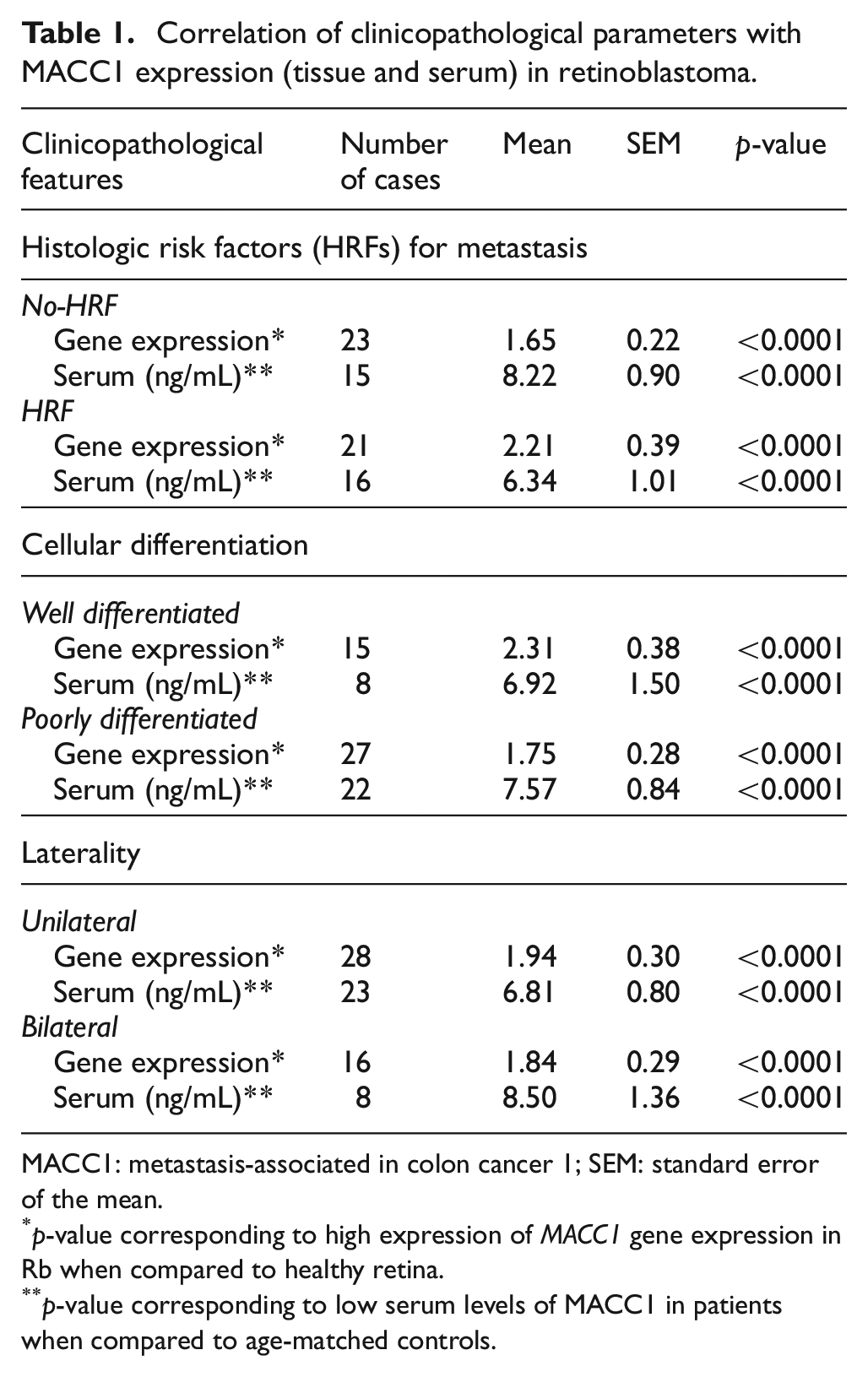
MACC1: metastasis-associated in colon cancer 1; SEM: standard error of the mean.
p-value corresponding to high expression of MACC1 gene expression in Rb when compared to healthy retina.
p-value corresponding to low serum levels of MACC1 in patients when compared to age-matched controls.
Immunolocalization of MACC1 in Rb tissues
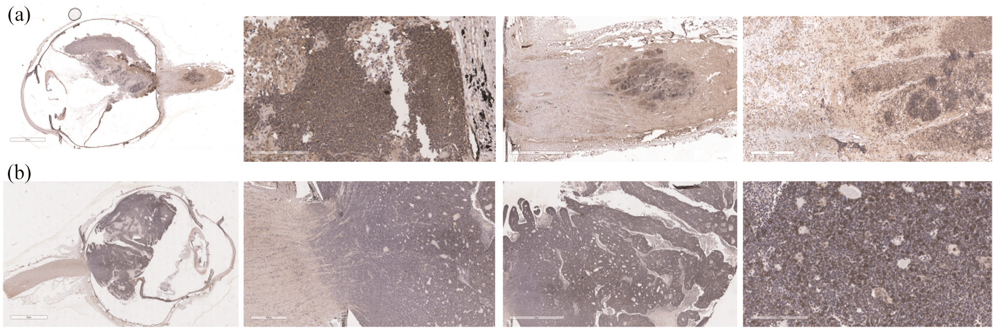
Immunolocalization showed MACC1 expression in the cytoplasm and with many cells showing nuclear membrane and nuclear positivity in HRF cases (Figure 2(a)); however, in No-HRF cases, the expression was predominantly cytoplasmic (Figure 2(b)). The expression varied from moderate to high in both HRF and No-HRF cases. Mean IRS scoring revealed that the MACC1 expression was significantly higher in cases with HRF than compared to No-HRF cases (p < 0.05) (Figure 3). Both nuclear and cytoplasmic MACC1 expressions were also observed in the Rb metastatic foci in the BM aspirates (Figure 4).
Immunolocalization of MACC1 in Rb tissues. Rb tumour cells show intense immunoreactivity to MACC1, with (a) HRF cases showing nuclear, membrane and cytoplasmic expression and (b) No-HRF cases showing predominantly cytoplasmic expression.

IRS score of MACC1 tissue immunolocalization.

Immunolocalization of MACC1 in Rb bone marrow positive cases: Cell block of the bone marrow aspirate with tumour cells (*) stained with haematoxylin and eosin under (a) low and (b) high magnifications. MACC1 positive tumour cells (*) within bone marrow under (c) low and (d) high magnifications.
MACC1 expression was noted as a nuclear staining in the healthy inner retinal layers, nuclei of limbal epithelial cells and cytoplasmic expression was observed within the healthy corneal epithelium, optic nerve, retinal pigment epithelium and choroid (Figure 5(a)–(e)).
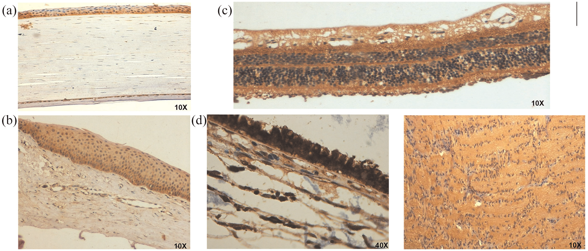
Immunolocalization of MACC1 in healthy ocular tissues: MACC1 expression was observed in healthy (a) corneal epithelium, (b) limbus, (c) neural retina, (d) retinal pigment epithelium (RPE) and choroid and (e) optic nerve (10×).
Immunolocalization of c-MET in Rb tissues
c-MET was highly expressed in all cases, with variable staining patterns, localized to cytoplasm, nuclear membrane and nucleus (Figure 6(a) and (b)). Staining pattern was predominantly nuclear in HRF cases whereas it was localized to cytoplasm in No-HRF cases. Mean IRS scoring revealed that the c-MET expression was significantly higher in cases with HRF than compared to No-HRF cases (p < 0.05) (Figure 7). Nuclear and cytoplasmic staining patterns of c-MET were also observed in the healthy inner retinal layers, nuclei of limbal epithelial cells and in the corneal epithelium, optic nerve, respectively, (Figure 6(c)–(f)).

Immunolocalization of c-MET in Rb tissues and other ocular tissues: (a) Rb cells expressed c-MET with the HRF cases showing nuclear, membrane and cytoplasmic expressions (10×) and (b) No-HRF cases showed less expression of c-MET with predominant cytoplasmic expression (10×, 20×). c-MET was also shown to be expressed in healthy (c) corneal epithelium, (d) limbus, (e) neural retina and (f) optic nerve (4×, 10×).

Semiquantitative IRS score of c-MET tissue immunolocalization.
Serum levels of MACC1 in retinoblastoma
Serum analysis of MACC1 protein level in Rb patients when compared to healthy age-matched control samples revealed that there is a significantly lower expression in Rb cases (7.25 ± 0.70 ng/mL) (p < 0.0001) (Figure 8(a)). HRF cases (6.34 ± 1.01 ng/mL) had lower expression of MACC1 protein when compared to the cases without HRF (8.22 ± 0.90 ng/mL) and healthy controls (14.69 ± 1.79 ng/mL) (p < 0.05) (Figure 8(a)). Interestingly, we also observed that the MACC1 serum levels were significantly lower (3.3 ± 0.7 ng/mL) in cases with proven metastasis compared to the control (p < 0.0001) and cases without metastasis (p < 0.0001) (Figure 8(b)). Clinicopathologic correlation revealed that higher MACC1 serum expression is significantly associated with poorly differentiated tissues and cases with No-HRF (Table 1).

MACC1 serum analysis in Rb: (a) HRF cases (n = 16) had lower expression of MACC1 protein when compared to No-HRF cases (n = 15, p < 0.05) and healthy age-matched controls (n = 5, p < 0.0001) and (b) MACC1 serum expressions in non-metastatic Rb cases (n = 31) and in cases with extraocular extension (n = 3) were found to be lower when compared to healthy age-matched control serum samples (n = 5, p < 0.0001).
Discussion
Retinoblastoma, one of the most common malignant ocular tumours, is known for its aggressive nature and rapid intra and extraocular spread.1,4,27 While the clinical and histologic prognostic factors are reasonably well identified, further insights into understanding the mechanisms of Rb tumour progression and metastasis would help pave the way in designing therapies. This study focused on exploring one such potential biomarker for metastasis first identified in colonic tumour, MACC1, which was also highly expressed in primary Rb tumours. This study reports the novel finding of MACC1 overexpression in Rb tumours, more so in cases with HRFs for metastasis than those without HRF. Immunolocalization of MACC1 and its downstream regulator c-MET revealed both cytoplasmic as well as nuclear staining; however, nuclear staining correlated with high risk factor for metastasis, hinting at its possible role in proliferation and invasion in Rb tumours.
MACC1 was first identified by Stein and co-workers in colon cancer who observed that MACC1 mRNA levels could be an independent prognostic indicator for the formation of metastasis and tumour relapse. 19 MACC1 gene expression was found to be extremely high in colon cancer tissue when compared to normal colon cells. 11 Our finding of high MACC1 gene expression in Rb primary tissues is in concordant with the findings of Stein et al., and other subsequent studies in different tumours.14–16,18,28 According to Stein et al., 11 c-MET is a transcriptional target of MACC1 gene and regulates HGF/c-MET signaling and consequently contributes to metastasis in colon cancer. Our analysis of the c-MET protein overexpression in Rb is in concordance with their findings. The results of our study suggest that high MACC1 expression is consistent with c-MET expression in primary Rb tissues. It also further hints at the crucial role of MACC1 in activating the c-MET pathway, which is initiated by MACC1 nuclear translocation, and thereby promoting cellular proliferation and metastasis as supported by data from several other cancer studies.
While all the cases showed high expression of MACC1, it was significantly higher in HRF than No-HRF cases suggesting that it may have a role in invasion and proliferation thus reiterating the need for a longer follow-up. In a 10-year follow-up study in colorectal cancer, primary tumours that showed higher expression of MACC1 eventually developed distant metastases than those tumours with low expression. 29 A concordant result was also observed in hepatocellular carcinoma (HCC), where MACC1 expression was high in vascular invasive HCC. 30 Another observation which is not clearly understood (may or may not affect prognosis) is that MACC1 gene expression was significantly higher in primary cases with differentiated Rb cells when compared to poorly differentiated cases. Similar findings were reported by Kawamura et al., 31 where a higher MACC1 mRNA expression was noted in rectal cancer patients with differentiated tumour cells. Stein et al. 21 observed that in primary colon cancer tumours, MACC1 was mainly found in the cytoplasm, whereas nuclear MACC1 expression was observed in tumours of individuals who subsequently developed distant metastases. Our data concur with their observation as increased nuclear MACC1 expression was seen in Rb cases with HRF and also within the BM metastases.15,32–35
We also checked for other key pathways deregulated with MACC1 gene expression using the previously studied microarray data of Rb primary tumours (n = 11). It was noted that there is an upregulation of several downstream molecules of c-MET signaling such as c-Jun N-terminal kinase (JNK) pathway (CRK-2.12, JNK1-2.02 fold) and mitogen-activated protein kinase (MAPK) pathway (GAB1-1.68, PIK3C2B-3.3, SHP2-1.48, MEK4-2.55 fold). 25
An interesting observation of this study was the high expression of MACC1 in normal ocular cells such as the neural retina, retinal pigment epithelium, limbal and corneal epithelium. Dunlevy and Koppelman 23 also showed similar expression of MACC1 protein in the extracts from these normal ocular tissues. A comprehensive analysis of MACC1 expression in a large array of normal tissues has shown a range of its expression with the highest in the intestine, trachea, pituitary gland, stomach and kidney. 21 Moderate to low expression was observed in the pancreas, BM, ovary, lung, liver, heart and mammary tissues. This evidence strongly suggests that MACC1 has a normal role in the growth and proliferation of cells and in neoplastic tissue, the overexpression of this protein leads to abnormal proliferation and promotes metastasis. Although the scope of this study did not include other childhood tumours, it is possible that being a childhood tumour, there could be an inherent overexpression of this marker in the ocular tissues studied.
One of the intriguing findings in this study is that the serum expression of MACC1 was observed to be significantly lower in primary Rb patients when compared to the healthy controls. Serum MACC1 expression in three cases with clinically proven extraocular metastasis also showed significantly lower expression when compared to primary Rb and control samples. This is in contrast to the studies in colonic tumours which highlight the reliability of using MACC1 as a serum marker for predicting metastasis. Interestingly, Tan and co-workers showed that in benign breast cancer patients, the serum MACC1 levels were similar to the healthy controls and lower than that of cases with metastasis suggesting that pathway may be activated after the metastatic cascade is triggered. 36 Wang et al. 37 showed that the plasma mRNA levels of MACC1 in patients with benign non-small cell lung cancer (NSCLC) were similar to that of healthy controls whereas the cases with proven metastasis, the levels were significantly higher. These studies and the results from our study partially hint at the temporal expression of MACC1 during the course of tumorigenesis and metastasis. Interestingly, our analysis revealed that in age-matched controls (healthy paediatric serum samples), MACC1 levels were significantly higher when compared to the normal adult serum samples. We believe that this rise in MACC1 serum concentration could be associated with other normal functions of this protein such as cell growth, stem cell maintenance and development.19,21
The overexpression of MACC1 in primary Rb tumours, especially in cases with HRF and poorly differentiated tissues, hints at the activation of MACC1/c-MET pathway for promoting metastasis within the tissue. To the best of our knowledge, this is the first report that identifies a differential regulation of MACC1 pathway in Rb tumour tissues and serum during tumorigenesis and metastasis. We believe that analysing the serum MACC1 levels in a larger cohort would add more value to the findings in the future and define the use of MACC1 as a prognostic marker in Rb.
In conclusion, this study reports the novel finding of constitutive overexpression of MACC1 and c-MET in Rb primary tissues suggesting its role in proliferation and possibly in invasion. While the tissue overexpression implies it to be a promising marker for predicting metastasis, the serum levels in this study do not support it to be a prognostic clinical marker. Further studies, specifically with regard to the downstream molecules in MACC1/c-MET pathway, may throw light on its association with proliferation and metastasis.
Supplemental Material
sj-xlsx-1-tub-10.1177_1010428320975973 – Supplemental material for Overexpression of metastasis-associated in colon cancer 1 in retinoblastoma
Supplemental material, sj-xlsx-1-tub-10.1177_1010428320975973 for Overexpression of metastasis-associated in colon cancer 1 in retinoblastoma by Rohini M Nair, Varsha Prabhu, Radhika Manukonda, Dilip K Mishra, Swathi Kaliki and Geeta K Vemuganti in Tumor Biology
Footnotes
Acknowledgements
The authors thank Asian Institute of Gastroenterology (AIG) for their collaboration and for providing technical support (control samples). The authors also thank Sreedhar Boyenpally and Chenchu Naidu G (LVPEI) for providing the technical support in the collection, processing and generation of retinoblastoma histological sections.
Author contributions
The study was conceptualized by G.K.V., and the experiments were designed by R.M.N., V.P. and G.K.V. The experiments were conducted by R.M.N., V.P. and R.M. Validation of experiments and data interpretation were done by R.M.N., V.P., R.M. and G.K.V. Clinical and histological data analyses were done by R.M.N., V.P., R.M., D.K.M., S.K., and G.K.V. All the authors contributed to the discussion of the results and approval of the final manuscript.
Declaration of conflicting interests
The author(s) declared no potential conflicts of interest with respect to the research, authorship, and/or publication of this article.
Funding
The author(s) disclosed receipt of the following financial support for the research, authorship, and/or publication of this article: This study was supported by academic research grants from the DST-PURSE (University of Hyderabad), UPE-II (University of Hyderabad), UGC-UKIERI (Thematic Partnership 2017–2020) and Hyderabad Eye Research Foundation (HERF). R.M.N. was supported by the UGC, Government of India (Senior Research Fellowship), and V.P. and R.M. are supported by the UGC-UKIERI grant. The funding body had no role in the design of the study, collection, analysis, and interpretation of data and in writing the manuscript.
Supplemental material
Supplemental material for this article is available online.
References
Supplementary Material
Please find the following supplemental material available below.
For Open Access articles published under a Creative Commons License, all supplemental material carries the same license as the article it is associated with.
For non-Open Access articles published, all supplemental material carries a non-exclusive license, and permission requests for re-use of supplemental material or any part of supplemental material shall be sent directly to the copyright owner as specified in the copyright notice associated with the article.