
Abstract
In this study, we evaluated the cytotoxic activity and antiproliferative potency of novel octahydropyrazin[2,1-a:5,4-a′]diisoquinoline derivatives (
Introduction
Apoptosis is a significant form of cell death, which is crucial to maintain tissue homeostasis and eliminate harmful cells from an organism. The dysfunctions in apoptotic mechanisms may contribute to the development of human diseases such as autoimmune diseases, neurodegeneration, or cancer. A new challenge in cancer research is searching for effective therapeutics, which can modulate the apoptotic pathways, activate the cascade of caspases, or inhibit the anti-apoptotic molecules. Camptothecins and novel isoquinolines target eukaryotic type IB topoisomerases (Top1), whereas human type IIA topoisomerases (Top2α and Top2β) are the targets of the widely used anticancer agents such as etoposide, anthracyclines (doxorubicin, daunorubicin), and mitoxantrone.1,2 Camptothecin was the first topoisomerase inhibitor, and its antitumor activity was observed in colon, lung, pancreas, ovarian, and breast cancers. Its derivatives, topotecan and irinotecan, represent important drugs, which are also used in chemotherapy, but they have several restrictions. 3 Etoposide is a semi-synthetic derivative of podophyllotoxin, which acts on topoisomerase II (topo II), an enzyme involved in DNA processing during its replication, transcription, and recombination. It is employed in a wide spectrum of cancers. 4 In vitro studies have shown that etoposide increases topo II–mediated DNA breakage primarily by inhibiting the ability of the enzyme to religate cleaved nucleic acid molecules. It induces apoptosis through the cytochrome c/Apaf-1/caspase-9 pathway as well as the Fas-mediated death signaling pathway.5–11 Researches are still looking for diverse promising experimental anticancer drugs with better activity and fewer side effects than camptothecin or etoposide. Recently, our team synthesized novel diisoquinoline derivatives. Their cytotoxic activity toward cultured human breast cancer cells correlated with topo I/II inhibitory properties. 12 Promising results were also obtained in other research teams. A novel class of tetrahydroisoquinoline derivatives was designed and synthesized as antitumor agents and evaluated for their in vitro and in vivo biological activities. The tubulin polymerization assay demonstrated that two compounds exhibited better inhibition rate. In the MCF-7-xenograft mouse model, the tumor weight was also decreased, so that compounds demonstrated potent in vivo efficacy. 13
Recently, Leal et al. tested novel synthetic tetrahydroisoquinoline alkaloid (PM01183). The authors proved that it induced apoptosis in several tested cell lines such as A549, HT29, HCT116, MDA-MB-231, and IGROV-1 and examined its cytotoxicity using a panel of 23 tumor cell lines that represented 11 relevant types of human cancer. Anti-tumor activity of PM01183 was also investigated. Treatment with PM01183 resulted in tumor growth inhibition in NCI-H460 lung, A2780 ovarian, HT29 colon, and HGC-27 gastric xenograft models compared with vehicle-treated animals. 14 Zheng et al. have synthesized a series of 3,4-dihydroisoquinoline derivatives and several 1,2,3,4-tetrahydroisoquinoline, isoquinoline analogues, and evaluated their cytotoxicity and tubulin polymerization inhibitory activity levels. The 3,4-dihydroisoquinoline compounds seemed to be potential tubulin polymerization inhibitors. 15 The promising results from in vitro and in vivo studies give a basis for searching novel isoquinoline alkaloid derivatives.
In this study, we examined the cytotoxicity and antiproliferative potential of seven novel octahydropyrazin[2,1-a:5,4-a′]diisoquinoline derivatives (

The chemical structures and International Union of Pure and Applied Chemistry (IUPAC) names of tested compounds: (
Materials and methods
Materials
The octahydropyrazin[2,1-a:5,4-a′]diisoquinoline derivatives (
MCF-7 and MDA-MB-231 cell culture
Estrogen receptor–positive and estrogen receptor–negative breast cancer MCF-7 and MDA-MB-231 cells were maintained in DMEM supplemented with 10% FBS, 2 mM glutamine, 50 U/mL penicillin, and 50 mg/mL streptomycin at 37°C in a humidified atmosphere containing 5% CO2. Sub-confluent cells were treated with 0.05% trypsin and 0.02% ethylenediaminetetraacetic acid (EDTA) in calcium-free phosphate-buffered saline (PBS), counted in a hemocytometer, and seeded in six-well plates (Nunc) in 2 mL of growth medium (DMEM without phenol red with 10% CPSR1). Cells, which reached about 80% of confluency, were used for the assays.
Cell viability assay
To examine the effect of the studied compounds
[3H]-thymidine incorporation assay
To examine the effect of the studied compounds
Annexin V binding assay
Apoptosis was determined assessing phosphatidylserine exposure by annexin V–FITC (fluorescein isothiocyanate) binding by means of the FITC Annexin V Apoptosis Detection Kit II according to the manufacturer’s instruction (BD Pharmingen, San Diego, CA, USA). Cells (10,000 events measured) were analyzed in a flow cytometer (BD FACSCanto II flow cytometer; BD Biosciences Systems, San Jose, CA, USA). Annexin V bound with high affinity to phosphatidylserine and thus could be used to identify cells in all stages of the programmed cell death. 20 Propidium iodide (PI) exclusively stained cells with a disrupted cell membrane and could be used to identify late apoptotic and dead cells. Cells cultured in a drug-free medium were used as controls. Optimal parameter settings were found using a positive control (cells incubated with 3% formaldehyde in buffer for 30 min on ice). Forward scatter (FS) and side scatter (SC) signals were detected on a logarithmic scale histogram. FITC was detected in the FL1 channel (FL1 539; threshold value 52). Analysis was performed using the BD FACSCanto II flow cytometer, and results were analyzed with FACSDiva software (both from BD Biosciences Systems).
Determination of mitochondrial membrane potential
Disruption of the mitochondrial membrane potential (MMP) was assessed using the lipophilic cationic probe 5,5′,6,6′-tetrachloro-1,1′,3,3′-tetraethylbenzimidazolcarbocyanine iodide (JC-1 MitoScreen Kit; BD Biosciences Systems) as described previously. 21 Briefly, unfixed cells were washed and resuspended in PBS supplemented with JC-1. Cells were then incubated for 15 min at room temperature in the dark, washed, and resuspended in PBS for immediate BD FACSCanto II flow cytometry analysis. The percentage of cells with disrupted MMP was calculated in the FACSDiva software (both from BD Biosciences Systems).
Caspase-3 enzymatic activity assay
Caspase-3 activity was investigated using the PE Active Caspase 3 Apoptosis Kit (BD Biosciences Systems). MCF-7 and MDA-MB-231 breast cancer cells were treated with the compounds
Caspase-8 enzymatic activity assay
Caspase-8 activity was measured using the FAM-FLICA Caspase 8 Kit (ImmunoChemistry Technologies, Bloomington, MN, USA) according to the manufacturer’s instructions. The human MCF-7 and MDA-MB-231 breast cancer cells were treated with the compounds
Caspase-9 enzymatic activity assay
Caspase-9 activity was measured using the FAM-FLICA Caspase 9 Kit (ImmunoChemistry Technologies) according to the manufacturer’s instructions. The MCF-7 and MDA-MB-231 human breast cancer cells were treated with the compounds
Caspase-10 enzymatic activity assay
Caspase-10 activity was measured using FLICA Caspase 10 Assay Kit (ImmunoChemistry Technologies) according to the manufacturer’s instructions. Briefly, cultured MCF-7 and MDA-MB-231 breast cancer cells (1 × 10⁶) were washed twice with cold PBS and resuspended in buffer. About 5 µL of diluted FLICA reagent and 2 µL of Hoechst 33342 were added to 93 µL of cell suspension and mixed by pipetting. The cells were incubated for 60 min at 37°C. After incubation, cells were washed twice with 400 µL apoptosis wash buffer and centrifuged at 300g. After last wash, cells were resuspended in 100 µL apoptosis wash buffer and supplemented with 10 µg/mL PI. Analysis was performed using the BD FACSCanto II flow cytometer, and results were analyzed with FACSDiva software (both from BD Biosciences Systems).
Statistical analysis
All numerical data are presented as mean ± standard deviation (SD) from three independent experiments. The statistical analysis was performed using GraphPad Prism Version 6.0 (San Diego, CA, USA). One-way analysis of variance (ANOVA) and Dunnett’s multiple comparisons test were performed to demonstrate the difference between untreated (control) cells and the different treatments. A statistically significant difference was defined at p < 0.05.
Results
Novel diisoquinoline derivatives decrease cell viability in dose-dependent manner in MCF-7 and MDA-MB-231 cells
Cytotoxicity was measured using tetrazolium salt by Carmichael’s method.
19
All tested compounds reduced cell viability in a dose-dependent manner, and the compounds rank in MCF-7 cells in the order

Viability of (a) MCF-7 and (b) MDA-MB-231 cells treated for 24 h with different concentrations of tested compounds (
Novel diisoquinoline derivatives inhibit the biosynthesis of DNA in dose-dependent manner in MCF-7 and MDA-MB-231 cells
To prove that all tested compounds have antiproliferative activity, we measured DNA synthesis by the inhibition of [3H]-thymidine incorporation in tested breast cancer cells. We observed that four compounds

Antiproliferative effects of tested compounds in cultured (a) MCF-7 and (b) MDA-MB-231 cells as measured by inhibition of [3H]-thymidine incorporation into DNA. Mean ± SD values from three independent experiments (n = 3) done in duplicate are presented.
Novel diisoquinoline derivatives induce programmed cell death in MCF-7 and MDA-MB-231 cells
Flow cytometry was used to detect early (annexin-V+/PI−) and late apoptotic cells (annexin-V+/PI+) and necrotic (annexin-V−/PI+) and viable cells (annexin-V−/PI−) after 24 h of incubation with the compounds
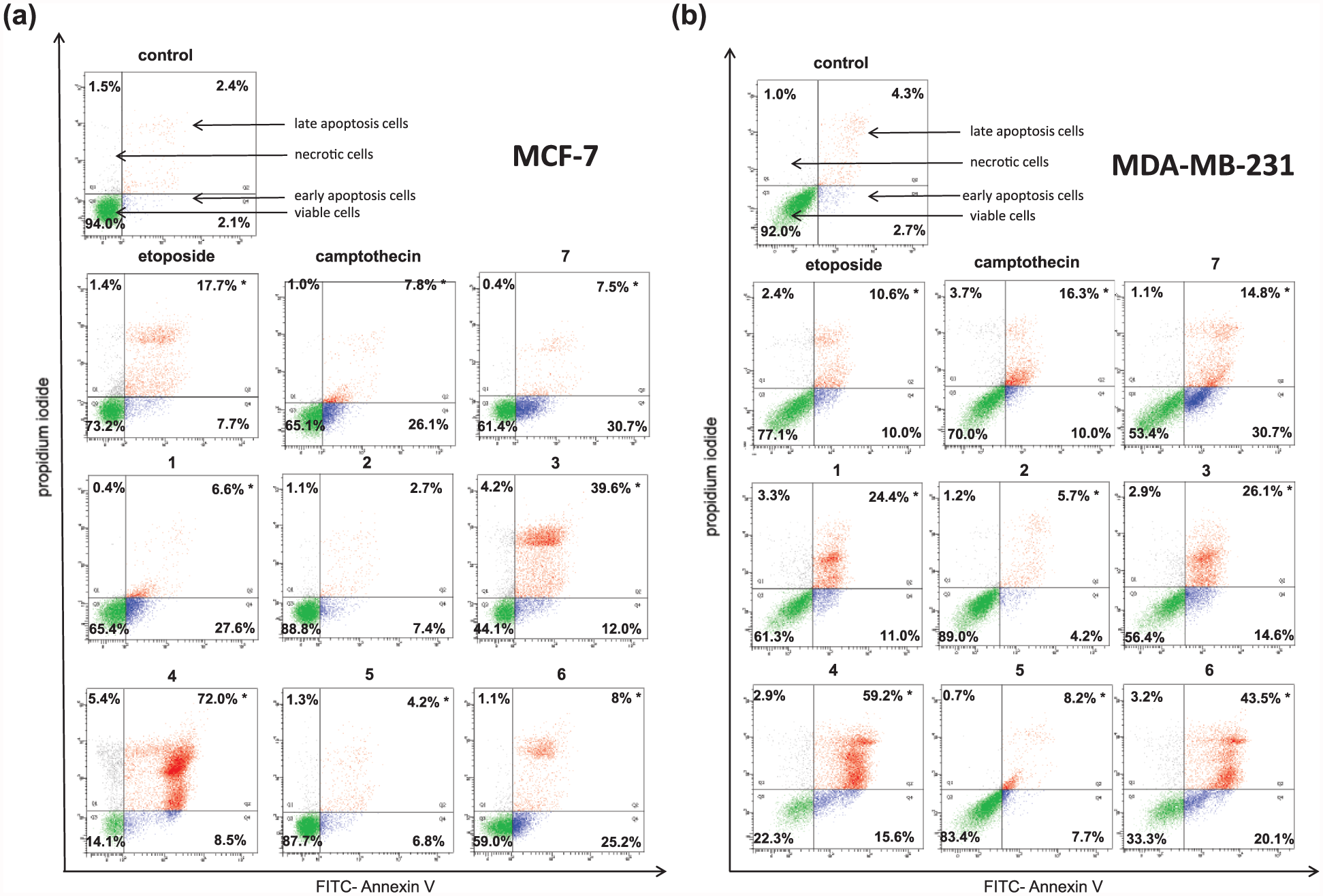
Flow cytometric analysis of (a) MCF-7 and (b) MDA-MB-231 breast cancer cells after incubation with etoposide, camptothecin, and compounds
Novel diisoquinoline derivatives lead to depolarization of MMP in MCF-7 and MDA-MB-231 cells
We have found that all tested compounds significantly decreased the MMP compared with control (p < 0.05, Figure 5). In control MCF-7 cells, we detected 4.3% cells with reduced MMP. After 24 h of incubation with camptothecin and etoposide, we detected 24% and 20.5% cells with reduced MMP. The compounds

Fluorescence of (a) MCF-7 and (b) MDA-MB-231 breast cancer cells treated for 24 h with 50 µM of etoposide, camptothecin, and compounds
Novel diisoquinoline derivatives activate initiator and executioner caspases in MCF-7 and MDA-MB-231 cells
Active caspase-8 activates effector/executioner caspases, which cause cell death by damage or destruction of the nucleus and other intracellular structures.
22
All tested compounds led to activation of caspase-8, which takes part in extrinsic death receptor pathway. The highest percentage of MCF-7 and MDA-MB-231 cells with active caspase-8 was observed after 24 h incubation with compound

Flow cytometric analysis of populations: (a) MCF-7 and (b) MDA-MB-231 breast cancer cells treated for 24 h with 50 µM of etoposide, camptothecin, and compounds
We have found that all tested compounds also activated caspase-9, which in turn activates executioner caspase-3 and initiates a caspase cascade, which eventually leads to demolition of the cell.
22
The strongest effect was observed after 24 h incubation with the compound

Flow cytometric analysis of populations: (a) MCF-7 and (b) MDA-MB-231 breast cancer cells treated for 24 h with 50 µM of etoposide, camptothecin, and compounds
Caspase-10 is another caspase, which is responsible for activation of executor caspase-3. We also proved that all tested diisoquinoline derivatives led to significant activation of initiator caspase-10 (p < 0.05). The compound

Flow cytometric analysis of populations: (a) MCF-7 and (b) MDA-MB-231 breast cancer cells treated for 24 h with 50 µM of etoposide, camptothecin, and compounds
The activation of initiator caspases 8, 9, and 10 led to activation of executor caspases. One of representatives is caspase-3. We noticed that tested compounds activated caspase-3 in MDA-MB-231 cells. The strongest compound was the compound

Flow cytometric analysis of populations: (a) MCF-7 and (b) MDA-MB-231 breast cancer cells treated for 24 h with 50 µM of etoposide, camptothecin, and compounds
Discussion
The balance between the process of proliferation and cell death ensures the maintenance of homeostasis.
23
Numerous studies were evaluated to check the effect of disorders in cell proliferation in the development of cancer. Cell death may be a result after activation of a range of mechanisms, including apoptosis. Disruption of programmed cell death may play a key role in the pathogenesis and progression of breast cancer.24–26 It is believed that the degree of induction of apoptosis in cancer cells and inhibition of cell proliferation influence on effectiveness of anticancer therapy.27,28 Apoptosis is a process that occurs strictly in order according to a certain pattern, which at the beginning leads to a contraction of a cell, activation of proteolytic enzymes, the concentration of nuclear chromatin, and fragmentation of the deoxyribonucleic acid. The second stage of apoptosis is cell breakage and apoptotic bodies with cell organelles occur. Next, these structures are recognized by macrophages and removed by phagocytosis. The first stage of apoptosis is a reversible process, but the second stage after DNA fragmentation is irreversible and damages cannot be repaired. Our team synthesized a group of novel octahydropyrazin[2,1-a:5,4-a′]diisoquinoline derivatives. Their cytotoxic and antiproliferative activity was checked in two estrogen receptor–positive and estrogen receptor–negative breast cancer cell lines. Their biological properties were compared with etoposide and camptothecin, which were used as references. The key element of the study was to confirm that novel diisoquinoline derivatives induce programmed cell death. Our team proved that all synthesized compounds induced apoptosis using annexin V binding assay. We observed that all agents influenced on an explicit increase of annexin V-FITC-positive cells. The significantly highest increase was detected after 24 h of incubation with the compound
Mitochondria are considered to play a very important role in programmed cell death. One of the parameters of mitochondria dysfunction is decrease of MMP. We have found that all tested compounds significantly decreased the MMP compared with control (p < 0.05). We observed the highest percentage of breast cancer cells with reduced MMP after 24 h incubation with compound
Apoptosis as a significant terminal pathway for cells is regulated by different executioner and regulatory molecules,
33
and their aberrant function is fundamental to the growth of tumors and the development of anticancer drug resistance. Therefore, apoptosis has become one of the prime molecular targets for drug discovery and development, particularly for diseases like cancer. Apoptosis is executed via the extrinsic and intrinsic apoptotic pathways. The extrinsic apoptotic pathway is associated with the attachment of extracellular ligands to the extracellular domain of transmembrane receptors, whereas the intrinsic apoptotic pathway is initiated by several intracellular stimuli.
34
Caspases represent key regulatory proteins in both apoptotic pathways.
35
Taking into account the position of caspases in apoptotic signaling cascades, caspases are divided into initiator (caspase-8, caspase-9, and caspase-10) and effector (caspase-3, caspase-6, and caspase-7) caspases. Initiator caspases of both pathways can catalyze the proteolytic maturation of effector caspases, which lead to activation of a caspase cascade and finally result in demolition of the cell.
34
To determine in more detail the pathway at which apoptosis goes through, the activity of caspases was monitored. We used the flow cytometry technique, where antibodies recognize the active forms of caspases. In our research, we demonstrated that all novel synthesized compounds
Researches are still looking for diverse promising experimental anticancer drugs that can modulate apoptotic pathways. Preclinical and clinical trials of some novel therapeutic agents that target apoptosis in cancer cells have been done or are still in progress. The promising class of compounds are agents which can activate the cascade of caspases. A number of drugs have been developed to synthetically activate caspases. One of the drugs, which is an activator of caspases, is apoptin. It exerts selective properties and only induces apoptosis in cancer cells not in normal cells.
37
The activators of caspases are also small-molecule caspase activators. They are characterized by the presence of the arginine–glycine–aspartate motif. These compounds induce auto-activation of procaspase-3 and, finally, can sensitize cancer cells to chemotherapeutic agents.
38
Thus, targeting caspase-8 expression may lead to promising therapeutic effects. The researches showed that 5-aza-2′-deoxycytidine (decitabine), a cytosine nucleoside analogue, boosts caspase-8 promoter availability, and provides a situation for binding of SP1 and ETS-like transcription factors.39,40 One of the possible targets is caspase-9, which is a key component of mitochondrial apoptotic pathway. For instance, fenretinide is synthetic cytotoxic retinoid, which activates caspase-9, down-regulates Bcl-2 and Mcl-1, and possesses no effect on Bax expression.
41
The preclinical studies are still in progress. Yamabe et al.
42
proved that also caspase-based gene therapy is a possible strategy in cancer treatment. They have shown that caspase-3 gene therapy, in presence of etoposide, can induce apoptosis and lead to reduction of tumor volume in an AH130 liver tumor model.
42
Another research studies demonstrated that a series of molecules including alpha-(trichloromethyl)-4-pyridineethanol (PETCM), gambogic acid, and the gambogic acid derivative MX-2060 efficiently activated caspase-3 in vitro.43–45 Huang et al. synthesized three novel transition metal complexes with isoquinoline derivatives, and their mechanism of action was characterized.
46
All complexes exhibited strong proliferation inhibition activity against various tested cancer cells with high selectivity to tumor and normal cells. One complex induced BEL-7404’s apoptotic death mediated by mitochondria pathway, which was proved by the assays of the MMP depolarization, poly(ADP-ribose) polymerase (PARP)-proteins’ cleavage; Bcl-2, p53, and p21 expression; and caspase family members’ activation.
46
Our study revealed that novel compounds in group of diisoquinoline derivatives
Conclusion
Our studies confirmed that new diisoquinoline derivatives (
Footnotes
Declaration of conflicting interests
The author(s) declared no potential conflicts of interest with respect to the research, authorship, and/or publication of this article.
Funding
The author(s) disclosed receipt of the following financial support for the research, authorship, and/or publication of this article: The authors are thankful to the National Science Centre (Grant DEC-2012/07/B/NZ7/04382) for financial support.