
Abstract
An accelerated atherosclerotic process is a significant contributor to the development of vascular complications in diabetes. Endothelial dysfunction and heightened platelet hyperactivity in diabetes exacerbate vasoconstriction and thrombus formation, thereby accelerating the atherosclerotic process. The platelet hyperreactivity associated with the prothrombotic state is characterised by alterations in platelet morphology and function. Platelet indices, such as the platelet volume, platelet distribution width, platelet-large cell ratio and plateletcrit, reflect these changes. These platelet indices could emerge as an important marker of glycaemic status and risk of complications. Several studies have demonstrated that mean platelet volume, platelet distribution width and platelet-large cell ratio increase in diabetes, especially if there are vascular complications. These tests are readily available and hold promise as cost-effective tools for assessing the risk of complications, especially in resource-limited settings. However, well-designed longitudinal studies are necessary to establish the clinical utility of platelet indices in assessing glycaemic status and the risk of complications.
Keywords
Introduction
Diabetes mellitus affects more than 537 million people globally.[1] The growing prevalence of diabetes and its complications, such as cardiovascular disease (CVD) and chronic kidney disease (CKD), are public health challenges. The most significant burden falls on regions such as Asia and Africa, with limited healthcare access.[1,2] Addressing the problem requires the promotion of healthier lifestyles, awareness and access to care through cost-effective innovations.
Hyperglycaemia in type 2 diabetes mellitus (T2DM) is associated with a chronic, low-grade inflammatory milieu that contributes to both the development and progression of the disease.[3] Endothelial dysfunction, another hallmark of T2DM, results from insulin resistance, oxidative stress, inflammation, and enhanced formation of advanced glycation end-products (AGEs). The endothelial damage reduces nitric oxide (NO) bioavailability, thereby impairing vasodilation and promoting vasoconstriction, platelet aggregation and inflammation.[4]
In diabetes mellitus, platelet activation contributes to atherothrombosis and macrovascular and microvascular complications.[5] The platelet activation and dysfunction are reflected in the platelet indices. Platelet indices, such as mean platelet volume (MPV) and platelet distribution width (PDW), are altered in diabetes.[6-8] Increased platelet turnover and activation linked to chronic inflammation and vascular complications leads to the generation of younger and more reactive platelets with altered platelet indices.[7,9,10]
Altered platelet indices could indicate poor glycaemic control and help predict the risk of complications. They have the potential to serve as additional markers alongside traditional measures. Several studies have assessed platelet indices as markers of glycaemic control and complications. If validated in larger trials, the platelet indices can complement and also serve as alternatives for current glycaemic monitoring techniques, especially in resource-limited settings.
Glycaemic Assessment: Challenges in Resource-limited Settings
T2DM is a complex metabolic disorder with altered glucose metabolism due to insulin deficiency and resistance. Hyperglycaemia leads to the induction of oxidative stress, formation of AGEs, activation of protein kinase C (PKC) and hexosamine pathways.[11] Adverse effects of hyperglycaemia and the successive cascades in metabolic dysregulation lead to complications of diabetes.
Information on current glycaemic status is necessary to strategise the approach to achieve glycaemic control. HbA1c is the accepted method for monitoring glycaemic control and reflects the average glycaemic status in the last 2-3 months. Elevated HbA1c levels are associated with a higher risk of diabetes complications. HbA1c data can be supplemented by information on diurnal glycaemic variation and hypoglycaemia by records of self-monitoring of blood glucose with glucose meters and continuous glucose monitoring systems.[12]
The HbA1c test, although the primary tool for assessing long-term glycaemic control, has several limitations. It may be inaccurate in individuals with conditions affecting red blood cell turnover, such as anaemia, hemoglobinopathies, recent blood transfusions, and CKD. Some haemoglobin variants may interfere with HbA1C results.[13] In individuals with certain haemoglobin disorders, such as sickle cell disease, HbA1C cannot be measured.[14]
In resource-limited settings, there is often a shortage of qualified healthcare professionals and adequate laboratory infrastructure. Assays certified by the National Glycohemoglobin Standardization Program (NGSP) and standardised to the Diabetes Control and Complications Trial (DCCT) reference may not be always available. Moreover, the cost and accessibility of laboratory services may be a hindrance in many countries where resources are limited.
Fructosamine and glycated albumin are alternative tests for monitoring glycaemic control and reflect average blood glucose levels over the past 2-4 weeks. The evidence linking the results of these tests to complications of diabetes is weaker compared to HbA1c. The expenses and availability of these tests limit their widespread use.[12]
Even routine tests indicated for diabetes management, including HbA1c, may be unaffordable to certain sections of society. These tests also require specialised equipment that may not be available everywhere. Innovative strategies to overcome the challenges in resource-limited regions are necessary. Affordable and simpler tests, adaptable to local conditions that require minimal infrastructure, could improve access, enhance adherence and offer practical solutions for glucose monitoring.
Platelet Pathophysiology in Diabetes
The functional changes that occur in platelets from diabetes increase their procoagulant tendency. This is worsened by the underlying chronic inflammation and endothelial dysfunction that augment platelet reactivity.[6]
Platelet Hyperreactivity
Platelet hyperreactivity in diabetes is characterised by an exaggerated response to activation stimuli, leading to increased aggregation and clot formation.[15] The heightened reactivity is driven by chronic hyperglycaemia, oxidative stress, glycation of platelet proteins and systemic inflammation.[6,7]
Hyperglycaemia plays a central role in platelet hyperreactivity by causing osmotic swelling, glycation of proteins and increasing platelet reactivity. Insulin reduces platelet activity by activating the insulin receptor substrate-1 (IRS-1) pathway, but in T2DM, insulin resistance impairs this process.[16] Oxidative stress exacerbates platelet aggregation through increased reactive oxygen species (ROS) and AGEs, leading to endothelial dysfunction and reduced NO production.[17] Inflammatory molecules, such as CD40L and toll-like receptors, contribute to platelet hyperreactivity by activating procoagulant pathways and promoting platelet-leukocyte aggregates.[18] Large platelets in diabetes express more glycoprotein (GPIb, GPIIb/IIIa) and P2Y12 receptors, which enhances the tendency towards aggregation.[19] Additionally, metabolic reprogramming driven by the functional plasticity of platelets adapts to changing energy needs, sustaining activation.[20] Altered platelet miRNAs, for example, reduced miR-223 and elevated miR-26b and miR-140, contribute to heightened platelet activity by targeting P2Y12 and P-selectin, which are involved in platelet aggregation and inflammation.[21]
Increased Platelet Turnover
In diabetes, platelet turnover is accelerated, leading to the production of larger and more reactive platelets. The ongoing hyperglycaemia induces activation and shortens the lifespan of platelets. The newly formed large platelets contain more granules and exhibit heightened reactivity. These hyperactive platelets are prone to aggregation and create the prothrombotic state observed in diabetes, increasing the risk of CVD. The rapid turnover of platelets also amplifies oxidative stress and inflammation, further aggravating vascular damage.[6,15]
Platelet Morphology and Dysfunction
Hyperglycaemia and metabolic dysfunction lead to hyperactive platelets that aggregate and release inflammatory proteins in response to sub-threshold stimuli. Nonenzymatic glycation of surface proteins decreases membrane fluidity and activates platelets.[15] Elevated blood glucose levels also induce osmotic swelling, leading to increased volume, heightened reactivity and a shortened lifespan, stimulating the recruitment of younger, larger platelets.[6,22]The larger, overactive platelets with pseudopodia release more prothrombotic factors.[23]
Endothelial Dysfunction and Platelets
Endothelial dysfunction contributes to platelet hyperreactivity and enhanced CVD risk in diabetes in a major way. Key factors contributing to endothelial dysfunction include activation of protein kinase C, overexpression of growth factors and cytokines and increased oxidative stress.[4,6] Under normal conditions, the endothelium regulates vascular tone and integrity by balancing the release of vasodilators such as NO, prostacyclin and bradykinin, as well as vasoconstrictors such as prostanoids, endothelin and angiotensin-II. However, in diabetes, hyperglycaemia, excess free fatty acids and insulin resistance create a vicious cycle of increased endothelial dysfunction, oxidative stress, inflammation and platelet hyperactivity. The inflammatory cascade disrupts endothelial function and promotes thrombosis.[22]
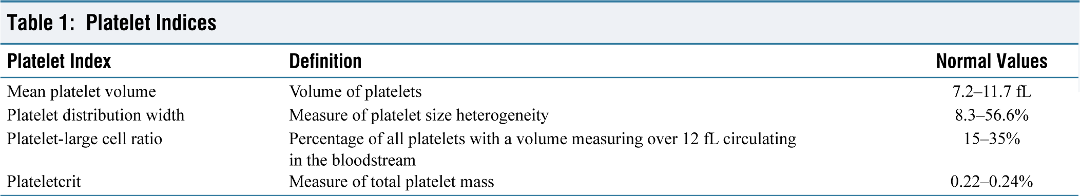
Platelet Indices
The changes in platelet morphology and function are reflected in the alterations in the platelet indices. Platelet parameters are routinely available in the laboratory blood cell counters. The indices include MPV, PDW, plateletcrit and platelet-large cell ratio (P-LCR).[24] The commonly used indices are presented in Table 1.[25]
Platelet Indices
Platelet Number
Studies demonstrate that platelet count is elevated in individuals with diabetes compared to healthy controls.[26] Total platelet count is a net result of platelet degradation and platelet production. Hyperreactive platelets in diabetes have a reduced lifespan, stimulating the release of younger platelets, and thereby increasing platelet numbers.
Mean Platelet Volume
The volume of platelets is denoted by MPV, which is also a marker of functionality. Increased MPV is seen in diabetes mellitus, metabolic disorders and stroke.[9] The increase in MPV is a result of the release of younger, larger and hyperreactive megakaryocytes found in diabetes. The newly-released platelets contain denser granules and higher amounts of β-thromboglobulin, serotonin and thromboxane A2 and are more thrombogenic. A greater risk of proliferative diabetic retinopathy and myocardial infarction is reported in individuals with high MPV.[27]
Platelet Distribution Width
PDW is a marker of anisocytosis and describes the variability in the size distribution of platelets.[24] Activation of platelets leads to changes in platelet morphology and pseudopodia formation. This results in heterogeneity in size, which is measured by PDW.[28] High values of PDW suggest the release of larger, reticulated platelets and indicate platelet hyperreactivity. The PDW may be a better predictor of platelet activation than MPV, as thrombocyte swelling does not affect it.[29]
Platelet-large Cell Ratio
The P-LCR measures the percentage of large platelets in the total platelet count. It is another marker for platelet hyperactivity, easily identified in automated blood counters.[24] The P-LCR is more susceptible to alterations in platelet size than MPV, despite their correlation. It correlated with microvascular complications in diabetes in a few studies.[9,30]
Plateletcrit
Plateletcrit is the percentage of blood volume occupied by the platelets. It provides an assessment of the circulating platelet mass.[25] This composite measure might offer more accurate insights compared to other platelet indices. It is an effective screening tool for detecting platelet quantitative abnormalities. There is insufficient evidence exploring the link between plateletcrit and complications of diabetes. An association between lower plateletcrit and diabetic peripheral neuropathy has been demonstrated.[31]
Platelet Indices as Marker of Deranged Glycaemic Status and Complications
Platelet hyperreactivity in uncontrolled diabetes was demonstrated decades ago in the form of increased urinary thromboxane A2/prostacyclin ratio. The ratio positively correlated with the degree of hyperglycaemia.[32] Additionally, platelet hyperreactivity, hyperaggregability, increased thrombogenesis and decreased fibrinolysis have been demonstrated in diabetes, irrespective of its type.[7]
Glycaemic Status
Deranged glycaemic control directly affects platelet quality and function, which is demonstrated in altered platelet indices. A case-control study found lower platelet counts and higher MPV in 300 individuals with diabetes compared to 200 controls.[33] A Framingham Heart Study analysis linked elevated MPV with diabetes, with a stronger effect in males. Among diabetes medications, insulin was associated with higher MPV levels, whereas lower levels were found in individuals using metformin and sulfonylureas.[34]
All indices were higher in individuals with diabetes than controls in another study. Additionally, the volume indices—MPV, PDW and P-LCR—were elevated in individuals with diabetes and HbA1C >7% compared to those with HbA1C ≤7%.[35] Similar trends of higher volume indices in individuals with HbA1C > 7% were observed in other cohorts also.[9,24,36] A meta-analysis of 39 studies demonstrated higher MPV in T2DM and impaired fasting glucose but not in metabolic syndrome. PDW was wider in T2DM, but platelet counts were similar.[37]
Microvascular Complication
Raised MPV, PDW and P-LCR have been linked to microvascular complications.[9,30,38,39] In a study by Buch et al., MPV correlated with complications such as retinopathy, nephropathy and diabetic foot. Additionally, PDW was higher in diabetic retinopathy and nephropathy, whereas P-LCR did not show any significant association with these complications.[33] In an Egyptian cohort, MPV >11.9 fL showed 80% sensitivity and 97.8% specificity for diagnosing microvascular complications, while PDW >16.9 fL had 74.5% sensitivity and 100% specificity.[40] Higher values of MPV and fibrinogen have also been linked to microvascular complications in other studies.[41] PDW and MPV were significantly elevated in men with erectile dysfunction and diabetes. These indices were higher in cases of vasculogenic erectile dysfunction compared to other types. However, the platelet count did not reveal any association.[42]
A few studies have failed to show an association between microvascular complications and platelet indices.[43,44] A probable explanation is the use of antiplatelet medications such as clopidogrel and aspirin. Clopidogrel causes irreversible blockade of the P2Y12 ADP receptor subtype on the platelet membrane, thereby inhibiting the initiation and cross-linking of platelets by fibrin. Inhibition of platelet activation could preserve platelet size and MPV.[45]
Macrovascular Complication
The link between CVD, such as coronary artery disease (CAD), stroke and peripheral artery disease (PAD), and platelet indices have been explored, but not as extensively as the association with microvascular diseases.[46-49] Higher MPV has been documented in the presence of CAD in individuals with diabetes.[47] An association between arterial stiffness and subclinical myocardial damage and elevated MPV was observed in individuals even with prediabetes and newly diagnosed T2DM.[46] Similarly, in a Japanese study, elevated MPV predicted arterial stiffness as measured by cardio-ankle vascular index and maximum intima-media thickness.[50]
Increased MPV and PDW occur with acute coronary syndromes, especially in ST-segment elevation myocardial infarction.[48] Higher MPV in the setting of acute thrombotic events such as myocardial infarction and cerebral thromboembolism in individuals with diabetes has also been documented.[51] MPV is independently associated with the presence of PAD.[49]
Clinical Application
Monitoring of platelet indices can serve as an affordable and non-invasive method to assess disease progression and glycaemic status in individuals with diabetes. Higher levels of mean MPV, PDW, P-LCR and plateletcrit are linked to inadequate glycaemic control and an increased risk of microvascular complications such as diabetic retinopathy, CKD and neuropathy. In addition, these indices may also help forecast macrovascular issues, such as CAD and stroke.
Platelet indices are emerging as promising markers for assessing the risk of complications in diabetes, particularly T2DM. These indices reflect platelet activation, which is triggered by hyperglycaemia, oxidative stress and inflammation, contributing to both microvascular and macrovascular complications.
Since they can be obtained through standard blood tests, platelet indices can be easily incorporated into clinical practice, especially in settings with limited resources. Their potential to anticipate complications might aid in strategising treatment choices. Nevertheless, further investigation is required to clarify their precise role.[30]
Future Research
Effective and accessible tests to predict the risk of complications in individuals with diabetes are urgently needed, especially in resource-limited settings. Tests to measure the inflammatory characteristics of platelets and platelet mitochondrial metabolic reprogramming remain an area for future work. Platelet miRNAs are highly expressed by circulating platelets and are promising biomarkers of T2DM that are likely to be investigated in future clinical studies. Additionally, the impact of comorbidities and medications on platelet indices could be another potential area of research. Large population-based studies on platelet indices across different ethnicities could help to establish diagnostic and prognostic cut-offs. Research focusing on the diagnostic significance of platelet indices can lead to better risk assessment and tailored treatment strategies.
Conclusion
Platelet hyperreactivity plays a crucial role in the development of complications associated with diabetes. Hyperglycaemia and metabolic dysregulation significantly contribute to this enhanced platelet activity. Indices such as elevated mean platelet volume (MPV), platelet distribution width (PDW) and platelet-large cell ratio (P-LCR) reflect this hyperreactive state. These parameters can serve as early predictors of vascular complications in T2DM. Assessment of these indices using routine laboratory counters is a practical and cost-effective method for screening, particularly in resource-limited settings. Further research is needed to explore the effectiveness of these parameters in predicting complication.
Footnotes
Declaration of conflicting interests
The authors declared no potential conflicts of interest with respect to the research, authorship and/or publication of this article.
Funding
The authors received no financial support for the research, authorship and/or publication of this article.
Institutional ethical committee approval number
Ethical approval was not applicable for this article, as this is a review article drafted from various research articles and not from patients directly.
Informed consent
Patient Consent was not applicable, as this is a review article compiled from various research articles and guidelines and not from patients directly.
Credit author statement
Concept and design: Saptarshi Bhattacharya and Sanjay Kara
Data collection: Sweekruti Jena and Nishant Raizada
Data analysis and interpretation: Sweekruti Jena and Saptarshi Bhattacharya
Manuscript drafting: Sweekruti Jena and Saptarshi Bhattacharya
Critical revision: Sanjay Kara and Nishant Raizada
Data availability
All data included in the manuscript.
Use of artificial intelligence
No.