
Abstract
Background
Cataracts remain the foremost cause of preventable blindness globally, with oxidative stress serving as a central contributor to their pathogenesis. Given the limited accessibility of surgical intervention in many populations, there is a pressing need for effective non-surgical therapeutic alternatives. We aimed to explore the protective role of piperitone against cataract development in experimental rodents.
Purpose
We aimed to explore the protective role of piperitone against cataract development in experimental rodents.
Materials and Methods
In the present study, cataracts were experimentally triggered in rat pups via a solitary subcutaneous dose of sodium selenite on post-natal day 12. Piperitone was administered at two different doses to evaluate its potential in mitigating cataractogenesis. Antioxidant capacity, oxidative stress markers, and indicators of lens protein oxidation were quantified to determine the compound’s antioxidative efficacy. Additionally, lens ascorbic acid levels and total calcium content were measured to assess their impact on lens clarity. Inducible nitric oxide synthase, cyclooxygenase-2, and nuclear factor kappa B were measured to investigate piperitone’s anti-inflammatory action. Furthermore, key components of the nuclear factor erythroid 2-related factor 2 (Nrf2) redox signaling pathway were evaluated to elucidate the underlying antioxidant mechanism. Lens opacification scores were recorded as a direct measure of cataract progression.
Results
The findings revealed that piperitone significantly enhanced antioxidant defenses, reduced oxidative damage and protein modification in the lens, and suppressed inflammatory signaling. It also modulated redox homeostasis via activation of the Nrf2 pathway and led to a marked reduction in lens opacity.
Conclusion
Collectively, our data support the therapeutic efficacy of piperitone as an antioxidant agent for the prevention or delay of cataract development.
Keywords
Introduction
Cataracts continue to represent the leading cause of preventable blindness globally, contributing to nearly 50% of all blindness cases (Canatan, 2024; Cvekl & Vijg, 2024; Lee & Afshari, 2017; Nartey, 2017). Epidemiological data reveal a significant global rise in cataract prevalence and are allied to the burden of disease metric, with a reported 58.45% increase in visual impairment attributable to cataracts over this period (Fang et al., 2022). Pediatric cataract incidence ranges from 0.32 to 22.9 per 10,000 children, with higher prevalence observed in high-income countries (Sheeladevi et al., 2016). Age-related lens opacification remains the predominant form of cataract, typically initiating between the ages of 45 and 50, although other etiologies such as trauma, metabolic disorders, and systemic diseases are also recognized (Richardson et al., 2020). Multiple risk factors contribute to cataractogenesis, including immutable biological traits and lifestyle-related factors such as tobacco use and ultraviolet radiation exposure (Iwundu et al., 2025).
Cataract surgical rates display significant global variation, ranging from as low as 36 to as high as 12,800 procedures per million individuals annually, with surgical coverage often falling below 50% in many regions (Hashemi et al., 2025). Despite notable advancements in surgical techniques and post-operative outcomes, disparities in access and treatment persist, particularly in low-resource settings (Rao et al., 2011). Socio-economic determinants, such as a lower Human Development Index, limited physician availability, and higher prevalence of uncorrected refractive errors, are strongly correlated with increased cataract-related disability-adjusted life years (DALYs) (Yang et al., 2021). Moreover, gender-based disparities remain evident, with females disproportionately affected, especially in regions with lower socioeconomic development (Fang et al., 2022). Although cataract surgery is currently the standard and most effective treatment, ongoing research efforts aim to identify non-surgical therapeutic alternatives, particularly for application in areas with limited access to ophthalmic surgical care (Chen et al., 2021).
The pathophysiology of cataractogenesis is multifactorial, with oxidative stress, a decline in redox-regulating enzymes, and age-related cellular degeneration playing central roles (Böhm et al., 2023). Among these, reactive oxygen species (ROS) are particularly critical in mediating lens damage and opacification. Oxidative stress occurs when the production of ROS surpasses the body’s ability to counteract them with antioxidant defenses, causing structural and functional damage in lens epithelial cells (Nita & Grzybowski, 2016). Clinical and experimental studies have reported significantly elevated lipid peroxidation byproducts and reduced antioxidant enzyme activity in individuals with cataracts compared to healthy controls (Kaur et al., 2012). The underlying mechanisms involve photochemical production of ROS such as superoxide radicals, which disrupt lens membrane integrity and impair the function of cation pumps, ultimately compromising lens transparency (Davies & Truscott, 2001). Elucidating these oxidative mechanisms provides a basis for the advancement of antioxidant-based clinical approaches addressing cataract prevention and management (Kulbay et al., 2024). Antioxidants, particularly vitamins C and E, have been shown to mitigate ROS-induced damage, with vitamin E demonstrating a capacity to slow cataract progression in pre-clinical models (Braakhuis et al., 2019; Matías-Pérez et al., 2024).
Recent studies have underscored the broad pharmacological potential of monoterpenes, key constituents of essential oils derived from various plant species. These compounds have demonstrated significant antimicrobial activity, primarily through disruption of microbial cell membrane integrity (Zielińska-Błajet & Feder-Kubis, 2020). Beyond their antimicrobial effects, monoterpenes exhibit anti-inflammatory, analgesic, and anti-cancer properties (da Silva et al., 2017; Machado et al., 2022; Sattayakhom et al., 2023). Extensive investigations have identified at least 32 monoterpenes with potential anti-inflammatory effects (de Cássia da Silveira e Sá et al., 2013). Among these, piperitone, a monoterpene ketone widely present in essential oils, has emerged as a bioactive molecule of interest with demonstrated antimicrobial (Abdolpour et al., 2007; Alsharif et al., 2024; Yaguchi et al., 2009), anti-fungal (Abdolpour et al., 2007), anti-trypanosomal (Sakirigui et al., 2016), and insect-modulating activities. We aimed to explore the protective role of piperitone against cataract development in experimental rodents.
Materials and Methods
Chemicals
Piperitone (purity: ≥90.0%), sodium selenite, and others were procured from Sigma-Aldrich, USA. All the kits for biochemical estimations were purchased from Abcam, Elabscience, and Cusabio, USA, respectively.
Animals
Nine-day-old, 24 healthy Sprague-Dawley rat pups, each weighing around 45 ± 5 g on the 11th post-natal day (PND), were selected for the experiment. The animals were kept under standardized laboratory conditions, with temperatures maintained between 20°C and 24°C and relative humidity set between 40% and 60%. A 12-h light/dark cycle was followed. They were provided with unlimited access to regular feed (Bio-Serv, USA) and water, which were refreshed daily. Before the experiment began, all rats were verified to possess normal and healthy vision.
Experimental Design
Rats were randomly distributed into four groups with six animals in each (n = 6). Group I functioned as the untreated normal control. Cataractogenesis was initiated in Groups II, III, and IV by administering a single subcutaneous dose of sodium selenite (2.46 mg/kg body weight) on PND 12. Group II acted as the cataract control group, receiving only sodium selenite. In contrast, Groups III and IV received intraperitoneal injections of piperitone at 10 and 20 mg/kg body weight, respectively, once daily from PND 11 to PND 17. On PND 12, piperitone was given 1 h before the sodium selenite injection in both treatment groups. All animals were anesthetized with chloral hydrate on PND 24, and cataract formation was subsequently assessed. Subsequently, animals were euthanized via CO2 inhalation, and the lenses were excised, snap-frozen, and stored at –70°C for subsequent biochemical assays.
Estimation of Antioxidants
Homogenization of the lens isolated from the experimental animals was carried out under rigorously controlled conditions to maintain experimental consistency and accuracy. Lens tissues were processed in an ice-cold Tris–HCl buffer (5 mM, pH 7.4) containing 2 mM ethylenediaminetetraacetic acid (EDTA), using a mechanical homogenizer operating at 1,500 rpm. The procedure included three complete oscillation cycles, each consisting of upward and downward strokes. Post-homogenization, the samples were centrifuged at 10,000 × g for 10 min at 4°C to eliminate residual cellular debris and intact cells. The clear supernatant was promptly collected and utilized for downstream biochemical assays. These included the determination of antioxidant enzyme activities such as superoxide dismutase (SOD), catalase, glutathione S-transferase (GST), glutathione peroxidase (GPx), glutathione reductase (GR), as well as the measurement of reduced glutathione (GSH) content. All enzymatic activities and GSH levels were quantified using validated protocols as outlined in the assay kits provided by Abcam.
Estimation of Oxidative Stress Mediators
Key biomarkers and mediators associated with oxidative stress, namely, total sulfhydryl groups, nitric oxide (NO), and malondialdehyde (MDA), were quantitatively analyzed in both untreated selenite-induced cataract model rats and those administered with piperitone. These parameters serve as critical indicators of oxidative damage and redox imbalance within the lens tissue. Quantification was carried out using commercially available colorimetric assay kits. Specifically, the total sulfhydryl content and NO levels were assessed utilizing kits obtained from Elabscience, while lipid peroxidation, as indicated by MDA levels, was measured using a colorimetric kit provided by Abcam, adhering strictly to the guidelines specified by the manufacturer, ensuring standardization and reproducibility of the results.
Estimation of Ascorbic Acid
The concentration of ascorbic acid in the lenses of both untreated selenite-induced cataract rats and those treated with piperitone was determined using a previously established method (Nakazawa et al., 2011). Briefly, lens tissues were homogenized in phosphate-buffered saline (PBS) and subsequently deproteinized with a 2.5% metaphosphoric acid solution. The homogenates were then centrifuged to obtain the supernatant, which was subjected to titration using 0.15 mg/mL 2,6-dichlorophenolindophenol (DCPIP). The final OD of the reaction mixture was quantified spectrophotometrically at 540 nm to quantify ascorbic acid content.
Estimation of Total Lens Calcium
Total calcium content in the lens tissue was quantified following a modified established protocol (Inomata et al., 1997). Initially, the dry weight of each lens was determined by incubating the samples at 100°C for 20 h. The lenses were then processed through acid extraction by incubating them with 0.2 mL of concentrated hydrochloric acid at room temperature for a period of 12–16 h. Following this, the extract volume was brought up to 1.0 mL using deionized water. To remove insoluble particles, the mixture was centrifuged at 10,000 rpm for 10 min. The calcium levels in the clear supernatant were subsequently determined using atomic absorption spectrophotometry.
Estimation of Inflammatory Markers
To examine the inflammatory response linked to selenite-induced cataract formation and assess the anti-inflammatory effects of piperitone, the expression of major pro-inflammatory markers, inducible nitric oxide synthase (iNOS), cyclooxygenase-2 (COX-2), and nuclear factor kappa B (NF-κB), was analyzed. These markers were quantified using enzyme-linked immunosorbent assay (ELISA) kits obtained from Cusabio. Briefly, the lens supernatant was added to specific antibodies precoated on ELISA plates. After incubation to allow antigen–antibody binding, wells were washed to remove unbound components. A biotinylated detection antibody was then added, followed by streptavidin-conjugated enzyme. Upon substrate addition, a colorimetric reaction ensued, and the absorbance was measured at a 450 nm wavelength using a microplate reader. The intensity of the color produced was directly proportional to the concentration of the target protein, allowing for quantitative analysis of inflammatory marker expression.
Assessment of Redox Signaling
The Nrf2 signaling molecules play a crucial role in mediating redox homeostasis during the process of cataract development. To evaluate the modulatory effect of piperitone on this pathway, we quantified the levels of heme oxygenase-1 (HO-1), NAD(P)H:quinone oxidoreductase 1 (NQO1), and nuclear factor erythroid 2-related factor 2 (Nrf2) in the tissue samples of the experimental animals. ELISA was employed for this purpose, utilizing commercially available kits obtained from Abcam. The procedures outlined in the manufacturer’s protocol were strictly adhered to during sample preparation, incubation, and washing steps. The absorbance of the enzymatic reaction was recorded at 450 nm using a microplate reader, and protein levels were determined using standard curves.
Assessment of Cataractogenesis
At the conclusion of the post-natal experimental period, mydriasis was induced in all rat pups using topical administration of 0.5% tropicamide and 2.5% phenylephrine hydrochloride. Cataract formation in both eyes was evaluated using a slit-lamp biomicroscope. Lens opacities were graded on a standardized six-point scale by a single examiner blinded to the treatment groups to minimize observational bias. The grading scale ranged from Grade 0 (completely transparent lens) to Grade 6 (mature, dense opacity involving the entire lens structure). Intermediate stages represented progressive morphological alterations, including subcapsular and nuclear changes such as scattered opacities, fiber swelling, and increasing nuclear density. For quantitative analysis, a cumulative cataract score was determined by calculating the proportion of animals within each opacity grade relative to the total number of animals assessed. To enhance the reliability and objectivity of the evaluation, two independent observers, also blinded to the experimental groups, independently verified the grading outcomes.
Statistical Analysis
Results are expressed as the mean along with the corresponding standard deviation (mean ± SD). Group comparisons were performed using one-way analysis of variance (ANOVA), followed by Dunnett’s multiple comparison post hoc test. Statistical analysis was carried out using GraphPad Prism software (version 8.3.0), with a p value less than .05 considered indicative of statistical significance.
Results
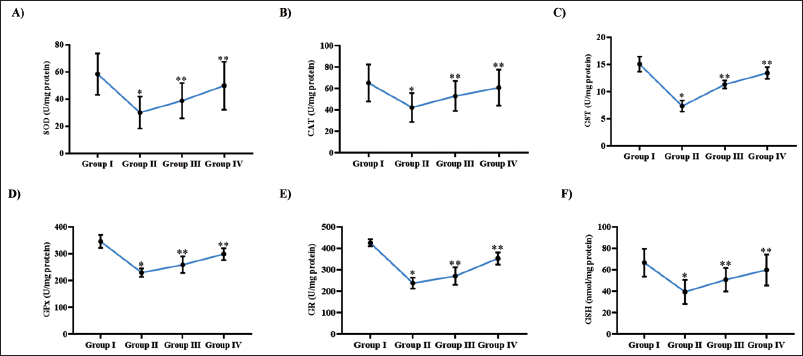
Piperitone Served as a Potent Enhancer of Antioxidant Defense Mechanisms in a Rat Model of Cataractogenesis
The antioxidant defense system, particularly the glutathione-dependent pathway, plays a pivotal role in preserving lens transparency and protecting against cataractogenesis. A substantial reduction in the levels of enzymatic and non-enzymatic antioxidants was identified in rats with selenite-induced cataracts compared to the untreated control group, signifying heightened oxidative stress linked with cataract formation. Cataract induction led to marked depletion of key antioxidant enzymes GPx, CAT, and SOD critical for the detoxification of hydrogen peroxide and superoxide radicals. Furthermore, selenite exposure also suppressed components of the glutathione metabolic cycle, as evidenced by diminished levels of reduced GSH, GR, and GST activities, all of which are essential for maintaining intracellular redox homeostasis and neutralizing toxic electrophilic compounds. Treatment with monoterpene ketone piperitone significantly restored antioxidant defenses in a dose-dependent manner. Administration of piperitone led to elevated levels of GPx, CAT, and SOD, enhancing the enzymatic detoxification of ROS. Additionally, piperitone effectively augmented the glutathione system by increasing GSH content and enhancing the activities of GR and GST (Figure 1).
Piperitone Served as a Potent Enhancer of Antioxidant Defense Mechanisms in a Rat Model of Cataractogenesis. Quantitative Analysis of Antioxidant Defense Parameters: (A) Catalase (CAT), (B) Superoxide Dismutase (SOD), (C) Glutathione S-Transferase (GST), (D) Glutathione Peroxidase (GPx), (E) Glutathione Reductase (GR), and (F) Reduced Glutathione (GSH) Levels in Lens Tissues From Control, Cataract-induced (Untreated), and Piperitone-treated (10 and 20 mg/kg) Groups. Data are Presented as Mean ± Standard Deviation (SD). Statistical Comparisons Among Groups Were Conducted Using One-way Analysis of Variance (ANOVA) Followed by Dunnett’s Post Hoc Test. Differences Were Considered Statistically Significant at p < .05.
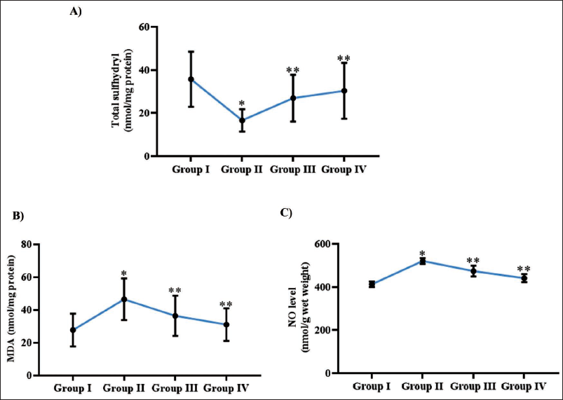
Piperitone Attenuated Oxidative Stress in a Rat Model of Cataractogenesis
Figure 2A illustrates the total sulfhydryl content, a recognized indicator of oxidative stress and lens protein oxidation. In the selenite-induced cataract group, a significant reduction in total sulfhydryl levels was observed (15.7 ± 0.004 nmol/mg protein) than the control group (37.5 ± 0.009 nmol/mg protein), indicating elevated oxidative damage. However, treatment with piperitone at doses of 10 and 20 mg/kg led to a dose-responsive restoration of sulfhydryl levels, increasing them to 22.3 ± 0.006 and 28.4 ± 0.005 nmol/mg protein, respectively. Figure 2B and 2C demonstrate the effect of piperitone on lipid peroxidation and nitrosative stress, as assessed by MDA and NO levels. The cataract-induced group exhibited markedly elevated MDA (43.9 ± 0.006 nmol/mg protein) and NO (567 ± 7.5 nmol/g wet weight) levels compared to control animals, which maintained significantly lower values (MDA: 23.4 ± 0.004 nmol/mg protein; NO: 410 ± 2.5 nmol/g wet weight). Piperitone administration resulted in a notable, dose-dependent reduction in both MDA and NO levels. At 10 mg/kg, MDA and NO levels were reduced to 37.6 ± 0.008 nmol/mg protein and 485 ± 3.5 nmol/g wet weight, respectively, while at 20 mg/kg, these values further declined to 35.4 ± 0.003 nmol/mg protein and 428 ± 9.5 nmol/g wet weight.
Piperitone Attenuated Oxidative Stress in a Rat Model of Cataractogenesis. Quantitative Analysis of Oxidative Stress Markers: (A) Total Sulfhydryl Content, (B) Malondialdehyde, (C) Nitric Oxide Levels in Lens Tissues From Control, Cataract-induced (Untreated), and Piperitone-treated (10 and 20 mg/kg) Groups. Data are Presented as Mean ± Standard Deviation (SD). Statistical Comparisons Among Groups Were Conducted Using One-way Analysis of Variance (ANOVA) Followed by Dunnett’s Post Hoc Test. Differences Were Considered Statistically Significant at p < .05.
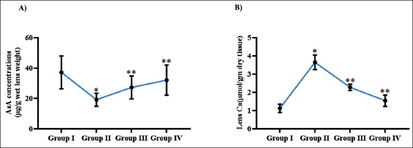
Piperitone Regulated Ascorbic Acid and Calcium Homeostasis in Lenses of Rat Model Cataractogenesis
The effect of piperitone treatment on lens transparency in cataract-induced animals was evaluated by quantifying ascorbic acid and calcium levels in the lenses, as shown in Figure 3. Induction of cataract caused a marked decline in ascorbic acid levels to 17.8 ± 0.06 µg per wet lens weight, alongside a significant elevation in total lens calcium content to 3.4 ± 0.00007 µmol/g dry tissue, compared to the control group. Administration of piperitone at 10 and 20 mg/kg demonstrated a dose-dependent restoration of ascorbic acid levels to 27.2 ± 0.04 µg and 28.4 ± 0.05 µg per wet lens weight, respectively. Simultaneously, the treatment significantly reduced calcium accumulation, with levels declining to 1.9 ± 0.00008 µmol/g and 1.4 ± 0.00006 µmol/g dry tissue for the 10 and 20 mg/kg doses. In the control group, ascorbic acid levels were maintained at 34.7 ± 0.09 µg per wet lens weight, while calcium levels remained low at 0.9 ± 0.00004 µmol/g dry tissue, indicating optimal lens homeostasis.
Piperitone Regulated Ascorbic Acid and Calcium Homeostasis in Lenses of Rat Model Cataractogenesis. Quantitative Analysis of (A) Ascorbic Acid, (B) Total Calcium Content in Lens Tissues From Control, Cataract-induced (Untreated), and Piperitone-treated (10 and 20 mg/kg) Groups. Data are Presented as Mean ± Standard Deviation (SD). Statistical Comparisons Among Groups Were Conducted Using One-way Analysis of Variance (ANOVA) Followed by Dunnett’s Post Hoc Test. Differences Were Considered Statistically Significant at p < .05.
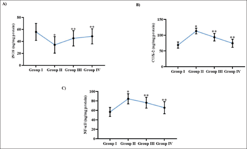
Piperitone Triggered Anti-inflammatory Effects in a Rat Model of Cataractogenesis
Figure 4 demonstrates the inflammatory attenuating effect of piperitone in the context of selenite-induced cataractogenesis. Administration of piperitone at 10 and 20 mg/kg led to a notable elevation in iNOS levels, reaching 38.9 ± 0.02 and 48.2 ± 0.05 ng/mg protein, respectively. In contrast, iNOS levels were substantially lowered in the untreated cataract group (34.3 ± 0.03 ng/mg protein), suggesting impaired inflammatory regulation. Meanwhile, inflammatory stimulators COX-2 and NF-κB were significantly elevated in the cataract-induced group, with levels reaching 101.4 ± 0.05 ng/mg protein for COX-2 and 82.5 ± 0.04 ng/mg protein for NF-κB, indicating heightened inflammatory activity associated with cataract formation. However, piperitone treatment effectively attenuated these markers in a dose-dependent manner. COX-2 levels were reduced to 89.6 ± 0.02 and 80.9 ± 0.03 ng/mg protein, while NF-κB levels declined to 79.4 ± 0.03 and 68.9 ± 0.04 ng/mg protein following 10 and 20 mg/kg piperitone administration, respectively. In the control group, baseline levels of iNOS, COX-2, and NF-κB were recorded at 57.8 ± 0.06, 67.2 ± 0.04, and 44.8 ± 0.09 ng/mg protein, respectively, reflecting normal inflammatory homeostasis.
Piperitone Triggered Anti-inflammatory Effects in a Rat Model of Cataractogenesis. Quantitative Analysis of Anti-inflammatory Markers: (A) Inducible Nitric Oxide Synthase (iNOS), (B) Cyclooxygenase-2 (COX-2), (C) Nuclear Factor Kappa B (NF-κB) Levels in Control, Cataract-induced (Untreated), and Piperitone-treated (10 and 20 mg/kg) Groups. Data are Presented as Mean ± Standard Deviation (SD). Statistical Comparisons Among Groups Were Conducted Using One-way Analysis of Variance (ANOVA) Followed by Dunnett’s Post Hoc Test. Differences Were Considered Statistically Significant at p < .05.
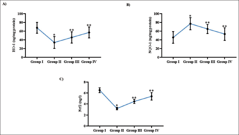
Piperitone Modulated Redox Signaling Pathways in a Rat Model of Cataractogenesis
Figure 5 illustrates the impact of piperitone on redox signaling pathways in selenite-induced cataractogenesis. Cataract-induced animals exhibited a significant downregulation of key antioxidant signaling molecules, with HO-1 and Nrf2 levels reduced to 32.5 ± 0.06 and 2.9 ± 0.0007 ng/mg protein, respectively, indicating compromised redox homeostasis. In contrast, treatment with piperitone at doses of 10 and 20 mg/kg led to a dose-dependent restoration of these markers. HO-1 levels increased to 42.4 ± 0.04 and 51.4 ± 0.03 ng/mg protein, while Nrf2 levels rose to 3.8 ± 0.0007 and 5.7 ± 0.0005 ng/mg protein, respectively, suggesting activation of the Nrf2-mediated antioxidant response pathway. The NQO1 expression regulated by Nrf2 signaling was significantly elevated in the untreated cataract group (76.8 ± 0.05 ng/mg protein), potentially as a compensatory response to oxidative stress. However, piperitone treatment reduced NQO1 to 62.4 ± 0.04 and 57.2 ± 0.04 ng/mg protein at 10 and 20 mg/kg, respectively, indicating modulation of redox signaling toward physiological levels. In the control group, HO-1 and Nrf2 levels were maintained at 69.6 ± 0.08 and 6.1 ± 0.0009 ng/mg protein, respectively, reflecting normal redox regulation, while NQO1 levels remained low at 43.2 ± 0.06 ng/mg protein.
Piperitone Modulated Redox Signaling Pathways in a Rat Model of Cataractogenesis. Quantitative Analysis of Redox Signaling Molecules: (A) Heme Oxygenase-1 (HO-1), (B) NAD(P)H:quinone Dehydrogenase 1 (NQO1), (C) Nuclear Factor Erythroid 2-related Factor 2 (Nrf2) Levels in Control, Cataract-induced (Untreated), and Piperitone-treated (10 and 20 mg/kg) Groups. Data are Presented as Mean ± Standard Deviation (SD). Statistical Comparisons Among Groups Were Conducted Using One-way Analysis of Variance (ANOVA) Followed by Dunnett’s Post Hoc Test. Differences Were Considered Statistically Significant at p < .05.
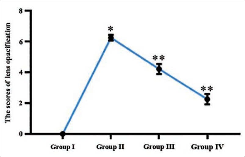
Piperitone Prevented Cataract Progression in a Rat Model of Cataractogenesis
Figure 6 presents the assessment of cataract induction and progression, quantified through lens opacification scores. Administration of selenite effectively induced cataractogenesis in the experimental animals, as evidenced by a significant increase in lens opacity. The untreated cataract group exhibited the highest opacification score, recorded at 5.9 ± 0.0008, indicating advanced cataract development. In contrast, the control group maintained complete lens clarity, with no detectable opacification. Piperitone administration resulted in a dose-responsive decline in lens opacity. Animals administered with low-dose piperitone exhibited a moderate decline in lens opacification (3.7 ± 0.0007), while a more pronounced safeguarding effect was evident at the 20 mg/kg dose, which significantly reduced the score to 1.7 ± 0.0007.
Piperitone Prevented Cataract Progression in a Rat Model of Cataractogenesis. Lens Opacification Scores of Control, Cataract-induced (Untreated), and Piperitone-treated (10 and 20 mg/kg) Groups. Data are Presented as Mean ± Standard Deviation (SD). Statistical Comparisons Among Groups were Conducted Using One-way Analysis of Variance (ANOVA) Followed by Dunnett’s Post Hoc Test. Differences were Considered Statistically Significant at p < .05.
Discussion
Cataractogenesis is predominantly driven by oxidative stress, which leads to protein denaturation and apoptotic death of lens epithelial cells (Zhang et al., 2023). ROS play a central role in the molecular mechanisms underlying cataract development, while antioxidant systems function as key modulators to mitigate oxidative damage (Lee et al., 2024). Among these systems, GSH serves as a principal intracellular antioxidant, acting synergistically with enzymatic antioxidants and non-enzymatic antioxidants. These agents collectively counteract the detrimental effects of ROS, including superoxide anions, hydrogen peroxide, and hydroxyl radical, thereby preserving lens integrity (Fan et al., 2017; Forman et al., 2009; Giblin, 2000).
GSH is particularly crucial in maintaining the redox stability of the lens by protecting crystallin proteins from oxidative modifications, which is essential for sustaining lens transparency. In age-related cataracts, oxidative stress contributes to the formation of protein-thiol mixed disulfides, leading to protein aggregation and subsequent lens opacification (Lou & Augusteyn, 2025). The intracellular steady-state concentration of GSH is maintained through a multifaceted network involving de novo synthesis, uptake from ocular humors, regeneration from oxidized glutathione (GSSG), efflux, and enzymatic degradation via γ-glutamyl transpeptidases. The tight regulation of glutathione-related pathways is essential for protecting the lens from oxidative damage mediated by ROS (Lou, 2022; Reddy, 1990).
In this context, we investigated the potential of piperitone to enhance antioxidant defenses, with a particular focus on modulating glutathione homeostasis in a rat model of cataract induction. Previous studies have shown that naturally derived antioxidants such as vitamins C and E and curcumin can effectively reduce lipid peroxidation and delay cataract progression (Thiagarajan & Manikandan, 2013). Consistent with these findings, our results indicate that piperitone exerts a protective effect by significantly modulating the glutathione redox system and restoring antioxidant levels that were markedly depleted in cataract-induced, untreated rats.
Sulfhydryl groups present in lens proteins and the antioxidant glutathione are essential for maintaining lens transparency by regulating redox homeostasis and preserving protein structural integrity. Under conditions of oxidative stress, these thiol groups undergo oxidation, leading to the formation of disulfide bonds and protein-thiol mixed disulfides, which promote protein aggregation and ultimately result in lens opacification (Jomova et al., 2023; Lou & Augusteyn, 2025). Ascorbic acid serves as a key antioxidant in the lens, providing protection against ultraviolet-induced oxidative damage. Moreover, it assists in the regeneration of compounds like vitamin E and GSH, thereby enhancing the overall antioxidant defense system. Age-associated decline in ascorbic acid levels has been correlated with increased cataract severity (Lim et al., 2020). In our study, rats exposed to sodium selenite exhibited a significant reduction in both ascorbic acid and sulfhydryl group levels, indicating oxidative stress and the onset of cataractogenesis. However, treatment with piperitone effectively restored the levels of ascorbic acid and sulfhydryl groups, suggesting its protective role in mitigating cataract development through enhancement of the lens’s antioxidant capacity.
Lipid peroxidation is recognized as a key pathogenic mechanism in cataractogenesis (Babizhayev, 2012; Kisić et al., 2009). In ocular tissues, ROS can initiate a series of harmful biochemical alterations, including the peroxidation of membrane lipids and extensive oxidative modifications to proteins. These changes can promote the aggregation and precipitation of intracellular proteins, contributing to cellular dysfunction (Boscia et al., 2000; Datiles & Kinoshita, 1998). Lipid peroxidation also correlates with increased cell membrane permeability, decreased cellular proliferation, and disturbances in lipid and protein homeostasis, underpinning its critical role in the development of age-related (senile) cataracts (Donma et al., 2002; Zoric et al., 2008). In the present study, administration of piperitone markedly enhanced antioxidant capacity, reduced oxidative stress, and suppressed lipid peroxidation in a rat model of cataractogenesis, highlighting its potential as a protective agent against the progression of cataracts.
NO serves a complex role in ocular physiology and pathology. While it is vital for maintaining normal visual functions and regulating ocular blood flow (Cantó et al., 2019), excessive NO synthesis, particularly through the induction of iNOS, can induce oxidative and nitrosative stress within the eye (Chiou, 2001). In the context of cataract development, increased NO levels facilitate the formation of reactive nitrogen species, such as peroxynitrite, which can inflict damage on lens proteins, compromise membrane integrity, and weaken antioxidant defenses. Studies using Shumiya cataract rats have shown that increased iNOS expression occurs early in cataract development, preceding calcium influx and calpain activation. Inhibiting iNOS with aminoguanidine effectively reduced lens opacity, highlighting NO’s significant role in cataractogenesis (Inomata et al., 2001). In this study, treatment with piperitone effectively reduced levels of NO in rats with cataracts. The observed decrease in lens calcium content following piperitone administration may be attributed to the downregulation of NO, which likely prevented abnormal calcium influx associated with cataractogenesis.
Treatment with piperitone in rats with selenite-induced cataracts significantly reduced the levels of NF-κB, a transcription factor whose activation has been linked to cataractogenesis through the promotion of inflammation and apoptotic pathways in lens epithelial cells (Sun et al., 2020; Shao et al., 2017). Oxidative stress can facilitate NF-κB translocation into the nucleus, leading to an increased inflammatory stimulating cytokine, thereby contributing to the development of cataracts (Liu et al., 2023). Conversely, COX-2, an enzyme that can be upregulated by NF-κB, exerts complex effects; some prostaglandins derived from COX-2 pathways inhibit NF-κB activity, whereas others, such as prostaglandin E2, can enhance NF-κB-mediated transcriptional activity (Poligone & Baldwin, 2001). Moreover, inhibition of histone deacetylases has been shown to mitigate cataract formation by suppressing NF-κB activation and reducing the expression of inflammatory mediators (Liu & Yin, 2019). In the present study, piperitone treatment also led to a decrease in COX-2 expression, further demonstrating its inflammatory attenuating property in the context of selenite-induced cataracts.
The Nrf2-Keap1 signaling is a central mediator of cellular defense mechanisms against redox imbalance, which is a principal factor in cataract pathogenesis (Cullinan & Diehl, 2004). Nrf2 orchestrates the transcription of numerous antioxidants and detoxifying enzymes, including glutathione-related enzymes, thioredoxin reductase, and HO-1 (Rushmore et al., 1991; Yu et al., 2010). Within the lens, this pathway sustains redox balance through an interconnected network involving glutathione and ascorbate systems (Bejarano et al., 2023). Age-related decline in Nrf2 activity correlates with diminished antioxidant defenses and heightened vulnerability to cataract formation (Liu et al., 2017). Experimental evidence indicates that silencing Nrf2 in human lens epithelial cells leads to increased protein aggregation, whereas activation of Nrf2 through antioxidant agents confers protection against oxidative stress-induced cataracts (Wu et al., 2008; Yang et al., 2015; Zheng et al., 2015). Additionally, the Nrf2/HO-1 axis exerts cytoprotective effects by inhibiting lipid peroxidation, caspase-3 activation, and tumor necrosis factor-alpha (TNF-α) expression (Nakagami, 2016; Zhou et al., 2017). These findings underscore the therapeutic potential of targeting Nrf2 to prevent and treat age-related or oxidative stress-induced cataracts. In our study, piperitone administration significantly reduced the levels of Nrf2, HO-1, and NQO1 in rats treated with selenite, thereby attenuating oxidative stress-induced cataractogenesis. The observed improvements in lens opacification scores further support the protective effect of piperitone against selenite-induced lens opacity.
In this study, we used the sodium selenite-induced cataract model in rat pups because this model recapitulates key aspects of human senile cataracts, including oxidative stress and rapid lens opacification. This model enables the investigation of molecular mechanisms underlying cataractogenesis, allowing for the exploration of potential therapeutic targets. The model’s translational relevance is supported by its ability to mimic oxidative stress-induced damage in human cataracts. Furthermore, our study has several limitations. These findings are preclinical and limited to an animal model, warranting further validation in clinical settings. Despite this, our data provide novel insights into piperitone’s anti-cataractogenic effects. Future studies should focus on translational research, exploring piperitone’s efficacy and safety in humans. If successful, piperitone could contribute to low-cost, accessible cataract therapies in resource-limited settings. Its potential as an affordable, non-surgical treatment option could significantly impact global eye health. Further research is needed to fully elucidate piperitone’s therapeutic potential and mechanisms of action. Additionally, investigating piperitone’s pharmacokinetics and pharmacodynamics will be crucial for clinical development.
Conclusion
The findings of this research demonstrate the broad-spectrum protective potential of piperitone against cataract development triggered by selenite in rats. Piperitone demonstrated potent antioxidant activity by enhancing glutathione homeostasis, preserving sulfhydryl groups, restoring ascorbic acid levels, and attenuating lipid peroxidation-key factors implicated in cataract formation. Additionally, piperitone exerted significant anti-inflammatory effects by downregulating inflammatory stimulators such as iNOS, NF-κB, and COX-2. Importantly, modulation of the Nrf2/Keap1 signaling pathway and its downstream targets, including HO-1 and NQO1, suggests a central role in the redox-mediated protection of lens tissues. The reduction in calcium accumulation and protein oxidative damage further substantiates its role in preserving lens clarity. This study highlights the potential of piperitone as a lead compound for the future development of non-surgical cataract interventions.
Footnotes
Abbreviations
COX-2: Cyclooxygenase-2; GPx: Glutathione peroxidase; GR: Glutathione reductase; GSH: Glutathione; GST: Glutathione-S-transferase; HO-1: Heme oxygenase-1; iNOS: Inducible nitric oxide synthase; MDA: Malondialdehyde; NF-κB: Nuclear factor kappa B; NO: Nitric oxide; NQO1: (NAD(P)H:quinone oxidoreductase 1; Nrf2: Nuclear factor erythroid 2-related factor 2; PND: Post-natal day; SOD: Superoxide dismutase.
Declaration of Conflict of Interests
The authors declared no potential conflicts of interest with respect to the research, authorship, and/or publication of this article.
Ethical Approval
This work has been approved by the Institutional Animal Ethical Committee of Xi’an Ai’er Ancient City Eye Hospital, Xi’an City, Shaanxi Province, China.
Funding
The authors received no financial support for the research, authorship, and/or publication of this article.