
Abstract
Background
Allergic rhinitis, a worldwide health issue, is defined by immunoglobulin E (IgE)-mediated inflammation of nasal mucosa, resulting in symptoms like sneezing and nasal obstruction. The increasing incidence of allergic rhinitis globally intensifies the burden on healthcare systems, requiring thorough methods for effective management and prevention.
Purpose
The current study evaluated the effects of corosolic acid against ovalbumin (OVA)-induced allergic rhinitis in mice.
Materials and Methods
Allergic rhinitis was triggered in mice with intraperitoneal injection of OVA (1 mg/mL) combined with aluminum hydroxide (20 mg/mL; 0.1 mL/mouse). Corosolic acid was treated at concentrations of 10 and 20 mg/kg, respectively. The analysis of nasal symptoms (nasal rubbings and sneezing) was conducted on the final day of the OVA challenge. The histamine and allergen-specific IgE concentrations in the experimental mice were evaluated utilizing kits. The inflammatory cytokine levels were evaluated in the mice utilizing assay kits.
Results
The treatment of corosolic acid in the OVA-induced mice led to a significant diminution in nasal rubbings and instances of sneezing. In allergic rhinitis-induced mice, corosolic acid treatment effectively reduced the histamine, OVA-specific IgE, and eosinophil infiltration levels. Furthermore, the corosolic acid treatment markedly decreased the inflammatory biomarker levels in the OVA-induced mice with allergic rhinitis.
Conclusion
In conclusion, the current data demonstrated that corosolic acid significantly reduced inflammatory and allergic reactions in OVA-challenged mice. These findings suggest that corosolic acid may be an effective therapeutic agent for treating and preventing allergic rhinitis.
Introduction
Allergic rhinitis, a global health issue affecting a substantial number of the population, is characterized by an immunoglobulin E (IgE)-mediated inflammation of the nasal mucosa to inhaled allergens. This condition, while not typically life-threatening, considerably impairs quality of life, impacting daily activities and academic performance, particularly in pediatric populations (Bousquet et al., 2020). Its classification, guided by chronicity and severity, considers both symptomatic presentation and the broader impact on an individual’s well-being. The global prevalence of allergic rhinitis is projected to be 10%–20%, impacting approximately 500 million individuals worldwide. This high prevalence underscores the significant burden on healthcare systems globally, necessitating effective management strategies (Katelaris et al., 2012). The pathogenesis of allergic rhinitis involves a complex interplay of genetic predispositions, environmental causes, and an impaired immune system. For instance, a notable rise in allergic illness incidence over recent decades suggests that environmental shifts, rather than purely genetic factors, are primary contributors to this trend (Bousquet et al., 2005). Specifically, the “hygiene hypothesis” posits that reduced exposure to diverse microbial agents early in life may lead to an immature immune system prone to aberrant IgE responses to common environmental allergens. This lack of early microbial exposure is hypothesized to hinder the normal development of the immune system, potentially resulting in an increased susceptibility to allergic conditions such as allergic rhinitis (Burte et al., 2015). Furthermore, allergic rhinitis has been shown to profoundly influence quality of life by disturbing sleep, disrupting autonomic regulation, and exacerbating inflammation in the lower airways and paranasal sinuses, alongside contributing to oxidative stress (Savouré et al., 2022).
Inflammation of the nasal mucosa, often accompanied by signs such as sneezing, rhinorrhea, itching, and congestion, defines allergic rhinitis. The underlying immunological mechanism involves an aberrant type 2 inflammatory response, primarily driven by IgE antibodies, which recognize innocuous environmental allergens as threats, initiating a cascade of inflammatory mediators (Watts et al., 2019). This response involves the activation of mast cells and basophils, leading to the rapid discharge of histamine, leukotrienes, and prostaglandins, which are responsible for the immediate hypersensitivity symptoms. Moreover, chronic exposure to allergens can lead to sustained inflammation, airway remodeling, and the influx of additional inflammatory cells, contributing to the persistent and often debilitating symptoms of allergic rhinitis (Iwasaki et al., 2021). The intricate nature of allergic diseases, marked by various triggers and individual variability, necessitates sophisticated methodologies for effective management. This complexity is further compounded by the heterogeneous nature of allergic diseases, which are increasingly recognized as encompassing distinct endotypes and phenotypes requiring personalized therapeutic approaches (Sy & Siracusa, 2016).
Despite advancements, current treatment paradigms for allergic rhinitis largely focus on symptomatic relief and allergen avoidance, with allergen-specific immunotherapy being the only disease-modifying intervention available. Though even with these established treatments, a substantial number of patients experience suboptimal control of their symptoms or adverse effects from pharmacotherapy, underscoring the ongoing need for novel and safer therapeutic alternatives (Drazdauskaitė et al., 2021). This persistent gap in therapeutic options highlights the potential for plant-based interventions, which may offer complementary or alternative strategies with reduced side effects and broad-spectrum anti-inflammatory properties. Natural bioactive compounds, extensively investigated for their immunomodulatory properties, offer therapeutic potential for various human diseases, including allergic conditions, often with fewer adverse effects compared to conventional pharmaceuticals (Liu et al., 2024; Moradi et al., 2024). Corosolic acid is a natural bioactive pentacyclic triterpenoid compound that occurs in numerous medicinal plants, including Lagerstroemia speciosa (banaba) and Eriobotrya japonica (loquat). Corosolic acid showed anti-inflammatory and anti-tumor (Pundalik et al., 2022), anti-diabetic (Miura et al., 2006), antioxidant (Yamaguchi et al., 2006), anti-angiogenic and anti-lymphangiogenic activities (Yoo et al., 2015), anti-osteoarthritis (Han et al., 2022), cardioprotective (Wang et al., 2020), hepatoprotective (Guo et al., 2016), and neuroprotective (Zhang et al., 2022) properties. Apart from these biological activities, its protective effects against allergic rhinitis have not been assessed yet. Therefore, our current study evaluated the effects of corosolic acid against ovalbumin (OVA)-induced allergic rhinitis in mice.
Materials and Methods
Chemicals
The primary chemicals, including corosolic acid and OVA, were sourced commercially from Sigma–Aldrich, USA. The biochemical markers were assessed utilizing test kits sourced from Elabscience and Abcam, USA.
Experimental Mice
Healthy BALB/c mice aged 4–6 weeks were utilized in this work. The mice were accommodated in sanitized polypropylene enclosures with free access to pellet food and drinking water. Mice were caged in a controlled laboratory environment with a temperature of 24°C ± 2°C, humidity of 50%–60%, and 12-h light and dark cycles. Each mouse was acclimatized for 1 week to the laboratory environment prior to the commencement of the study.
Experimental Groups and Treatment Procedures
Mice were grouped into five groups, each containing six mice (n = 6). Group I served as the control mice. The mice from Groups II–V received intraperitoneal injections of 1 mg/mL OVA combined with aluminum hydroxide (20 mg/mL) in saline (0.1 mL/mouse) on days 1, 3, 5, 7, 9, 11, and 13 to trigger sensitization. Mice were then treated with 60 mg/mL OVA in saline (20 µL/mouse) intranasally on days 20–30 to induce the allergic rhinitis. Group II mice were subjected only to OVA administration, while Groups III and IV mice were treated with 10 and 20 mg/kg of corosolic acid, respectively, in conjunction with OVA administration. The control was administered saline without undergoing an OVA challenge. Subsequent to the last OVA exposure, mice were killed, and samples were collected for further analysis. The nasal tissues were excised, processed, and eosinophil levels were evaluated using light microscopy. The blood samples were employed to prepare serum for subsequent biochemical analyses.
Analysis of Nasal Symptoms
The incidence of nasal rubs and sneezing behaviors in the experimental mice was recorded to evaluate nasal symptoms. Following the final OVA intranasal challenge, the frequencies of nasal symptoms were documented throughout a 10-min interval, with experimental circumstances assessed by blinded observers.
Collection of Nasal Lavage Fluid
Mice were administered 1% of pentobarbital (50 mg/kg) intramuscularly to induce anesthesia, thereafter undergoing a partial tracheotomy. The 22-gauge catheter was introduced through the nostrils, commencing at the tracheal opening. Then, it was irrigated with 3 mL of saline solution. Nasal lavage fluid (NALF) was obtained from the anterior portion of the nostril and centrifuged at 5,000 rpm for 10 min. Subsequently, it was preserved at −20°C for subsequent analyses.
Assessment of Inflammatory Biomarkers
The concentrations of OVA-specific IgE and histamine in the serum of the mice were quantified using commercial test kits (Elabscience, USA). The concentrations of inflammatory cytokines, including interleukin (IL)-4, IL-5, IL-6, IL-33, tumor necrosis factor-alpha (TNF-α), and eosinophil cationic protein (ECP), in the NALF samples from experimental mice were evaluated using commercial test kits (MyBioSource, USA). Each test was conducted in triplicate by strictly following the specifications of the manufacturers.
Statistical Analysis
A one-way analysis of variance (ANOVA) and Tukey’s post hoc assay were performed to assess the values derived from each assay. Values are statistically validated using GraphPad Prism software and portrayed as mean ± SD of triplicates, with p < .05 considered as significant.
Results
Effect of Corosolic Acid on the Nasal Symptoms in the Experimental Mice
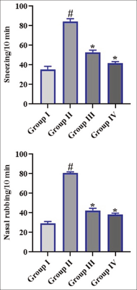
Figure 1 illustrates the incidences of nasal rubs and sneezing in the mice. The mice with OVA-induced allergic rhinitis demonstrated an increased incidence of nasal rubbing and sneezing. Whereas, the mice treated with 10 and 20 mg/kg of corosolic acid, respectively, exhibited a significant reduction in both nasal rubbings and sneezing incidences when compared with OVA-treated mice.
Effect of Corosolic Acid on Ovalbumin-specific Immunoglobulin E in Experimental Mice
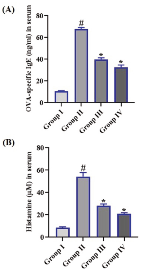
The concentrations of OVA-specific IgE in the serum were assessed, with results illustrated in Figure 2(A). The mice treated with OVA exhibited augmented serum levels of IgE when compared with the control. Interestingly, the 10 and 20 mg/kg of corosolic acid treatment markedly diminished the concentrations of IgE in the serum of OVA-induced mice, which supports the beneficial effects of corosolic acid.
Effect of Corosolic Acid on the Histamine Level in the Serum of Experimental Mice
The serum concentrations of histamine in the experimental mice are revealed in Figure 2(B). The increased level of histamine was observed in the serum of OVA-induced mice. Conversely, the treatment with 10 and 20 mg/kg of corosolic acid considerably decreased the serum histamine levels in the OVA-induced mice.
Effect of Corosolic Acid on Inflammatory Biomarkers in Nasal Lavage Fluid of Experimental Mice
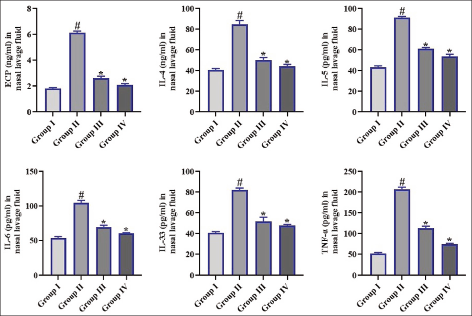
The concentrations of IL-4, IL-5, IL-6, IL-33, TNF-α, and ECP in NALF from the mice were assessed, with the outcomes illustrated in Figure 3. The mice with OVA-induced allergic rhinitis demonstrated elevated levels of IL-4, IL-5, IL-6, IL-33, TNF-α, and ECP in their NALF. Interestingly, the treatment of corosolic acid reduced the NALF levels of these inflammatory biomarkers in the OVA-induced mice.
Effect of Corosolic Acid on Eosinophil Counts in Nasal Tissues of Experimental Mice
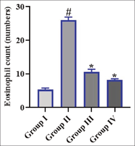
Figure 4 illustrates the levels of eosinophils in the nasal tissues of mice. A significant increase in the eosinophil count was noted in the nasal tissues of OVA-induced mice when compared with the control. However, the corosolic acid (10 and 20 mg/kg) treatment effectively reduced the eosinophil counts in the nasal tissues of OVA-induced mice.
Discussion
Allergic rhinitis, a global health concern, is characterized by IgE-mediated inflammation of the nasal mucosa, resulting in symptoms such as sneezing, rhinorrhea, and nasal obstruction. This prevalent allergic disease, affecting 10%–20% of the world population, considerably impairs patients’ quality of life and enforces major healthcare burdens. Its impact extends beyond mere discomfort, affecting daily activities, school performance in children, and overall productivity (Schuler Iv & Montejo, 2021). Furthermore, the rising prevalence of allergic rhinitis worldwide exacerbates the strain on healthcare systems, necessitating comprehensive strategies for effective management and prevention. The pathogenesis of allergic rhinitis involves a complex interplay of genetic predisposition, environmental causes, and an aberrant immune response. Specifically, it is defined by a type 2 helper T-cell immune response, resulting in the accumulation of allergen-specific IgE antibodies upon exposure to otherwise innocuous environmental allergens. Compounding these issues, B cells play an imperative role in the airway’s host-defense, tissue surveillance, and immune modulation, with their dysregulated function contributing to the exaggerated inflammatory states observed in allergic rhinitis (Siddiqui et al., 2022). The intricate cellular and molecular mechanisms underlying this immune pathology suggest the need for advanced therapies that specifically target the aberrant pathways without inducing systemic side effects. Current treatment modalities, while providing symptomatic relief, often fall short in addressing the underlying immunological dysregulation or present with undesirable systemic side effects, thus underscoring the urgent need for safer, more targeted interventions. Given these limitations, there is a growing interest in novel therapeutic approaches, particularly those that restore immune tolerance and mitigate allergic responses (Meng et al., 2020).
The analysis of nasal symptoms provides crucial insights into the efficacy of therapeutic interventions by quantifying the amelioration of cardinal allergic rhinitis signs, including sneezing and nasal rubbing. These observable behaviors serve as reliable in vivo biomarkers for assessing the severity of allergic responses and the subsequent reduction in inflammation following drug administration, directly correlating with clinical end points used in human trials (Ryu et al., 2020). Allergic rhinitis is defined by inflammation of nasal mucosa, manifesting as sneezing, itchiness, rhinorrhea, and obstruction. The precise measurement of these behavioral indicators in murine models allows for a direct evaluation of novel therapeutic agents. The evaluation of nasal symptoms in OVA-induced allergic rhinitis mouse models offers a standardized and reproducible method for preclinical drug development, enabling high-throughput screening of potential anti-allergic compounds (Sawaki et al., 2011). Moreover, such analyses facilitate the elucidation of underlying immunological mechanisms modulated by the tested compounds, providing valuable data on their pharmacological targets and modes of action. Therefore, the reduction in these overt symptoms reflects a dampening of the type 2 immune response that drives allergic rhinitis pathophysiology (Shinmei et al., 2009). In the present work, the incidence of nasal symptoms in the experimental mice was assessed. The OVA-induced mice exhibited an increased incidence of sneezing and nasal rubbing. However, the mice treated with corosolic acid revealed significantly reduced sneezing and nasal rubbing incidences. It was clear that corosolic acid treatment considerably mitigated the cardinal allergic rhinitis symptoms in the OVA-induced mice.
The pathophysiology of allergic rhinitis is intricate, involving a confluence of genetic predispositions, environmental factors, and the immune system’s dysregulation. Central to this dysregulation is the sensitization to specific allergens, such as OVA, which triggers the accumulation of allergen-specific IgE and subsequent histamine release, key mediators in the inflammatory cascade. IgE sensitization has markedly increased over recent decades and is an essential cause in the onset of allergic diseases (Takhar et al., 2005). This sensitization, specifically involving IgE antibodies directed against allergens like OVA, is a hallmark of Type I hypersensitivity reactions, where mast cells and basophils play pivotal roles in mediating the immediate allergic response. Upon subsequent exposure to the cognate allergen, these IgE-sensitized cells degranulate, releasing a multitude of preformed and newly synthesized mediators, including histamine, which is a primary effector molecule in the early phase of allergic rhinitis (Rondón et al., 2009). Histamine, historically recognized for its role in acute inflammation and immediate hypersensitivity, also influences chronic inflammatory processes and modulates various facets of the immune response. The complex interplay between IgE and histamine extends beyond acute reactions, significantly contributing to the prolonged pathophysiological changes observed in chronic allergic inflammation, including tissue remodeling (Taylor-Clark, 2010). Furthermore, the interaction between IgE and histamine extends to influencing the activation and recruitment of other inflammatory cells, like eosinophils, which are crucial for sustaining chronic allergic inflammation and tissue damage. This intricate relationship highlights that IgE-mediated mast cell activation and subsequent histamine release not only precipitate immediate symptoms but also orchestrate the complex cellular events that drive the persistent inflammatory state characteristic of allergic rhinitis (Bachert, 2002). The present findings evidenced that the OVA-challenged mice exhibited augmented serum concentrations of both histamine and IgE when compared with the control. However, the corosolic acid treatment markedly diminished the concentrations of IgE and histamine in the serum of OVA-induced mice.
Allergic rhinitis is a pervasive immunological disorder characterized by an aberrant inflammatory response primarily driven by IgE-dependent mechanisms. The complex pathogenesis of allergic rhinitis involves a multifactorial interplay of dysregulated immune system, leading to chronic inflammation of the nasal mucosa. This inflammatory cascade is largely orchestrated by various cytokines, which serve as crucial mediators in initiating and perpetuating the allergic response, ultimately contributing to the clinical manifestations of the disease (Li et al., 2023). Specifically, a hallmark of allergic rhinitis involves a type 2 immune response, characterized by the activation of Th2 cells, innate lymphoid cells, eosinophils, and mast cells, which collectively orchestrate the release of specific inflammatory mediators. Among these, key inflammatory cytokines like IL-4, IL-5, IL-6, IL-33, and TNF-α play pivotal roles in exacerbating the allergic inflammatory process (Albloushi & Al-Ahmad, 2023). These cytokines contribute to the influx of inflammatory cells, enhance IgE production, and promote airway hyperresponsiveness, thereby amplifying the allergic cascade. The late-phase IgE-mediated reactions are also significant contributors to the chronic aspects of allergic diseases, although the precise mechanisms underlying these sustained inflammatory responses remain an active area of investigation. Indeed, the intricate cellular and molecular mechanisms underlying type 2 immunity have been extensively investigated in the context of various allergic diseases (Bao & Zhu, 2022). Understanding the intricate roles of these cytokines is essential for developing targeted therapeutic strategies intended to alleviate the debilitating symptoms and enhance the quality of life for patients. In this study, we observed that the mice with OVA-induced allergic rhinitis illustrated augmented IL-4, IL-5, IL-6, IL-33, TNF-α, and ECP levels in their NALF. Captivatingly, the administration of corosolic acid considerably diminished the NALF levels of these inflammatory biomarkers in the OVA-challenged mice. These findings highlight that corosolic acid mitigated the nasal inflammatory reactions in the OVA-challenged mice.
Allergic rhinitis, a chronic inflammatory disease, is characterized by a complex interplay of immune cells and mediators. The pathophysiology of allergic rhinitis involves a cascade of immunological events, with antigen-specific IgE production and subsequent activation of mast cells, basophils, and eosinophils playing pivotal roles in initiating and propagating the inflammatory response. Specifically, eosinophils, specialized immune cells implicated in both protective and pathological processes, contribute significantly to the inflammatory milieu observed in allergic rhinitis (Gevaert et al., 2022). A critical aspect of this pathogenesis involves the substantial infiltration of eosinophils into nasal tissues, which is a hallmark of allergic inflammation and correlates with disease severity. These infiltrations directly contribute to tissue damage and the perpetuation of chronic inflammation within the nasal mucosa, thereby exacerbating the clinical manifestations of allergic rhinitis. The migration and activation of eosinophils are orchestrated by specific chemokines and cytokines, leading to the degranulation of cytotoxic proteins and lipid mediators that perpetuate the inflammatory cascade (Lombardi et al., 2022). Eosinophils, while distinct in lineage, often cooperate with mast cells and basophils, responding to similar cytokines and chemokines to amplify the allergic response. This coordinated cellular recruitment culminates in the chronic inflammatory state observed in allergic rhinitis, driven by the synergistic actions of these immune cells. The sustained presence and activation of eosinophils in nasal tissues contribute significantly to the structural remodeling and functional impairment characteristic of chronic allergic rhinitis (Xu et al., 2024). Therefore, mitigating inflammatory cell infiltrations, specifically eosinophil infiltrations, is essential to halt the progression of allergic rhinitis. The present results revealed an increased eosinophil infiltration into the nasal tissues of the OVA-challenged mice. Interestingly, corosolic acid treatment markedly decreased eosinophil infiltration into the nasal tissues of OVA-challenged mice. These findings highlight that the corosolic acid considerably reduced the inflammatory cell infiltrations, thereby facilitating the culmination of allergic rhinitis progression.
Our study has several limitations. First, the small sample size may impact the generalizability of our results. Additionally, we did not conduct a detailed mechanistic pathway analysis to elucidate the precise molecular mechanisms underlying corosolic acid’s therapeutic effects. The use of a single animal model (OVA-induced allergic rhinitis) may limit the applicability of our findings to other models or human populations. Furthermore, we only evaluated a limited dose range and treatment duration and did not assess long-term efficacy or compare corosolic acid’s effects with existing standard therapies. Our future studies will aim to address these limitations and provide a more comprehensive understanding of corosolic acid’s therapeutic potential for allergic rhinitis.
Conclusion
In conclusion, the current data demonstrated that corosolic acid significantly reduced inflammatory and allergic responses in OVA-induced mice. The administration of corosolic acid demonstrated a significant reduction in nasal symptoms, OVA-specific IgE, histamine level, eosinophil infiltrations, and inflammatory marker levels in the OVA-challenged mice. These findings suggest that corosolic acid may be an effective therapeutic agent for treating and preventing allergic rhinitis. While these findings are promising, further research is necessary to fully elucidate the mechanisms of action, optimal dosing strategies, and long-term safety profile. Clinical trials will be essential to confirm its efficacy and safety in humans, ultimately determining its potential as a novel therapeutic option for allergic rhinitis.
Summary
Allergic rhinitis is a global health issue affecting a substantial number of the population, characterized by an IgE-mediated inflammation of the nasal mucosa to inhaled allergens. Natural bioactive compounds, extensively investigated for their immunomodulatory properties, offer therapeutic potential for various human diseases, including allergic conditions. The current data demonstrated that corosolic acid significantly reduced inflammatory and allergic responses in OVA-induced mice. The present study suggests that corosolic acid may be an effective therapeutic agent for treating and preventing allergic rhinitis.
Abbreviations
ECP: Eosinophil cationic protein; Ig: Immunoglobulin; NALF: Nasal lavage fluid; OVA: Ovalbumin.
Footnotes
Declaration of Conflict of Interests
The authors declared no potential conflicts of interest with respect to the research, authorship, and/or publication of this article.
Ethical Approval
This project was approved by the Animal Care Committee of the Affiliated Hospital of Hebei University (no. HBU-2023-071).
Funding
The authors received no financial support for the research, authorship, and/or publication of this article.
Informed Consent
Not applicable.