
Abstract
Background
Drug-induced liver toxicity is a major clinical challenge, being the primary cause of abrupt liver failure. This condition is intensified by an older population that consumes more drugs and the increasing hazards associated with unknown drug consumption.
Purpose
This work was undertaken to study the beneficial values of ligustilide against acetaminophen-induced liver damage in rats.
Materials and Methods
The experimental rats were administered 640 mg/kg of acetaminophen to induce experimental liver toxicity and successively treated with ligustilide. Upon concluding the treatments, the concentrations of liver function marker enzyme activities, albumin, protein, and total/direct bilirubin were assessed using respective test kits. The concentrations of antioxidants, inflammatory cytokines, and lipid profile markers were assessed using the commercial test kits. The liver tissues acquired from the experimental rats were employed for a histological study.
Results
The ligustilide treatment significantly normalized the changes in hepatic function marker enzymes, elevated albumin and protein concentrations, and reduced total/direct bilirubin contents in the rats with acetaminophen-induced hepatic toxicity. Furthermore, the ligustilide treatment resulted in increased antioxidant concentrations and significantly decreased inflammatory cytokine levels. In addition, the ligustilide considerably mitigated the dyslipidemia condition in the experimental rats. The outcomes of the histological study of hepatic tissues further confirmed the hepatoprotective efficacy of ligustilide.
Conclusion
The findings of this work demonstrate the hepatoprotective effects of ligustilide against acetaminophen-induced liver toxicity in rats. Consequently, ligustilide can serve as a viable treatment option for addressing drug-induced liver toxicity.
Introduction
Xenobiotics, a term encompassing a wide range of foreign substances, have become an increasingly significant area of concern, particularly when it comes to their potential to induce liver toxicity. Drug-induced hepatotoxicity is a primary cause of acute liver failure in numerous countries, presenting a considerable therapeutic challenge. Xenobiotics are known as any chemical substances existing in an organism that are not normally synthesized or anticipated to occur within the body. These foreign compounds can be introduced into the body through various means, including pharmaceutical drugs, environmental pollutants, and dietary supplements. The liver, being the principal organ for the metabolism and elimination of xenobiotics, is especially vulnerable to their detrimental effects (Zhang et al., 2022). Drug-induced hepatic damage is a well-recognized form of xenobiotic-induced liver toxicity. The mechanisms underlying drug-induced hepatic toxicity can be defined as either intrinsic or idiosyncratic. Intrinsic liver toxicity, which is dose-dependent and predictable, is typically caused by the direct effect of drugs on hepatocytes. Conversely, idiosyncratic toxicity is unpredictable, often occurring at therapeutic doses, and is thought to involve complex interactions among the drug, the host’s genetic and environmental factors, and the immune system (Devarbhavi & Philips, 2024).
Drug-induced hepatic toxicity is a substantial public health issue, including over 50% of acute hepatotoxicity cases globally. Furthermore, it is anticipated that over 75% of idiosyncratic medication reactions lead to liver transplantation or mortality. The prevalence of xenobiotic-induced hepatic injury is also high, with various environmental pollutants and dietary supplements being implicated in liver damage (Hoppmann et al., 2020). The burden of xenobiotics and drug-induced liver toxicity is substantial, both in terms of individual and societal impact. Patients suffering from these conditions often experience significant morbidity, with some cases progressing to liver failure (Massart et al., 2022).
The mechanisms of drug-induced liver damage are intricate and diverse, encompassing numerous cellular and molecular pathways. Toxicity can be assessed experimentally by providing the molecule at escalating levels, alongside metabolic inducers, inhibitors, or toxicity enhancers, while depleting defensive systems, or by co-administering the medicine with a known harmful agent. Acetaminophen, a commonly utilized over-the-counter painkiller and antipyretic, is the most extensively researched drug for drug-induced hepatotoxicity (Hosack et al., 2023). The etiology of drug-induced hepatic toxicity frequently arises from drug metabolism, leading to the accumulation of reactive metabolites that can directly harm hepatocytes or elicit immunological responses, ultimately causing liver damage (Teschke & Danan, 2023). While the mechanisms of drug-induced hepatic toxicity can be studied in vitro, animal models, particularly those using rats, have been extensively utilized to investigate the pathophysiology and potential interventions. These models provide the assessment of the impacts of several drugs on hepatic function, in addition to investigating the fundamental cellular and molecular processes implicated in the onset of hepatic damage (Teschke & Danan, 2021).
The optimal approach for managing drug-induced liver toxicity involves prompt identification, cessation of the causative agents, supportive care, and a comprehensive understanding of the anticipated natural progression. While the current treatment options for drug-induced liver toxicity, such as supportive care and withholding the inciting agents, have their challenges, the applications of plant bioactive compounds to treat this condition have been increasingly explored (Kamel et al., 2023). Plant-derived compounds have shown promising results in preclinical and clinical studies, with some exhibiting hepatoprotective, anti-inflammatory, and antioxidant properties that may alleviate the deleterious effects of drug-induced hepatic damage (Li et al., 2023). Ligustilide is a naturally occurring bioactive benzoquinone derivative compound mostly present in the Apium graveolens (wild celery) and Angelica sinensis (Yang et al., 2019). Extensive previous studies have already demonstrated the various biological properties of the liqustilide, including osteogenesis (Yang et al., 2019), neuroprotective (Ren et al., 2020), anti-aging (Zhu et al., 2020), anti-tumor (Yin et al., 2023), anti-sepsis (Li et al., 2023), and anti-depressive (Ma et al., 2021) effects, and also promoted endometrial angiogenesis and the pregnancy outcomes in rats (He et al., 2024). However, there are no scientific studies to support its therapeutic roles against drug-induced liver toxicity. Consequently, this study was undertaken to explore the beneficial values of ligustilide against acetaminophen-induced hepatic toxicity in rats.
Materials and Methods
Chemicals
The acetaminophen (purity >98% high-performance liquid chromatography (HPLC), CAS number: 103-90-2, Cat. A7085, Mw 151.16, Sigma-Aldrich) suspension was prepared by dissolving acetaminophen in warm phosphate-buffered saline (PBS, pH 7.2). Acetaminophen-induced acute liver injury was established by intragastric administration of a toxic dose of 2 g/kg body weight of acetaminophen suspension. The newly prepared acetaminophen suspension was used in each experiment. Moreover, ligustilide (purity >98% HPLC, CAS number: 4431-01-0, Cat. SMB00400, Mw 190.24, Sigma-Aldrich) was dissolved in a 3% (w/v) solution of polysorbate 80 (TW-80). After all the animals were sacrificed, aspartate transaminase (AST), alanine transaminase (ALT), and glutathione (GSH) were measured in rats using the assay kit to determine the toxicity of acetaminophen-administered rats. The commercial diagnostic kits for biochemical studies were obtained from Elabscience, Abcam, and MyBioSource, USA.
Experimental Design of Drug Treatments
A total of 24 male albino Wistar rats weighing 180–200 g were obtained from the institutional animal facility. The rats were housed in polypropylene cages under standard laboratory conditions (temperature 22 ± 2°C, relative humidity 50%–60%, and a 12-h light/dark cycle) with ad libitum access to a standard pellet diet and water. After a 1-week acclimatization period, the rats were randomly divided into four experimental groups (n = 6 per group) and received the following treatments for 14 consecutive days:
Group I (normal control): Received 0.5 mL of 0.05% Tween 80 diluted in 0.9% NaCl orally, along with their standard diet.
Group II (acetaminophen-induced toxicity): Received acetaminophen at a dose of 640 mg/kg orally for 14 consecutive days to induce liver toxicity.
Group III (low-dose ligustilide treatment): Received acetaminophen (640 mg/kg) along with ligustilide (50 mg/kg, orally) for 14 consecutive days.
Group IV (high-dose ligustilide treatment): Received acetaminophen (640 mg/kg) along with ligustilide (100 mg/kg, orally) for 14 consecutive days.
Blood samples were collected and preserved for biochemistry analyses at the end of the treatment period. Additionally, hepatic tissues were rapidly excised, rinsed in an ice-cold buffer, and processed for further experimental evaluation.
Analysis of Liver Function Markers
Following euthanasia, liver tissues were promptly excised, rinsed with ice-cold saline, and homogenized in a phosphate buffer solution (pH 7.4, 1:10 w/v) using a homogenizer on ice to prevent enzymatic degradation. The homogenates were centrifuged at 15,000 rpm for 20 min at 4°C, and the resulting supernatant was collected for biochemical analysis. Additionally, blood samples were obtained via cardiac puncture and centrifuged at 3,000 rpm for 10 min at 4°C to obtain serum. Hepatic function markers, including ALT, AST, and alkaline phosphatase (ALP), were quantified using commercial enzyme-linked immunosorbent assay (ELISA) kits according to the manufacturer’s protocol (Elabscience, USA).
Analysis of Albumin, Total Protein, and Bilirubin Levels
Serum levels of albumin, total protein, and total/direct bilirubin were measured using colorimetric assay kits (Elabscience, USA). Each assay was conducted in triplicate, and absorbance readings were taken using a microplate reader (model: SPECTRONIC 200 UV–Vis spectrophotometer, Thermo Fisher Scientific, USA). Standard calibration curves were generated for each parameter to ensure accuracy.
Analysis of Antioxidant Markers
The hepatic concentrations of catalase (CAT), GSH, superoxide dismutase (SOD), and glutathione-S-transferase (GST) were assessed using commercial assay kits (MyBioSource, USA). The liver tissue homogenates were prepared in ice-cold phosphate buffer (0.1 M, pH 7.4), centrifuged at 12,000 rpm for 15 min at 4°C, and the supernatant was used for analysis. Enzyme activity levels were measured spectrophotometrically at their respective absorbance wavelengths, following the manufacturer’s instructions.
Lipid Marker Levels
Serum concentrations of high-density lipoprotein (HDL), low-density lipoprotein (LDL), total cholesterol (TC), and triglycerides (TG) were determined using commercial lipid profiling kits (MyBioSource, USA). The assays were conducted in triplicate using an automated biochemical analyzer (model: Hitachi Automatic Analyzer 7600-210, Hitachi, Tokyo, Japan), and results were compared against standard calibration curves.
Analysis of Inflammatory Cytokines
The concentrations of interleukin-1 beta (IL-1β) and tumor necrosis factor-alpha (TNF-α) in both serum and liver homogenates were quantified using sandwich ELISA kits (Abcam, USA). Samples were diluted appropriately to fall within the linear range of the assay. The cytokine concentrations were determined from a standard curve generated using known concentrations of recombinant cytokines, and all assays were performed in triplicate to ensure reproducibility.
Histopathological Analysis
Liver tissues were fixed in 10% neutral buffered formalin for 24 h, followed by sequential dehydration using graded ethanol (70%, 80%, 90%, and absolute ethanol). The tissues were then embedded in paraffin and sectioned at a thickness of 5 µm using a rotary microtome. The sections were stained with hematoxylin and eosin (H&E) and examined under a light microscope to assess histopathological changes.
Statistical Analysis
All experimental data were analyzed using GraphPad Prism (version 20). Results were expressed as mean ± standard deviation (SD) of triplicate measurements. Group comparisons were performed using one-way analysis of variance (ANOVA) followed by Tukey’s post hoc test. A p value of <.05 was considered statistically significant.
Results
Effect of Ligustilide on Hepatic Marker Enzymes in Experimental Rats
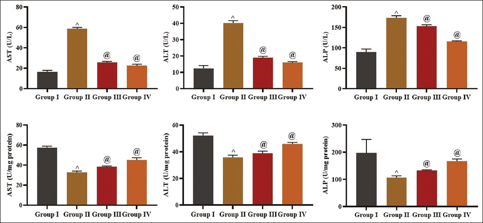
The alterations in liver marker enzymes in the serum and liver tissues of rats were examined, with the results illustrated in Figure 1. The acetaminophen-induced liver toxicity rats exhibited elevated ALT, AST, and ALP in their serum and subsequent reduction in these enzyme activities in their hepatic tissues. However, the 50 and 100 mg/kg of ligustilide treatment significantly diminished these enzyme activities in the serum and enhanced these enzymes in the hepatic tissues of the rats with acetaminophen-induced hepatic toxicity (Figure 1).
Effect of Ligustilide on the Liver Marker Enzymes in Experimental Rats. Each Bar Denotes the Mean ± SD of Three Independent Assays, Analyzed Using One-Way Analysis of Variance (ANOVA) and Tukey’s Post Hoc Tests. “^” Denotes That Data Significantly Differed at p < .01 from the Control Group (Group I); “@” Signifies That Data Significantly Differed at p < .05 from the Acetaminophen-induced Group (Group II).
Effect of Ligustilide on the Albumin, Total Protein, and Total/Direct Bilirubin in Experimental Rats
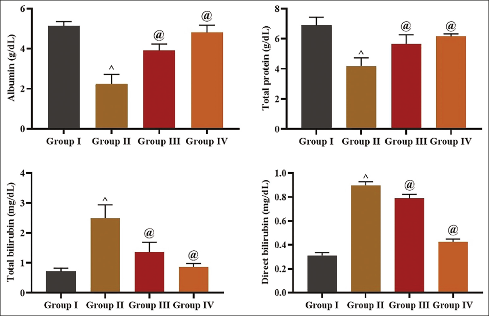
Figure 2 illustrates the albumin, protein, and total/direct bilirubin levels in the serum of experimental rats. The diminished albumin and protein concentrations and increased total/direct bilirubins were seen in the acetaminophen-induced liver toxicity rats about the control. Fascinatingly, 50 and 100 mg/kg of ligustilide significantly enhanced the albumin and protein contents and reduced the total/direct bilirubin in the serum of rats with acetaminophen-induced liver toxicity.
Effect of Ligustilide on the Albumin, Total Protein, and Bilirubin Levels in the Experimental Rats. Each Bar Denotes the Mean ± SD of Three Independent Assays, Analyzed Using One-Way Analysis of Variance (ANOVA) and Tukey’s Post Hoc Tests. “^” Denotes That Data Significantly Differed at p < .01 from the Control Group (Group I); “@” Signifies That Data Significantly Differed at p < .05 from the Acetaminophen-induced Group (Group II).
Effect of Ligustilide on Antioxidant Concentrations in the Liver Tissues of Experimental Rats
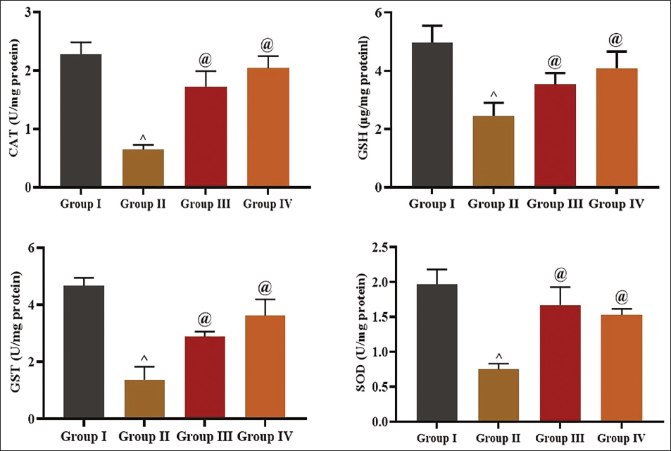
The concentrations of antioxidants, including GSH, CAT, SOD, and GST, in the hepatic tissue homogenates of the rats were assessed, and the data are presented in Figure 3. The drastically reduced concentrations of the antioxidants in liver tissue homogenates were observed in rats with acetaminophen-induced hepatic toxicity. Meanwhile, 50 and 100 mg/kg of ligustilide considerably increased all those antioxidant concentrations in the hepatic tissue homogenates of the acetaminophen-induced liver toxicity rats, which highlights its antioxidant properties.
Effect of Ligustilide on the Antioxidant Concentrations in the Liver Tissues of the Experimental Rats. Each Bar Denotes the Mean ± SD of Three Independent Assays, Analyzed Using One-Way Analysis of Variance (ANOVA) and Tukey’s Post Hoc Tests. “^” Denotes That Data Significantly Differed at p < .01 from the Control Group (Group I); “@” Signifies That Data Significantly Differed at p < .05 from the Acetaminophen-induced Group (Group II).
Effect of Ligustilide on Lipid Markers in Serum of Experimental Rats
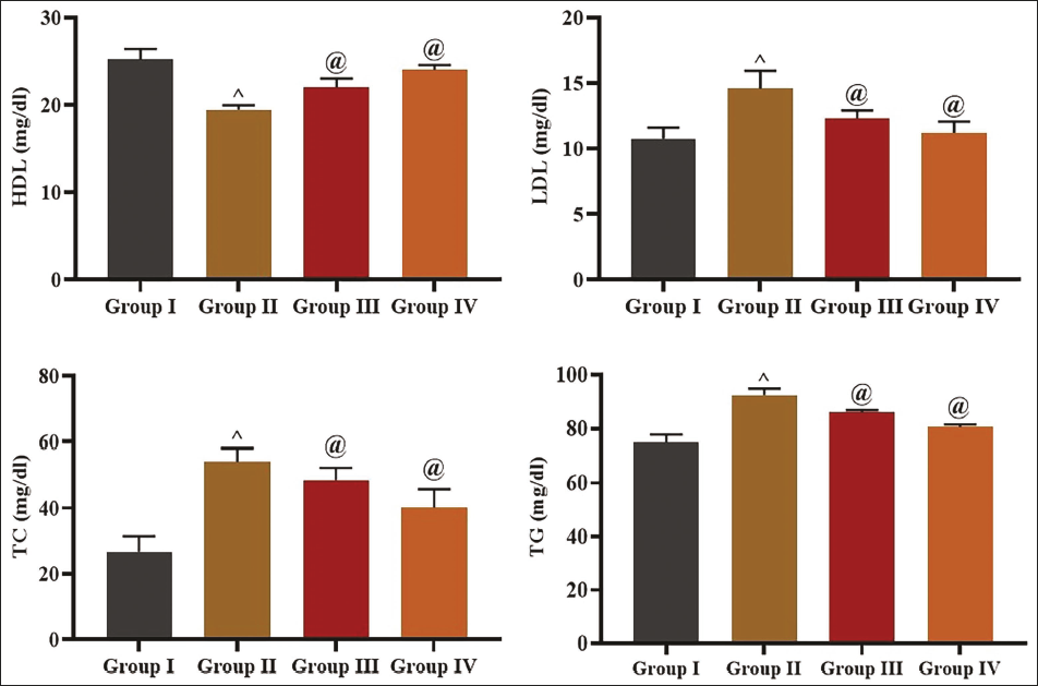
Figure 4 displays the lipid biomarkers in the serum of experimental rats. A considerable augmentation in LDL, TC, and TG and a subsequent reduction in HDL concentration were found in the serum of acetaminophen-induced liver toxicity rats. Captivatingly, 50 and 100 mg/kg of ligustilide remarkably diminished the LDL, TC, and TG concentrations and elevated the HDL in the serum of rats with liver toxicity.
Effect of Ligustilide on the Lipid Marker Levels in the Serum of Experimental Rats. Each Bar Denotes the Mean ± SD of Three Independent Assays, Analyzed Using One-Way Analysis of Variance (ANOVA) and Tukey’s Post Hoc Tests. “^” Denotes That Data Significantly Differed at p < .01 from the Control Group (Group I); “@” Signifies That Data Significantly Differed at p < .05 from the Acetaminophen-induced Group (Group II).
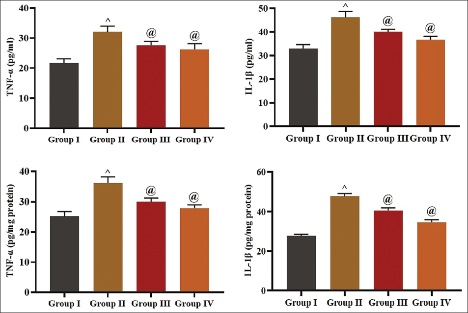
Effect of Ligustilide on Inflammatory Cytokines in Experimental Rats
The inflammatory markers were evaluated in both serum and hepatic tissue homogenates, and the findings are illustrated in Figure 5. Significantly increased concentrations of both IL-1β and TNF-α were noted in the serum and liver tissues of the rats with acetaminophen-induced liver toxicity. Nonetheless, ligustilide treatment at 50 and 100 mg/kg significantly diminished these cytokine concentrations in both the serum and liver tissues of the rats with acetaminophen-induced hepatic toxicity, which proves its anti-inflammatory properties.
Effect of Ligustilide on the Inflammatory Cytokine Levels in the Experimental Rats. Each Bar Denotes the Mean ± SD of Three Independent Assays, Analyzed Using One-Way Analysis of Variance (ANOVA) and Tukey’s Post Hoc Tests. “^” Denotes That Data Significantly Differed at p < .01 from the Control Group (Group I); “@” Signifies That Data Significantly Differed at p < .05 from the Acetaminophen-induced Group (Group II).
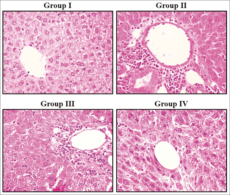
Effect of Ligustilide on Liver Histology of the Experimental Rats
Figure 6 illustrates the outcomes of the histopathological study of the hepatic tissues of experimental rats. The liver from the control group illustrated no inflammatory signs or hepatic injury and displayed the typical cellular configurations. Contrastingly, the liver tissues of rats with acetaminophen-induced liver toxicity displayed centrilobular hemorrhagic necrosis, inflammation, and infiltration of inflammatory cells when compared with control. The histological changes in the hepatic tissues of acetaminophen-induced rats were dramatically reduced by ligustilide.
Effect of Ligustilide on the Liver Histology of the Experimental Rats. Group I: The Liver Tissues from the Control Rats Exhibited No Signs of Inflammation or Hepatic Injury and Displayed the Typical Cellular Configurations. Group II: The Liver Tissues of Rats With Acetaminophen-induced Liver Toxicity Displayed Centrilobular Hemorrhagic Necrosis, Inflammation, and Infiltration of Inflammatory Cells. Groups III and IV: The Histological Changes in the Liver Tissues of Acetaminophen-induced Rats were Dramatically Reduced by the Ligustilide Treatment at Dosages of 50 and 100 mg/kg, Respectively.
Discussion
Drug-induced hepatic damage poses a serious public health issue, being a primary cause of acute liver failure and a notable factor in the market removal of approved pharmaceuticals. The mechanisms underlying drug-induced liver toxicity are complex and involve various pathways, including the disruption of cellular organelles, oxidative stress, and immune-mediated responses (Kaden et al., 2023). The liver is a key organ that significantly contributes to the metabolism and elimination of numerous drugs and xenobiotics. When the liver is exposed to potentially hepatotoxic substances, it can experience various forms of injury, ranging from mild and reversible changes to severe and irreversible damage (Rana et al., 2021). AST and ALT are two of the most commonly utilized indicators for identifying liver damage. These enzymes are often found in the hepatocyte’s cytoplasm, and their release into the bloodstream signifies cellular damage or necrosis. In instances of drug-induced liver injury, elevated levels of AST and ALT are frequently noted since the breakdown of hepatocyte membranes or the promotion of cell death results in the release of these enzymes into the bloodstream (Thakur et al., 2024). In addition to AST and ALT, ALP is another important marker of liver function. ALP is an enzyme involved in the transport of various molecules, including drugs and their metabolites, across cell membranes. Elevated ALP levels are often associated with cholestatic liver injury, which can occur when drugs or their metabolites impair the bile flow or disrupt the function of the bile ducts. The assessment of liver marker enzymes is an essential method for identifying drug-induced hepatic toxicity (Weber & Gerbes, 2022). In this work, the acetaminophen-induced liver toxicity in rats demonstrated elevated AST, ALP, and ALT activities in their serum and subsequent reduction in these enzyme activities in their liver tissues. Remarkably, the ligustilide treatment diminished these enzymes in the serum and enhanced these enzyme activities in the hepatic tissues of the rats with acetaminophen-induced hepatic toxicity. These findings suggested that ligustilide can inhibit drug-induced hepatic toxicity in experimental rats.
Drug-induced hepatic toxicity is a major problem in healthcare, as it can result in severe consequences and sometimes fatality. Monitoring and early detection of liver damage are crucial for the management of drug-induced hepatotoxicity. One important approach to assessing liver health is the analysis of specific serum biomarker levels (Tanaka et al., 2024). Albumin, a key serum protein, is an important marker of liver function. Reduced albumin levels can indicate impaired liver synthetic capacity, a hallmark of drug-induced liver injury. Total protein, which includes albumin and other proteins produced by the liver, can also be altered in the context of drug-induced hepatotoxicity (Benić et al., 2022). Reductions in these levels can signify impaired hepatic function and potential liver damage. Total bilirubin and direct bilirubin measurements provide insights into the liver’s bile-producing and bile-secreting capabilities. Elevated levels of these biomarkers may suggest the presence of hepatic damage due to drug toxicity (Castillo-Castañeda et al., 2024). Serum biomarker analysis can be a useful tool in the assessment of drug-induced hepatotoxicity. Routine monitoring of these biomarkers can help identify any abnormalities and implement the required therapy to alleviate the risk of further hepatic toxicity and ensure the safety of patients undergoing drug therapy (Huang et al., 2023). The current results demonstrated diminished albumin and protein levels and increased total/direct bilirubin in the serum of rats with acetaminophen-induced hepatotoxicity. Remarkably, the treatment of ligustilide significantly enhanced the albumin and protein contents and reduced total/direct bilirubin in the serum of rats with hepatic toxicity.
Drug-induced hepatotoxicity represents a significant public health issue, with oxidative stress being pivotal in its development. The liver, as the principal organ for the detoxification and metabolism of xenobiotics, is especially vulnerable to chemically induced oxidative damage (Li et al., 2015). Increasing data highlight that oxidative stress is closely related, creating a vicious cycle that exacerbates hepatic diseases. ROS and reactive nitrogen species (RNS) are generated during normal cellular processes in the liver, but under physiological conditions, their production is tightly regulated by endogenous antioxidant systems. However, when this balance is disrupted, leading to an excess of oxidants over antioxidants, oxidative stress can cause direct cell injury and activate various signaling pathways, ultimately contributing to liver damage through mechanisms such as nuclear receptor activation, autophagy, mitochondrial dysfunction, and cell death (Allameh et al., 2023). The pathways involved in drug-induced liver toxicity are complex and not fully elucidated, but oxidative stress is considered a key player (Mooli et al., 2022). GSH, GST, SOD, and CAT are critical antioxidants that play a key role in the liver’s protection against oxidative stress. Evaluating the levels of these antioxidants in the liver tissues during drug-induced toxicity can provide valuable insights into the underlying pathophysiology and potential therapeutic interventions. The antioxidant defense system, which includes the enzyme system and low molecular weight antioxidants, is responsible for maintaining the balance between oxidant and antioxidant production (Park et al., 2023). GSH is a critical antioxidant that plays an essential role in the detoxification of xenobiotics and the neutralization of ROS. GST, an enzyme that catalyzes the conjugation of GSH with electrophilic compounds, is also essential in the liver’s protection against oxidative stress. Similarly, SOD and CAT are key enzymes that neutralize superoxide radicals and hydrogen peroxide, respectively, thereby protecting the liver from oxidative injury (Zhao et al., 2024). The analysis of antioxidant enzyme levels in the liver during drug-induced toxicity can provide valuable data about the cellular processes implicated in the onset of hepatotoxicity. By understanding the interplay between drug metabolism, ROS generation, and the antioxidant defense system, researchers can develop more effective strategies for the management of drug-induced hepatic toxicity (Zhang et al., 2023). The results of this work clearly showed the drastically reduced concentrations of the antioxidants in the hepatic tissues of rats with acetaminophen-induced liver toxicity. Interestingly, the treatment with ligustilide significantly increased the antioxidant concentrations in the hepatic tissue homogenates of the acetaminophen-induced liver toxicity rats, which evidences its antioxidant properties.
Regular monitoring and recognizing drug-induced hepatotoxicity are crucial to prevent severe liver damage and acute hepatic failure. One important aspect of this monitoring is the analysis of lipid profile markers in the serum of patients (Greco et al., 2024). Lipids such as fatty acids have been implicated in cellular lipotoxicity, leading to apoptosis, defective insulin signaling, endoplasmic reticulum stress, and other downstream pathways. Liver pathology can influence medication metabolism, modifying efficacy and adverse effects, whereas drug metabolites may induce hepatic injury (Steinmann et al., 2023). The molecular pathways of drug-induced liver damage are intricate and frequently entail idiosyncratic reactions. Intracellular organelles and their functions are often the principal targets of hepatotoxicity, involving not just hepatocytes but also other hepatic cell types. Specific pharmacological characteristics, including elevated lipophilicity and substantial daily dosage, are linked to a considerable risk of drug-induced liver impairment (Sharma et al., 2023). The examination of lipid profile markers can yield significant insights into the onset and progression of drug-induced liver damage. TC and TG levels can be used as indicators of overall lipid metabolism. In contrast, LDL and HDL levels can provide information about the transport and distribution of lipids within the body. Alterations in these lipid markers may reflect underlying changes in liver function and can be used to monitor the impact of drug-induced hepatotoxicity (Habibullah et al., 2024). Briefly, the role of lipid profiles in the pathophysiology of drug-induced liver injury is a complex and multifaceted process. Understanding the interplay between lipid metabolism, drug metabolism, and cellular signaling pathways is crucial in elucidating the underlying mechanisms of this important clinical challenge (Zhang et al., 2021). In this work, the results demonstrated augmented LDL, TC, and TG and a subsequent reduction in HDL concentrations in the serum of rats with acetaminophen-induced liver toxicity. Fascinatingly, the ligustilide significantly reduced the LDL, TC, and TG concentrations and elevated the HDL in the serum of rats with liver toxicity. These findings evidenced that ligustilide can ameliorate dyslipidemia in rats with hepatic toxicity. Lisinine, a liver compound, has been found to reduce liver enzyme levels and tissue damage from acetaminophen overdose, reverse inflammation, oxidative stress, and cell apoptosis gene alterations, and mitigate acetaminophen-induced hepatotoxicity in mice through various pathways (Suo et al., 2025). A previous study found that WEEPAS, a bioactive component from Angelica sinensis (Oliv.) Diels, can protect mice from ethanol-induced liver tissue injury by reducing fat deposition and inhibiting oxidative stress response. FA, the main bioactive component, was found to regulate AMPK and PI3K/AKT signaling pathways, thereby reducing ethanol-induced liver tissue injury and lipid metabolism disorders in HepG2 cells and mice (Lu & Wang, 2025). The KEAP1/NRF2 pathway is crucial in oxidative stress response, controlling antioxidant and detoxifying genes. Finding modulators of this pathway and targeting ARE genes is essential for developing effective antioxidant agents. Natural products have been identified as potential drug candidates for supplementation, providing antioxidant capacity to human cells (Culletta et al., 2024).
Understanding the fundamental mechanisms of drug-induced liver damage is essential for formulating preventative and treatment strategies. A pivotal cause in the progression of drug-induced hepatic damage is the influence of inflammation and inflammatory cytokines. Inflammation is a common response to various insults, including drug-induced hepatic damage, and can participate in the progression and severity of the hepatic damage (Vachliotis & Polyzos, 2023). Idiosyncratic drug-induced hepatic toxicity is a significant issue, and the underlying processes are not fully understood. Increasing evidence suggests that the majority of idiosyncratic drug-induced liver injuries are immune-mediated and attributable to reactive metabolites. A reactive metabolite can bind to proteins, rendering them foreign and triggering an immunological response that may cause liver toxicity. In drug-induced liver damage, TNF-α and IL-1β are pivotal in the onset of the condition. Cytokines may be generated by several cell types, like Kupffer cells, hepatocytes, and invading immune cells, in reaction to drug-induced hepatic toxicity (Clària et al., 2023). TNF-α is a pro-inflammatory cytokine that can trigger apoptosis and necrosis in hepatocytes, leading to cell death and liver damage. IL-1β, another critical inflammatory cytokine, can activate signaling pathways that induce further inflammation and contribute to the onset of hepatotoxicity. The release of these inflammatory cytokines can lead to a self-perpetuating cycle of inflammation and liver damage, with the potential for the development of more severe and potentially fatal liver failure (Arroyo et al., 2021). It has been highlighted that the elevated levels of TNF-α and IL-1β in the serum and hepatic tissues of patients with drug-induced liver toxicity suggest their potential as biomarkers for the condition (Casulleras et al., 2020; Tiegs & Horst, 2022).
Understanding the role of inflammation and inflammatory cytokines in the pathophysiology of drug-induced hepatic toxicity is essential to the advancement of effective prevention and treatment strategies. The present findings evidenced the significantly increased concentrations of inflammatory cytokines in the serum and liver tissues of the rats with acetaminophen-induced hepatotoxicity. Fascinatingly, the treatment of ligustilide significantly diminished these cytokine concentrations in both the serum and liver tissues of the rats with acetaminophen-induced hepatic toxicity.
Conclusion
The findings of this work reveal the hepatoprotective effects of ligustilide against acetaminophen-induced hepatotoxicity in rats. The ligustilide treatment considerably regulated the liver marker enzyme activities, elevated antioxidant concentrations, and diminished inflammatory cytokines in acetaminophen-induced rats. Furthermore, the ligustilide treatment also regulated the dyslipidemia in the experimental rats. The hepatoprotective properties of the ligustilide are also supported by the findings of histopathological analysis. Consequently, ligustilide can serve as a viable treatment option for addressing drug-induced liver toxicity. Furthermore, it is advisable to conduct more experiments to achieve a precise understanding of the therapeutic values of ligustilide on drug-induced liver damage.
Abbreviations
IL-1β: Interleukin-1 beta; IL-6: Interleukin-6; ROS: Reactive oxygen species; SOD: Superoxide dismutase; TNF-α: Tumor necrosis factor-alpha.
Footnotes
Acknowledgment
The authors would like to thank the Affiliated Hospital of Hebei University, Baoding, China, for their support.
Declaration of Conflicting Interests
The authors declared no potential conflicts of interest with respect to the research, authorship, and/or publication of this article.
Ethical Approval
The protocols for animal experiments were approved by the Institutional Animal Ethics Committee, The First Central Clinical College of Tianjin Medical University, Tianjin 300190, China.
Funding
The authors received no financial support for the research, authorship, and/or publication of this article.
Informed Consent
Not applicable