
Abstract
Background
Diabetic retinopathy (DR) is the foremost microvascular problem that causes drastic visual impairment in diabetes patients. Hyperglycemia-triggered reaction cascade of inflammation and oxidative stress constitute the DR pathogenesis. The existing treatment options are not completely satisfactory.
Materials and Methods
We investigated the cell viability by 3-[4,5-dimethylthiazol-2-yl]-2,5 diphenyl tetrazolium bromide (MTT) assay, inflammatory mediators, lactate dehydrogenase (LDH), superoxide dismutase, glutathione, and malonaldehyde (MDA) levels by ELISA and qRT-PCR assay, protein expression of Nrf2 and heme oxygenase-1 (HO-1) by western blotting assay were analyzed.
Results
According to our research, nerolidol (NRD) increases the proliferation and antioxidant activity of human retinal endothelial cells (HRECs) by inducing Nrf2/HO-1 signaling, while attenuating MDA, an oxidative stress marker, LDH, and inflammatory mediators. These outcomes suggest that a substantial reaction of inflammation and oxidative stress injury happened in DR, which might be correlated to the instigation of the signaling Nrf2/HO-1.
Conclusion
NRD effectively suppresses oxidative stress and inflammation in HG-induced HRECs. The primary mechanism of NRD on DR may be linked to the activation of the Nrf2/HO-1 pathway and may give a useful medicine for DR treatment.
Introduction
Diabetes mellitus (DM) is the world’s leading public health burden, with 27 definitely developing and rising over the next few decades As a severe impediment of DM, diabetic retinopathy (DR) affects the loss of vision and extremely worsens their life worth (Teo et al., 2021). Approximately, 35.4% of DM patients exhibit various kinds of DR (Lee et al., 2015). Hyperglycemia (HG) mostly contributes to the development of DR (Kang & Yang, 2020), and excess blood sugar provides atherosclerotic plate formation, which inflames the retinal vessels (Robison et al., 1985). Recently, several influences have been found to be related to DR, including advanced glycation end products (AGEs), inflammation, and oxidative stress. Oxidative stress possesses a central deed in the incidence and progression of DR, even if the pathophysiology of DR has not been completely elucidated (Kowluru, 2013). The retina is extremely vulnerable to oxidative stress owing to the features of high consumption of oxygen, glucose oxidation, and strong coverage of light (Catala, 2011; Nabavi et al., 2015). Non-enzymatic addition or monosaccharides glycosylation in proteins enhances the auto-oxidation response under a high glucose level environment that produces numerous reactive oxygen species (ROS) (Itoh et al., 1999; Lapolla et al., 2005). The weakening of the protective antioxidants and upgraded oxidative stress are the central providers of HG-facilitated damage (Cao et al., 2014). Currently, the fundamental remedial method for DR is a modification of microvascular difficulties, comprising laser photocoagulation, vitrectomy, intravitreal drug healing, and VEGF pills (Chatziralli & Loewenstein, 2021; Wang & Lo, 2018). As the occurrence of DM rises, innovative approaches are required for the prevention or cure of DR in earlier phases. Thus, exploring further effective natural compounds based on accepting mechanisms of action in DR is instantly required.
Recently, plant-based bioactive natural constituents have been tested in clinical research for DR treatment as a result of their low side effects (Ai et al., 2020). Nerolidol (NRD) is sesquiterpene alcohol, which is sequestered as essential oils from aromatic floras, such as neroli, ginger, lemongrass, tea tree, and lavender (Azzi et al., 2018). NRD exerts numerous pharmacological activities comprising anti-inflammatory, antioxidant, anti-cancer, and apoptotic effects (Chan et al., 2016). Recently, NRD has been exposed to relieve oxidative stress, inflammation, and apoptosis in cardiotoxicity driven by chemotherapeutic mediator cyclophosphamide (Iqubal et al., 2019). It has also been recognized that NRD blocked the inflammatory reaction in LPS-stimulated ALI by the modulation of antioxidants and the AMPK/Nrf2/(HO)-1 signaling pathway (Ni et al., 2019). Furthermore, NRD has been revealed to be valuable as an antitumor compound owing to its efficacy in targeting cell survival and proliferation molecules (Biazi et al., 2017), which act as a chemosensitizer in many malignancies (Ambroz et al., 2015; Hanušová et al., 2017). Nevertheless, to the finest of our understanding, the influence of NRD on DR treatment is left uncertain.
Nrf2 dissociates from Keap1 and moves into the nucleus, where it causes the transcription of genes involved in antioxidant defense, including superoxide dismutase, (SOD) and Heme Oxygenase-1 (HO-1) to reduce ROS accumulation during stressful situations. When ROS is primarily elevated, Nrf2 stimulation could act as a compensatory mechanism to counterbalance ROS amassing, but with an unceasing upsurge in ROS, the Nrf2–ARE system will be overcome, subsequently with a sustained rise in ROS. According to accumulating evidence, the diabetic retina has higher transcript levels of Nrf2 and Keap1, but Nrf2 fixes more to Keap1 and less to DNA, which leads to the transcription of the antioxidant enzyme being lessened and more assisting the antioxidant defense system. (Li, Li, et al., 2019; Zhong et al., 2013). It has previously been reported that NRD decreases inflammation in LPS-stimulated ALI by activating antioxidant status in the AMPK/Nrf2/HO-1 signaling pathway; however, there are no data on this regard regarding the therapeutic use of NRD for DR sequelae (Ni et al., 2019). Hence, we assessed the antioxidant and anti-inflammatory efficacy of NRD in an in vitro DR model through the regulation of Nrf2/HO-1 signaling.
Materials and Methods
Chemicals
NRD and biochemicals were obtained from Gibco (CA, USA). LDH, GSH, SOD, and MDA assay kits were procured from Ese-Bio (Shanghai, China). Cell culture reagents and ELISA assay kits were provided by Elabscience Biotechnology (Wuhan, China). The western blot primary antibodies and HRP-conjugated secondary antibodies were acquired from Beyotime Biotechnology, USA. The Nuclear Extraction kit and ELISA kit were bought from Elabscience Biotechnology (Wuhan, China).
Cell Culturing
For in vitro study, human retinal endothelial cells (HRECs) were bought from Shanghai Aiyan Biotechnology Co., Ltd. (Shanghai, China). These were cultured to the endothelial cell media, comprising FBS (10%), penicillin/streptomycin (1%), and glucose (5.5 mmol/L) was used as the group normal glucose (NG), in a 5% CO2 atmosphere at 37°C. To imitate DR, NG was substituted with 25 mmol/L glucose used as high glucose (HG).
Determination of Cell Viability Test
High glucose-prompted HRECs were grown in 96-well plates (2 × 10³) for one day and subsequently preserved with diverse quantities of NRD (Control, 5, 10, 15, 20, and 30 µM) for an extra day. Afterwards, 3-[4,5-dimethylthiazol-2-yl]-2,5 diphenyl tetrazolium bromide (MTT) (50 µL) was added and kept for 2 hour at 37°C. Determination of the optical density was estimated at 570 nm by a microplate reader (BioTek Instruments, Inc., Winooski, VT, USA).
Assay of Inflammatory Mediators, LDH, SOD, GSH, and MDA Levels
The intensities of inflammatory mediators (IL-6, IL-1β, and TNF-α), LDH, and oxidative stress-associated indicators (glutathione (GSH), SOD, and MDA) in HG-activated HRECs were estimated by ELISA assay kits according to the consistent company’s guidelines.
Determination of Inflammatory Mediators Levels by qRT-PCR Assay
An expression level of inflammatory mediators (mRNA) comprising IL-6, IL-1β, and TNF-α has been identified employing RT-qPCR. Briefly, whole RNA was sequestered on the HG-stimulated HRECs and was employed for the synthesis of cDNA and the analysis of RT-qPCR was done. According to the 2–∇∇Ct method, the mRNA expression levels were calculated, and GAPDH was employed as an internal control.
Nuclear Isolation Assay
A nuclear extraction kit was used to isolate subcellular nuclei from the HG-stimulated HRECs. Concisely, the HRECs were homogenized in HEPES buffer (20 mM), comprising MgCl2 (1.5 mM), phenylmethylsulphonyl fluoride (1 mM), KCl (10 mM), leupeptin (10 µg/mL), aprotinin (10 µg/mL), sucrose (250 mM), EDTA (0.5 mM), and EGTA (0.5 mM), to make the fraction of cytosol. Then, homogenate, the resultant solution, was centrifuged at 750 g for 10 minutes at 4°C to isolate the cytosol and nuclei.
Estimation of Nrf2 Action
The activity of Nrf2 was evaluated by a Nrf2 DNA-Binding TransAM® ELISA kit affording to the company’s directions. Shortly, 96-well plates were pre-coated with an antioxidant response element sequence oligonucleotide, and then treated with 15 µg of nuclear protein. One hour later, anti-Nrf2 and secondary antibodies were treated. These were measured by a spectrophotometer at 450 and 655 nm.
Analysis of Nrf2/HO-1 Protein Expression Levels by Western Blotting
HG-prompted HRECs were grown for 24 hour and the cell lysates were prepared. The measurement of protein content was performed by the usage of the Protein BCA Assay Kit (Pierce Chemical Co, TX, USA). Succeeding electrophoretically dispersed the proteins by 10% SDS-PAGE and moved onto PVDF membranes. Then, the film was blocked by using a probe overnight at 4°C and primary antibodies Nrf2/HO-1 and Laminin 1:1,000 dilutions, preserved overnight at 4°C, were administered; subsequently, secondary antibodies (1:5,000) were added and preserved for 1 hour. Internal control is Lamin-B/β-actin. ECL detection kit (Bio-Rad, CA, USA) was used for immunoblotting.
Statistical Examination
Data were conveyed as mean ± SD. A statistical study was accomplished by GraphPad Prism software version 8.0.1 which was employed to perform an analysis of variance (ANOVA) and subsequently Duncan’s test. The statistically significant value is considered as p < 0.05.
Results
NRD Stimulates HG-activated HRECs Viability
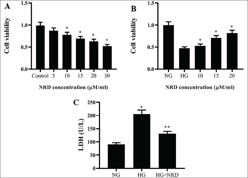
The HRECs proliferation was estimated in the MTT test with different quantities (Control, 5, 10, 15, 20, and 30 µM/ml) of NRD (Figure 1(A)). We established that HRECs proliferation elevated in the low dose of NRD (5, 10, and 15 µM), then started to reduce (p < 0.05) at NRD 20 µM, and again alleviated (p < 0.05) at NRD (40 µM), which proposed that NRD (10 and 15 µM) may be the harmless dose reasonably. Afterwards, this study established that HG-administration pointedly mitigated (p < 0.05) the HRECs viability (Figure 1(B)). Administration of NRD (5 and 15 µM) moderately attenuated the constraining action of HG on HRECs proliferation (p < 0.05). In view that there had no substantial changes among the actions of these two NRD (5 and 15 µM) quantities on HG-activated HRECs viability, 15 µM NRD was selected for further analysis. Endothelial dysfunction marker namely LDH activity is shown in Figure 1(C). Treatment with HG on HRECs revealed that the level of LDH was enhanced. Conversely, administration of NRD controlled expressively (p < 0.05) the discharge of LDH produced by HG.
NRD Stimulates HG-activated HRECs Viability. (A & B) HRECs Were Added with Different Quantities (5–40 µM) of NRD for 24 hour. MTT Test Was Used to Measure the Cell Viability. (C) The LDH Level of HG-induced HRECs Was Determined. The Results Were Displayed as Mean ± SD and the Significance is Considered as *p < 0.05 Against Untreated Control.
Influence of NRD on the Antioxidant Level and MDA Affected by HG-activated HRECs
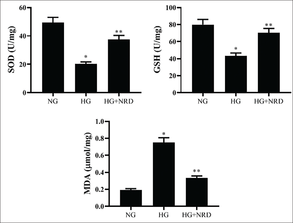
Under an oxidative stress environment, HG abnormally prevented the discharge of GSH and SOD, while elevating (p < 0.05) the MDA levels. Stimulatingly, NRD administration considerably elevated (p < 0.05) the SOD and GSH, whereas diminished the level of MDA, thereby reducing the oxidative stress in HG-stimulated HRECs (Figure 2).
NRD Reduces Oxidative Stress Factors and Enhanced Antioxidant Status in HG-activated HRECs. The Results Were Displayed as Mean ± SD and the Significance is Considered as *p < 0.05 Against Untreated Control.
NRD Averts the Level of Inflammatory Mediators Increased by HG-driven HRECs
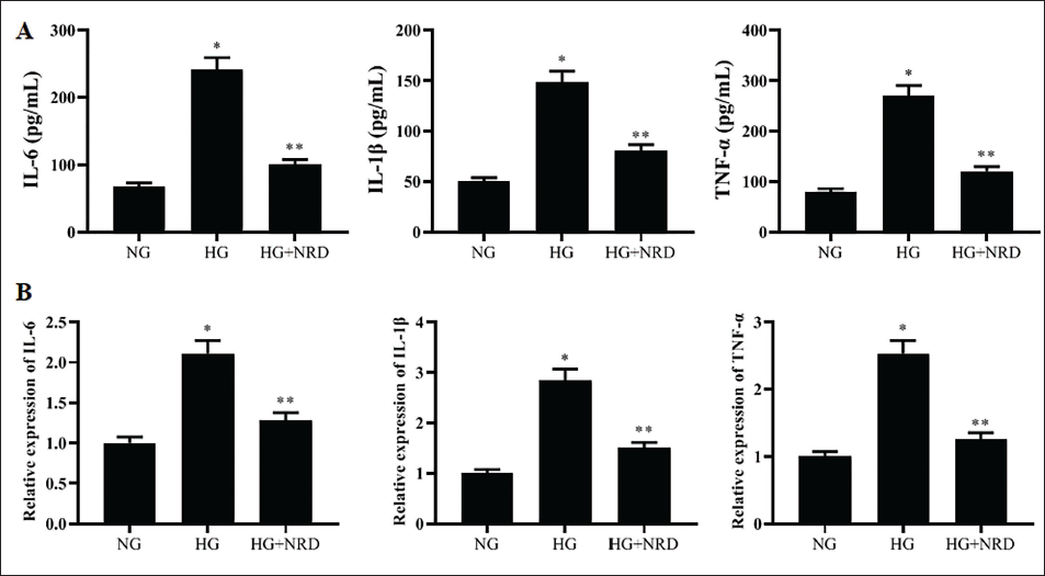
Inflammatory mediators have a profound action on the development of DR. So, we examined the action of NRD on inflammatory cytokines levels (Figure 3(A)) and their relative expression (Figure 3(B)). HG considerably augmented (p < 0.05) the cytokine intensities and relative expression mRNA levels of IL-6, IL-1β, and TNF-α in HRECs; however, NRD usage could reduce these cytokines levels and their relative expression levels.
NRD Curbs Inflammation in HG-stimulated HRECs. (A) The Inflammatory Mediators (IL-1β, IL-6, and TNF-α) Level in HG-activated HRECs Was Assessed by ELISA. (B) The Relative Levels of mRNA of Inflammatory Mediators (IL-1β, IL-6, and TNF-α) in HG-activated HRECs Was Evaluated by RT-qPCR. The Results Were Displayed as Mean ± SD and the Significance is Considered as *p < 0.05 Against Untreated Control.
NRD Ameliorates Nuclear Nrf2 and Their Relative Protein Expression Level
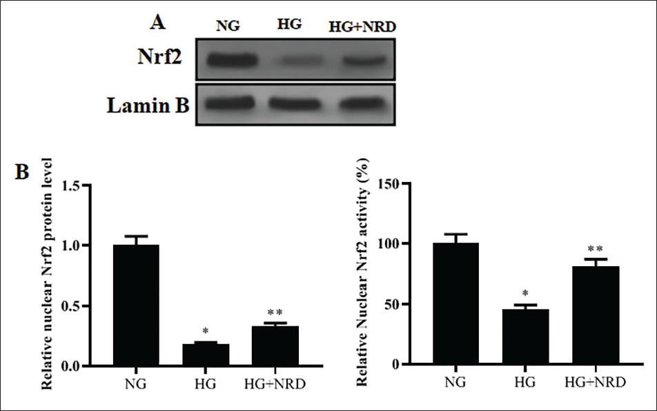
Nrf2/HO-1 signaling requires the pathogenesis of DR. Remarkably, we established that HG administration repressed the Nrf2 protein expression and its relative Nrf2 protein level which was considerably inverted (p < 0.05) by the treatment of NRD (Figure 4A). Similarly, relative nuclear Nrf2 activity was reduced in HG-activated HRECs nuclei; the binding DNA action of Nrf2 was too subnormal in the HG, while it was stimulated by NRD administration (Figure 4B).
NRD Ameliorates the Nuclear Nrf2 Levels in HG-activated HRECs. (A) The Nuclear Nrf2 Level Expression Protein in HG-triggered HRECs Was Evaluated by Western Blot. (B) The Nuclear Nrf2 Action Was Assessed in HG-stimulated HRECs. The Data Were Exposed as Mean ± SD and the Significance Is Considered as *p < 0.05 Against Untreated Control.
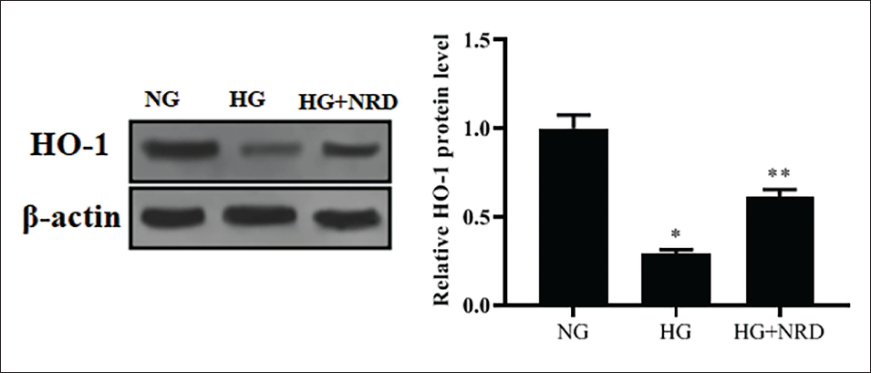
NRD Improves Nuclear HO-1 and Its Relative Protein Expression Level
The nuclear HO-1 and its relative protein level were downregulated by the HG
NRD Ameliorates the Nuclear HO-1 Levels in HG-activated HRECs. The Nuclear HO-1 Protein Expression Level in HG-triggered HRECs Was Evaluated by Western Blot. The Data Were Displayed as Mean ± SD and the Significance Is Considered as *p < 0.05 Against Untreated Control.
Discussion
DR is a well-known microvascular complication allied with DM that causes blindness. DR affects the value of patients’ life nature extremely and executes a substantial encumbrance on relations (Teo et al., 2021). The pathogenesis of DR has not been completely established to date. Accumulating evidence suggested that the DR disorder is attentively connected to disorders containing neuropathy, inflammation, and vasculopathy amid which, the oxidative stress and immune reactions facilitated by the Nrf2/HO-1 pathway exhibit a vital role (Kowluru, 2013; Zhong et al., 2013). Nrf2 is a significant transcriptional controller that stimulates the transcriptional expression of numerous antioxidant stress-linked protein genes containing catalase, heme oxygenase-1, and glutathione S-transferase, to auxiliary to eradicate a huge amount of oxidative stress byproducts and inciting elements in the physique, diminishing the impairment of organs and tissues owing to inflammation. The medical management of DR is still inspiring, and there is no acceptable medication to defend against DR (Chatziralli & Loewenstein, 2021; Wang & Lo, 2018). Thus, exploring more effective and suitable mediators for DR management is instantly desired. Here, we found for the primary phase that NRD reduces DR efficiently suppressing inflammation and oxidative stress through promoting the Nrf2/HO-1 pathway.
NRD is a renowned sesquiterpene bioactive compound that originates from essential oils isolated from plants and flowers (Azzi et al., 2018). As well, NRD has numerous pharmacological actions, including anti-inflammatory, antioxidant, and anticancer activities (Chan et al., 2016). Prior research unveiled that NRD represses LPS-stimulated acute kidney inflammation models in rats (Zhang et al., 2017). Besides, NRD moderates the creation of pro-inflammatory intermediaries in LPS-triggered peritoneal macrophages (Fonsêca et al., 2016). Notably, these findings specified that inflammatory responses could be inverted by NRD. It has been documented that NRD is uncovered to relieve oxidative stress, inflammation, and apoptosis in cardiotoxicity driven by chemotherapeutic mediator cyclophosphamide (Iqubal et al., 2019). Recently, NRD has been revealed to be valuable as an antitumor compound owing to its efficacy in targeting cell survival and proliferation molecules (Biazi et al., 2017), which act as a chemosensitizer in many malignancies (Ambroz et al., 2015; Hanušová et al., 2017). Furthermore, it was recognized that NRD curbed the inflammatory response in LPS-stimulated ALI by the modulation of antioxidants and the AMPK/Nrf2/(HO)-1 signaling pathway (Ni et al., 2019). Nevertheless, to the greatest of our understanding, the action of NRD on DR treatment is left uncertain. In our study, NRD could enhance HG-triggered HRECs viability, elevate antioxidant status, and upregulated Nrf2/(HO)-1 protein levels, whereas reduced the endothelial dysfunction marker LDH and inflammatory mediators as compared to the HG group.
Several reports demonstrated that inflammation and oxidative stress have a profound action on DR development (Catala, 2011; Kowluru, 2013; Nabavi et al., 2015). The inflammation occurs in the vascular region, and several cytokines comprising IL-6, TNF-α, and IL-1β were recognized (Williams et al., 2013). Collecting evidence points out that the development of DR is associated with inflammation, which is a central controller of DR (Semeraro et al., 2015). The elevated generation of inflammatory intermediaries is the chief source of DR progression and evolution. Numerous biochemical and pathological retinal difficulties are linked with DM, which is dependable on inflammation (Joussen et al., 2004). Innumerable documents have established that the augmented adhesion of leukocytes to the retinal vessels in the trial model of DM is an early incident in DR development (Joussen et al., 2001). HG can make oxidative stress mostly change the polyol and hexosamine path stimulation, the overacting of the isoforms level of protein kinase-C, and the gathering AGEs (Kowluru, 2013; Robison et al., 1985). Alternatively, HG suppresses the anti-oxidant defense organization by epigenetic alteration, subsequent in an imbalance among the foraging and formation of ROS (Cao et al., 2014). Eventually, extreme amassing of ROS prompts lipid peroxidation, mitochondrial dysfunction, inflammation, and apoptosis leading to structural/functional variations in the retina (Cao et al., 2014; Itoh et al., 1999; Lapolla et al., 2005). Therefore, it is crucial to explore and illuminate the oxidative stress-correlated action of DR, which might offer manifold possible beneficial goals to improve effective DR remedies.
Additionally, oxidative stress also contributed to the process of DR pathology (Cao et al., 2014). The significant indicators of oxidative stress are MDA, GSH, and SOD levels. The lipid peroxidation product of MDA is interrelated with oxidative stress. Prior documents have exposed that for MDA, an oxidative stress index concentration is pointedly elevated, and that of GSH and SOD are considerably reduced in DM, signifying that diabetic rats are under the state of oxidative stress. Thus, the regulation of the oxidative stress impairment and inflammatory reaction is the central method to accomplish DR. Our findings presented that TNF-α, IL-1β, IL-6, MDA, and LDH levels were reduced, while GSH and SOD levels were elevated in the HC+NRD group, and Nrf2 and HO-1 proteins were upregulated signifying that substantial response of inflammation and oxidative stress injury happened in DR, which may perhaps correlate with the instigation of Nrf2/HO-1 pathway. Vinholes et al. (2014) suggested that NRD is a good antioxidant that exerts protection against oxidative damage. Another study conducted and reported that cis-NRD mediated its strong antioxidant activity in protecting the hepatocytes through the inhibition of lipid peroxidation induced by tert-BuOOH, thereby 1 mM of cis-NRD resulted in MDA reduction.
Chan et al. (2016) described that the dosage of NRD was safe, and translated from animal to clinical studies to evaluate its efficacy. NRD has a great potential to be used as a new chemical and therapeutic drug in the field of agriculture and medicine, respectively, and sufficient baseline information is available for guiding future works and commercial exploitation. Further in their study, NRD dermal LD50 values were found higher than 2,000 mg/kg body weight, indicating low acute toxicity via the transdermal route, but in oral administration, the oral LD50 values of NRD in rats and mice were higher than 2,000 mg/kg body weight (>5,000 and 9,976 mg/kg body weight, respectively), indicating low acute toxicity via the oral route.
Conclusion
In conclusion, NRD efficiently restrains the oxidative stress and inflammation response in HG-induced HRECs. The principal mechanism of NRD on DR might be connected to the triggering of the Nrf2/HO-1 pathway and may provide a constructive medication for DR treatment.
Limitations of the study
NRD effectively suppresses oxidative stress and inflammation in HG-induced HRECs. The primary mechanism of NRD on DR may be linked to the activation of the Nrf2/HO-1 pathway and may give a useful medicine for DR treatment. We used a small sample size but in future, we will use a large sample size in future studies; also, NRD safety and side effects were not clearly explained, but we need to study full toxicity effects.
Abbreviations
DM: Diabetes mellitus; DR: Diabetic retinopathy; AGEs: Advanced glycation end products; ROS: Reactive oxygen species; NRD: Nerolidol; SOD: Superoxide dismutase; HO-1: Heme Oxygenase-1; MDA: Malonaldehyde; MTT: 3-[4,5-dimethylthiazol-2-yl]-2,5 diphenyl tetrazolium bromide; LDH: Lactate dehydrogenase; GSH: Glutathione; ELISA: Enzyme-linked immunosorbent assay; Nrf-2: Nuclear factor erythroid 2-related factor 2; AMPK: AMP-activated protein kinase.
Footnotes
Declaration of Conflicting Interests
The authors declared no potential conflicts of interest with respect to the research, authorship, and/or publication of this article.
Ethics Approval and Consent to Participate
Not applicable.
Funding
The author received no financial support for the research, authorship, and/or publication of this article.