
Abstract
Background
In ayurvedic practice, the Guduchi (Tinospora cordifolia (Willd.) Miers) stem is used as a Medhya drug for its beneficial effects on memory improvement.
Objectives
The current study was planned to explore the Medhya properties of the Guduchi stem extract by observing its ameliorating effect on AlCl3-induced neurotoxicity in rats that acted as a chronic model of memory loss.
Materials and Methods
The aqueous extract of the Guduchi stem was prepared per the Ayurvedic Pharmacopoeia of India and administered to the AlCl3-treated Wistar rats for 42 days. The biochemical assessment of the brain tissues of the treated animals was done by the acetylcholinesterase (AChE) inhibition assay, protein expression, and oxidative stress assays, namely lipid peroxidation, reduced glutathione, superoxide dismutase, and catalase assay. The neurobehavioral assessment was done using the elevated plus maze (EPM) test.
Results
The EPM test revealed that treatment with Guduchi extract showed marked improvement of memory status in rats along with reduced oxidative stress, and a marked modulation of the AChE inhibition and expression of AChE tubulin proteins.
Conclusion
The results substantiate the Medhya properties of the Guduchi. Detailed investigations are required to be carried out to explore the precise mechanism of the neuroprotective action of the Guduchi stem extract against the AlCl3-induced neurotoxicity in rats.
Introduction
Dementia is a condition that chronically and progressively affects memory and learning and consequently affects thinking and comprehension. The most likely cause of dementia is Alzheimer’s disease, a neurodegenerative condition (
Ayurveda, the ancient system of medicine is being practiced in India for centuries. As a time-tested system of medicine, Ayurveda can serve as a rich and potential source for newer therapies in modern medical research. As per the Ayurveda postulation, Smṛtināsha (loss of memory) is the prodromal symptom of jarā (aging) exhibited when Smṛti (memory) is vitiated by rajas (passion) and tamas (obscurity). 8 As per the Ayurveda, the term Medha broadly implies intellect, memory, and cognition. The classical Ayurvedic text Charak Samhita has described a group of drugs called Medhya that affect the Medha. 9 Mandukaparni (Centella asiatica Linn.), Yashtimadhu (Glycyrrhiza glabra Linn.), Guduchi (Tinospora cordifolia), and Shankhapushpi (Convolvulus pluricaulis Choisy) are the four plants that form the typical Medhya drug group. 10 Since the Medhya drugs are also known to affect memory and are readily available, they possess the potential to act as an alternative and complementary therapy to the existing modern medicine therapies for the management of dementia, an outcome of neurodegenerative diseases like Alzheimer’s disease. However, a perusal of the literature shows ample scope for research on dementia management using Medhya drugs due to want of robust data.
Guduchi (Tinospora cordifolia (Willd.) Miers) also known as Giloy or Amrita is a versatile herbal plant that is readily available and commonly used. As per the Ayurveda literature, aqueous extractives of the Guduchi stem are indicated for consumption for its Medhya effects. 9 Although Guduchi is used extensively in Ayurveda as Medhya drug, few published reports on experimental studies of Tinospora cordifolia using acute and short-term memory loss models are available.11–13 However, there is a paucity of well-designed experimental data on its therapeutic effects in chronic neurodegenerative disease models with special reference to memory. Repeated dose administration of aluminum chloride is known to cause memory loss and cognitive deficits in experimental rats by activating the AChE enzyme.3, 14 The model shows chronic neurodegeneration in the form of memory loss, similar to Alzheimer’s disease. Therefore, the aluminum chloride-induced model in rats was selected to explore the memory-improving effect of Guduchi.
Materials and Methods
Crude Drug and Extract
The stem of Guduchi was collected from the local market of Pune and was authenticated by the botanist of the Regional Ayurveda Research Institute, Pune. Physico-chemical properties such as loss on drying, ash value, and extractive values of the dried sample of the Guduchi stem were studied as per the Ayurvedic Pharmacopoeia of India, 15 to assess the quality of the crude drug.
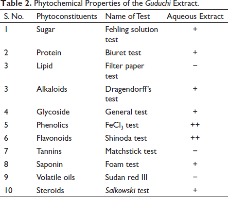
The aqueous extract of the Guduchi stem was prepared as per the method given in the Ayurvedic Pharmacopoeia of India. 15 Dried Guduchi stems of 30 g were powdered and macerated with 600 mL of distilled water containing 1% chloroform for 24 hr with intermediate shaking at room temperature. The solution was then filtered and dried at 40°C to obtain the powdered extract. The same cycle was repeated until the complete exhaustion of the crude drugs. The extract was subjected to preliminary phytochemical screening for detecting the presence of various phytochemicals, namely carbohydrates (Fehling test), proteins (Biuret test), tannins (Matchstick test), steroids (Salkowski test), glycosides, saponins (Foam test), alkaloids (Dragendorff’s test) as per the standard methods prescribed. 16
LC-MS Analysis of the Extract
Guduchi extract was analyzed by the liquid chromatography-triple-quadrupole tandem mass spectrometry (LC-MS/MS) with electrospray ionization (LC-ESI-MS/MS) technique using an Ultra High Definition (UHD) Accurate-Mass 6538 Q-TOF LCMS system (Agilent Technologies, CA, USA) with an Infinity Lab Poroshell 120SB-C18 analytical column (3.0 × 100 mm2, 2.7 µm). The mobile phases were water, acetonitrile, and formic acid (H2O:ACN:FA = 90:9.9:0.1) in a gradient mode (5%–97%). The injection volume was 20.00 µL, and the column temperature was maintained at 40°C. Parameters for analysis were set using positive ion mode with spectra acquired over a mass range from m/z 100 to 1,700 for MS and from m/z 50 to 1,700 for MS/SM; data were acquired at a 2 GHz extended dynamic range with narrow isolation width. The MS/MS data were analyzed using quantitative analysis software (Ver B.10.0, Agilent Technologies, CA, USA), and the compounds were identified using a commercially available licensed METLIN metabolite PCDL library. The accuracy of the confirmation of the compounds was established on their error of less than 5 ppm and the MS/MS fragment matching.
Animal Welfare
The in vivo study of the Guduchi extract was conducted in the experimental animal facility of the Indian Institute of Science Education and Research (IISER), Pune. A total of 36 healthy, disease/specific pathogen free (SPF) male Wistar rats (Crl: WI Charles River, USA strain) of 8–10 weeks were obtained from the animal breeding facility of the IISER, Pune. The animals were housed in a barrier-maintained SPF facility. The animals were acclimatized for 7 days. The health examination of all the animals was performed during the acclimatization period. Animals were housed in polypropylene cages (42.5 × 26.5 × 18.5 cm3) with lids and corn cob bedding. The room temperature and relative humidity were maintained at 22°C
Experimental Design and Treatment
The animals were randomly distributed into four groups (Groups 1, 2, 3, and 4) to make the mean body weight of all groups nearly equal before the start of dosing. Each group contained nine animals. Group 1 animals were administered purified water orally for the entire study period of 42 days and kept as normal control (NC). Aluminum chloride (Merck, Germany) was used to induce neurotoxicity in experimental rats. Aluminum chloride solution in water (100 mg/kg), which was used to be prepared fresh every day, was orally administered for 42 days to the rats of Groups 2, 3, and 4. 14 Donepezil HCl (Eisai Pharmaceuticals India Pvt. Ltd, Visakhapatnam, Batch No. AR1901) was used as a standard drug. Group 2 was kept as disease control (DC), whereas Group 3, which received Donepezil hydrochloride @ 1 mg/kg orally for 42 days 1 hr after the administration of aluminum chloride, was kept as standard control (SC). 14 In literature, researchers have frequently used doses of Tinospora cordifolia extracts between 100 and 200 mg/kg in various shorter duration (up to 15 days) in vivo cognition, depression and memory deficit models in rodents.12, 17–19 Hence, based on the available data from previous studies, Group 4 was treated with an aqueous extract of Guduchi (GT) at the dose rate of 200 mg/kg, 1 hr after the administration of aluminum chloride for 42 days. Every day, the required quantity of the dried Guduchi extract powders based on the latest body weights of the animals in the groups was taken and dissolved in pure water and administered to the animals orally. The concentration of the extract solutions administered was 20 mg/mL. Stainless steel gavage needle was used for oral administration to the rats.
The animals were observed daily for general clinical signs and changes in the skin, fur, eyes, mucus membrane, the occurrence of secretions and excretions, behavior, morbidity, and mortality twice daily.
Elevated Plus Maze
Neurobehavioral changes with reference to the memory due to the treatment were assessed using the elevated plus maze (EPM) test on days 20 and 42 of the experiment. An EPM with walls that were 40 cm high and with closed arms that were traversed by open arms was used. The middle square was 10 × 10 cm2 in size. The maze was kept 50 cm (2 inches) above the ground. During the acquisition stage, the animal was positioned at the end of the arm facing away from the center square. Initial transfer latency (ITL), which measures how long it takes an animal to transition from an open arm to a closed arm, was measured. Each animal was given 20 s to explore the maze after the ITL was recorded before being returned to its home cage.
Following treatment, retention transfer latencies (RTLs) on day 42 and ITLs on day 20 were noted.14, 20
Animal Sacrifice and Organ Collection
Animals were humanely sacrificed after 42 days using the CO2 asphyxia method of euthanasia. The liver, kidney, and brain were collected and weighed after the sacrifice. Brain tissues were collected for biochemical assays as per the standard methods described under each assay. The liver, kidney, and brain were collected in 10% neutral buffered formalin for the histopathological study.
Biochemical Assessment
Estimation of AChE from Brain
The hippocampus and cortex regions of the collected brain tissues were separated to estimate AChE activity. The cortex and hippocampus of the isolated brain were separated immediately and collected in different centrifuge tubes, weighed, and homogenized in an ice-cold phosphate buffer solution. The homogenates were then stored in a −20°C deep freezer until the assay was carried out. The assay was conducted according to the method described by Ellman14, 21 using the colorimetric AChE assay kit (Amplite, AAT Bioquest, USA) as per the procedure provided with the kit. The assay buffer was composed of the sodium phosphate buffer solution (0.1 M, pH 8.0), 0.1% BSA, a working solution of dithiobis-nitrobenzoic acid, and a working solution of acetylthiocholine iodide. The assay mixture was composed of 50 µL tissue sample homogenate and an equal quantity of the assay buffer.
Serially diluted AChE of 50 µL was mixed with 50 µL of the assay buffer to perform the SC assay. The change in the absorbance of the assay mixture was measured for 2 min at 30 s intervals at 410 nm using the enzyme-linked immunosorbent assay (ELISA) Plate Reader (Epoch, Biotek, USA, software version 5.1.11). The observations were expressed as the percentage inhibition of AChE.
Lipid Peroxidation, Reduced Glutathione, and Catalase and Superoxide Dismutase Assay from the Brain
The hippocampus and cortex regions of the collected brain tissues were separated to assess oxidative stress parameters. The isolated brain was separated immediately and collected in a centrifuge tube, weighed, and homogenized in an ice-cold phosphate buffer solution. The homogenate was then stored in a −20°C deep freezer till the assay was performed. The extent of lipid peroxidation (LPO) was measured in terms of malondialdehyde (MDA) production using the method described by Ohkawa et al. 22 The reduced glutathione (GSH) level was measured using the method described by Ellman. 23 The catalase assay was performed following the standard method. 24 Superoxide dismutase (SOD) activity was estimated according to the method described by Ukeda et al. 25 using the colorimetric SOD assay kit (Amplite, AAT Bioquest, USA) as per the procedure provided with the kit.
Proteomic Analysis of the Brain
The hippocampus of three animals from each group was collected in the separate centrifuge tubes, weighed, and homogenized in an ice-cold phosphate buffer solution containing the protease inhibitor tablet. The samples were further processed using the ProteaseMAX surfactant (Promega Inc., USA) following the procedure provided along with the product (
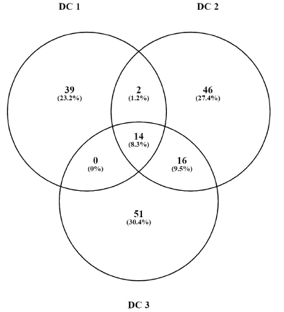
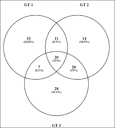
The procedure involved the precipitation of the protein from the samples, followed by its quantitation using the Bradford reagent using the ELISA plate reader. The precipitated proteins were digested using the ProteaseMAXTM surfactant, trypsin, and iodoacetamide in the NH4HCO3 buffer solution. In the last step, excessive protein digestion was stopped using formic acid. The mixture was then centrifuged at 12,000 rpm at 4°C for 20 min. The supernatant was thereafter collected from the tubes in LC-MS vials. The samples were analyzed using the UHD Accurate Mass 6538 Q-TOF LCMS system (Agilent Technologies, CA, USA) with a Zorbax extend C-18 analytical column (4.6 × 150 mm2 particle size, 5 µm) and a mobile phase made of water, acetonitrile, and formic acid (H2O:ACN:FA = 90:9.9:0.1). The data obtained after analysis using Agilent Spectrum Mill MS Proteomics Workbench LC-MS software revealed the m/z ratio, abundance, and Uniprot ID of detected proteins. Proteins with more than three distinct peptides and significant abundance (more than 105 Da) were considered. The Venn diagram of commonly expressed proteins is shown in Figures 1–3. The proteins that were commonly expressed were identified and their abundance was compared using the paired t-test. Possible protein–protein interaction was studied using a string database related to text-mining, experiments, databases, co-expression, neighborhood gene fusion, and co-occurrence. 28
Proteins Commonly Expressed in all Three Animals in DC.
Proteins Commonly Expressed in all Three Animals in GT.
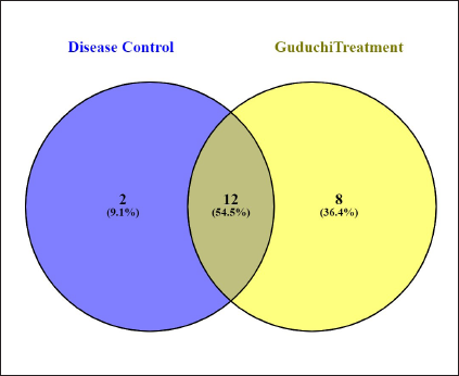
Proteins Commonly Expressed in DC and GT.
Statistical Analysis
Groupwise means of weekly absolute body weights, body weight gain, feed consumption, absolute organ weights, relative organ weights to body weight, antioxidant assay, and AChE assay were statistically analyzed by applying one-way analysis of variance (ANOVA) using SPSS 16.0 software. Elevated plus maze test data were analyzed by two-way ANOVA, followed by Bonferroni’s test, and the significance level was determined within and between groups using Graph Pad Prism V5.0 software.
Histopathology
Brain, kidney, and liver samples were trimmed, processed in ascending grades of alcohol and xylene, embedded in paraffin blocks, sectioned, and stained with routine hematoxylin and eosin, and subjected to histopathology evaluation.
Results
Crude Drug and Extract
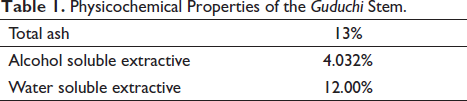
Physico-chemical properties such as a loss on drying, ash value, and extractive values of the Guduchi stem were within limits as described in the Ayurvedic Pharmacopoeia of India, 15 (Table 1), which confirmed the quality of the crude drug. The extraction yield was found to be 12.99%. The extract was dark brown with solid consistency. The results of phytochemical screening of Guduchi extract showed the remarkable presence of carbohydrates, glycosides, alkaloids, phenolics, and flavonoids in the aqueous extract (Table 2).
Physico-chemical Properties of the Guduchi Stem.
Phytochemical Properties of the Guduchi Extract.
LC-MS Analysis of Extracts
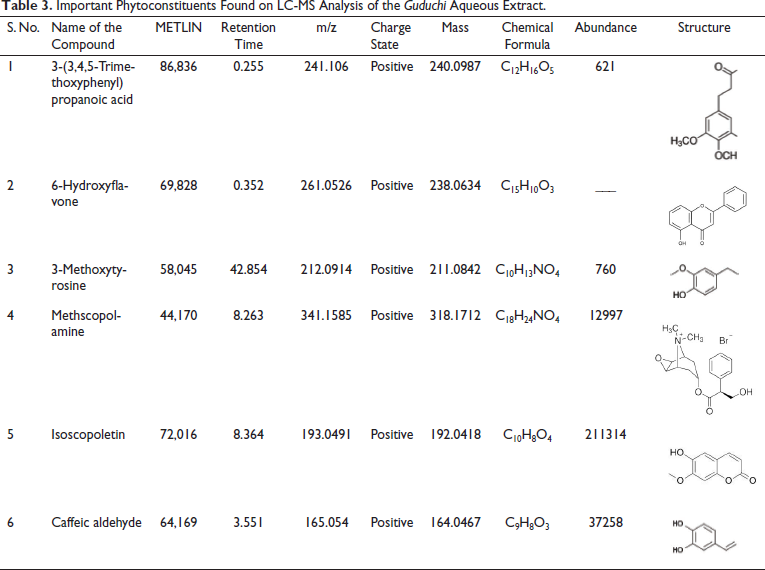
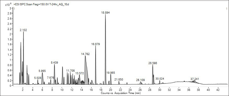
The LC-MS chromatogram of the aqueous extract of the Guduchi stem is represented in Figure 4. LC-MS analysis revealed the identification of six important phytoconstituents in the extract after integration with the METLIN metabolite PCDL library, 29 as shown in Table 3.
Important Phytoconstituents Found on LC-MS Analysis of the Guduchi Aqueous Extract.
Chromatogram of LC-MS Analysis of the Guduchi Aqueous Extract.
Clinical Signs, Mortality, and Other In-life Phase Observations
The cage-side observation showed no significant change in the clinical signs of the animals across the groups. No significant change in feed consumption and the body weight gain was found between the groups.
Elevated Plus Maze
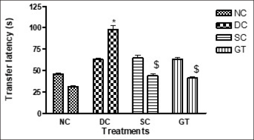
In the EPM test, ITL on day 20 was significantly increased in the DC group compared with other groups, including the NC group. Furthermore, the ITL in the SC and GT groups was comparable with the DC group. However, on day 42, RTL in the SC and GT groups was significantly reduced compared with the DC group. Therefore, the treatment with the Guduchi extract showed comparable results to that of donepezil hydrochloride treatment in improving memory (Figure 5).
Elevated Plus Maze Test.
Biochemical Assessments
AChE Assay
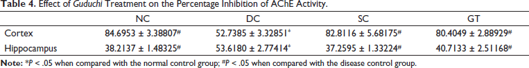
Cortical inhibition of AChE in the DC group was significantly lower than in other groups, including the NC group. Therefore, treatments in the SC and GT groups induced significant inhibition of cortical AChE in comparison with that in the DC group. Moreover, the AChE inhibition in the GT and SC groups was comparable with that in the NC group. The hippocampal AChE inhibition in the DC group was significantly higher than that in other groups, including the NC group. Thus, treatment with donepezil in the SC group and with the Guduchi extract in the GT group induced significantly more activity of hippocampal AChE than that in the DC group (Table 4).
Effect of Guduchi Treatment on the Percentage Inhibition of AChE Activity.
Oxidative Stress Assessment
The cortical and hippocampal MDA production in the DC group was significantly higher than in other groups, including the NC group. Thus, the treatment in the SC and GT groups led to a significantly decreased production of MDA in the cortex and hippocampus compared with that in DC group (Table 5).
Effect of Guduchi Treatment on Oxidative Stress Parameters in the Cortex.
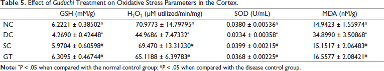
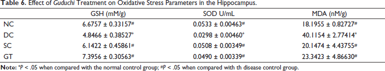
The cortical and hippocampal GSH and SOD concentration in the DC group were significantly lower than that in other groups, including the NC group. Thus, the treatment in the SC and GT groups led to a significant increase in the concentration of GSH and SOD in the cortex and hippocampus compared with that in the DC group. Similarly, the H2O2 concentration was found to be increased in the cortex in the SC and GT groups, which can be attributed to the treatment (Table 6). Overall oxidative stress was found to be reduced in rats treated with donepezil (SC) and Guduchi (GT).
Effect of Guduchi Treatment on Oxidative Stress Parameters in the Hippocampus.
Histopathology
Histopathological evaluation of the brain, kidney, and liver revealed that no aluminum chloride treatment was performed in the DC group compared with the NC group. No histomorphological changes were observed in the SC and GT groups as well. The results were consistent with earlier reports.30, 31
Proteomic Analysis
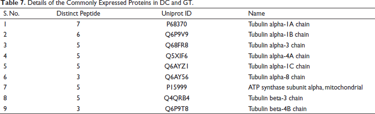
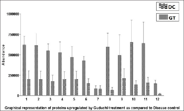
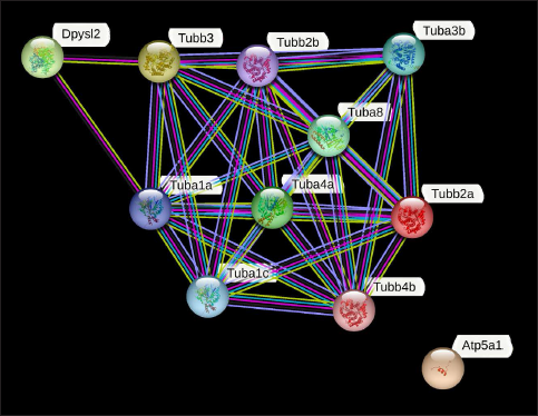
The data showed the expression of 12 proteins, which were commonly expressed in the DC and GT groups. The details of the proteins are given in Table 7. The groupwise abundance of proteins and their graphical representation are presented in Figure 6, which shows the upregulation in the GT. Both α and β tubulins were the major proteins expressed. Furthermore, protein–protein interaction was predicted using the string database that indicated that proteins are highly interrelated and co-expressed. 28 The string plot is presented in Figure 7.
Details of the Commonly Expressed Proteins in DC and GT.
Graphical Representation of Abundance of Commonly Expressed Proteins in DC and GT.
String Plot of Proteins Commonly Expressed in DC and GT Indicating that Proteins are Highly Interrelated.
Discussion
In vivo neuroprotective activity of aqueous extract of Guduchi was explored using the aluminum chloride-induced neurotoxicity model in male Wistar Rats. The aqueous extracts of Guduchi at the dose of 200 mg/kg body weight were administered to the rats for 42 days. It was followed by memory assessment using the Elevated plus maze test and sacrifice of the animals to collect the brain for antioxidant activity assay, AChE inhibition assay, and histopathology evaluation.
The results of qualitative and quantitative phytochemical analysis of the Guduchi stem were in line with the standards mentioned in the Ayurvedic Pharmacopoeia of India, thus confirming the quality standard of the crude drug Guduchi stem and its extract. Additionally, the preliminary signature data for LC-MS analysis of the aqueous was generated, which needs to be further validated. Earlier, the LC-MS analysis of alcoholic extracts Tinospora cordifolia was performed.32–34 However, the above-said phytochemical analysis of the aqueous extract of Guduchi stem prepared by the ayurvedic method should serve as baseline data for future validation and standardization studies. The phytochemical studies of the extract revealed the presence of various compounds known to affect the functions of the brain through different mechanisms. 6-Hydroxyflavone, a flavonoid found in GT, is reported to have anxiolytic-like effect in the elevated plus-maze test in mice. 35 Interestingly, another compound found in GT, 3-methoxytyrosine is a metabolite of L-DOPA, a well-known drug used to treat Parkinson’s disease. 36 Methscopolamine is a well-known anti-ACh drug, 37 whereas isoscopoletin belongs to a group of coumarin compounds that have shown anti-Alzheimer’s disease activity in molecular docking studies. 38 Caffeic aldehyde has reportedly reduced neurodegeneration and improved locomotor activity in an experimental study in mice. 39 Hence, the amelioration of the AlCl3-induced memory loss by the GT may be attributed to the presence of neuromodulatory phytochemicals.
Acetylcholine is the principal neurotransmitter in the nervous system, which plays a vital role in various functions such as memory, learning, and coordination of motor activity. 40 The marked decrease in the level of ACh is an important pathophysiological change observed in various neurodegenerative diseases such as Alzheimer’s disease and Parkinson’s disease.2, 3 High levels of aluminium have been reported in the brains of AD patients.3, 7, 41, 42 Aluminum is acetylcholinotoxic in experimental animals and causes apoptotic loss of neurons, eventually resulting in AD. 43 The decrease in the level of ACh is also associated with damage to the cholinergic neurons, possibly due to increased oxidative stress in the brain.3, 44 Thioacetylcholine is hydrolyzed by the enzyme AChE, which stops the transmission of nerve impulses. In diseases like Alzheimer’s disease, where large amounts of AChE are seen in AD patients’ brains, the degree of memory loss is significantly correlated with the decline of cholinergic transmission. 14 In the present study, a significant increase in AChE activity due to its lower inhibition in the cortex was observed in the Aluminum chloride-treated group compared to the NC group, which is in line with published literature. 14 The present study also showed the significantly decreased activity of hippocampal AChE due to higher inhibition in the Aluminum chloride treated group when compared to the NC group. The results were found to be as per the literature published. 45 Treatment with Guduchi extract in the present study showed significantly decreased AChE activity in the cerebral cortex compared to DC and similar to NC and SC, which is in line with the earlier published reports. 46 Agarwal et al. have suggested that the Guduchi extract treatment in rats leads to cognitive enhancement by increasing the availability of ACh. 11 Treatment with Guduchi showed significantly increased AChE activity in the hippocampus compared to DC, which may be attributed to the reversal of AlCl3-induced changes in the hippocampus. 45
EPM test assesses memory-related behavior by measuring transfer latency from open arm to close.20, 47 The aluminum chloride-induced neurotoxicity leads to progressive memory loss, which is reflected in the increased RTL compared to the ITL, as rats could not memorize and recollect the path on the maze. The present study showed that AlCl3 administration to the rats resulted in progressive deterioration of memory and consequent neurobehavioral deficits. Therefore, rats of the DC group showed an increase in RTL compared to ITL and a significant increase in RTL compared to NC. Therefore, the results of the EPM test in the neurotoxicity DC group show that the model was established as reported in earlier reports.14, 20, 30 Treatment with Guduchi showed a significant decrease in RTL compared to the DC. This data indicates a significant memory improvement and cognitive behaviour of rats compared to aluminum chloride treated groups. The published literature substantiates the finding. As per the classic Ayurveda texts, the juice of the Guduchi is used as Medhya, i.e., which may be implied to cause improvement in memory and intellect. 9 Further, as per the published literature, aqueous extract of Guduchi at the dose of 100 mg/kg for 15 days enhances memory and cognition in cyclosporin-treated experimental rats 11 Guduchi extract has also shown significant cognition improvement when administered at 200 mg/kg in experimental rats. 12 A study on acute sleep-deprived Wistar rats has shown that treatment of ethanolic extract of the Tinospora cordifolia ameliorates anxiety and cognition. 13 Guduchi choorna (whole plant powder) was one of the Medhya rasayanas used in a clinical study which has shown a significant effect on improvement in memory of human subjects. 48 A randomized, double-blind placebo control study has shown that Tinospora cordifolia treatment in combination with yoga helps to reduce mental stress in patients. 49
A decrease in catalase, SOD, GSH, and an increase in MDA/LPO level, as well as neuronal damage in the cortex and hippocampus regions of the brain, were all signs indicating a considerable increase in oxidative stress in the brain following aluminium chloride therapy in the current study. According to earlier research, aluminium stimulates and activates LPO when there is iron present in the brain. 50 Aluminium enhances iron-based oxidation in the brain, which alters iron homeostasis mainly via the Fenton reaction, increasing iron-induced oxidative injury. 51 Aluminium is reportedly responsible for modulation in brain amyloidosis through oxidative damage. 52 An increase in oxidative stress due to aluminium chloride treatment in rats leads to exhaustion of the body’s various endogenous antioxidant enzymes such as catalase, SOD, and GSH resulting in their decreased concentrations in the tissues. 53 Thus, the findings of antioxidant assays in DC are as per the reported literature. In the present study, treatment with an aqueous extract of Guduchi significantly reduced oxidative stress which was reflected in increased catalase, SOD, and GSH levels and decreased MDA/LPO levels in the hippocampus and cortex region compared to aluminium chloride-treated DC Group. The results of the Guduchi treatment on oxidative stress are in line with published literature.10, 13, 54, 55
These results of the proteomic study revealed the increased expression of tubulin proteins in GT, which are involved in the generation, migration, and differentiation of neurons. 56 α and β–tubulins are reported to play a significant role in the survival and positioning of neurons. 57 α- and β-tubulin heterodimers form microtubules which are required for axonal transport of molecular motors in neurons. 58 Yashtimadhu is one of the Medhya drugs, whose root extract treatment, on IMR 32 cell lines, has revealed that it ameliorates the rotenone-induced apoptosis and hyperphosphorylation of ERK-1/2 by preventing mitochondrial oxidative stress and apoptosis. 59 However, perusal of literature shows that there is a scarcity of data regarding neuronal protein expression due to Guduchi extract treatment. Thus, the treatment with Guduchi aqueous extract has shown its effect on multiple parameters such as modulation of the inhibitory activity, decreased oxidative stress, and increased expression of tubulin proteins which might have led to the amelioration of memory loss caused by the AlCl3 neurotoxicity. However, the data obtained are preliminary, and therefore further detailed investigations are required to explore the precise role of the different proteins and their possible neuroprotective action in the AlCl3-induced memory loss model in the presence of Guduchi treatment.
The concept of Medhya has a broad understanding in an ayurvedic context. According to the classical ayurvedic texts of “Charaka Samhita” the Medhya activity promotes intellect (Dhi) retention power (Dhriti) and memory (Smriti). 9 In the present study, the functional changes exhibited by rats treated with the Guduchi extract show an improvement in memory and cognition which are consistent with the concept of Medhya in reference to intellect (Dhi) retention power (Dhriti) and memory (Smriti).
Conclusion
The results suggest that Guduchi aqueous extract exhibited amelioration of AlCl3 induced neurotoxicity in rats by the neuroprotective action, which may be attributed to the prevention of oxidative damage to brain tissue and modulation of AChE activity in the cerebrum and hippocampus. Moreover, the role of the expression of the tubulin proteins in neuroprotection needs to be validated. The observed beneficial biochemical changes and memory improvement substantiates the Medhya property of the Guduchi in the context of Ayurveda.
Summary
The present article reports the study of an aqueous extract of the Guduchi (Tinospora cordifolia) stem that is an ayurvedic preparation, on experimental model of memory loss in rats. The drug is used in the ayurvedic system of medicine, which is an Indian system of traditional medicine. According to Ayurveda, the drug has Medhya properties that also lead to memory improvement. The present study showed the beneficial effect of the Guduchi extract in the experimental model of dementia. The findings are helpful to validate the drug properties mentioned in the Ayurveda and will be beneficial in promoting the test drug Guduchi as complementary treatment in modern therapies used to manage the dementia.
Footnotes
Acknowledgement
The authors are thankful to the Director General, Central Council for Research in Ayurvedic Sciences, New Delhi for the funding support.
Declaration of Conflicting Interests
The authors declared no potential conflicts of interest with respect to the research, authorship and/or publication of this article.
Ethical Statement
The approval for the conduct of the animal experimentation study was obtained from the Institutional Animal Ethics Committee vide approval number IAEC/2019_3/05 (dated: October 15, 2019).
Funding
The study was carried out under the Intra Mural Research Scheme of the Central Council for Research in Ayurvedic Sciences, New Delhi vide project sanction no. 3-110//2019-CCRAS/Admin./IMR4/3750 (dated: September 30, 2019).