
Abstract
Biological sex determination is challenging in forensic sciences, especially if the traditional dental measures are not present due to loss of teeth or damage. Intercanine and intermolar measurements have been widely used, but are limited in such a context. This cross-sectional study was aimed at evaluating the validity of intra-incisor-canine width as a sex identifying instrument, as well as to test its reliability in association with intercanine and intermolar width. Sixty adults (30 men, 30 women; mean age 21.5 ± 2.1 years) with Angle Class I occlusion were included in the sample. Maxillary dental impressions and bite marks were obtained. Traditional intercanine and intermolar widths, along with those of the new bilateral incisor-to-canine distances, were measured using standard odontometric measures. Statistical tests involving Student’s t-tests, Pearson correlations, and logistic regression were used. There was significant sexual dimorphism in intermolar width, both in dental casts (p = .009). Maxillary incisors to canines showed a significant difference between sexes bilaterally in dental casts and in bite marks (p < .001). Binary logistic regression analysis showed that the odds of being male increased by 2.67 times (95% CI: 1.41–5.05) for every 1 mm increase in incisor-to-canine width, with a classification accuracy of 91.7% for sex determination. The incisor-to-canine width of the maxillary arch can be an adjunctive method to sex identification in forensic dentistry when the traditional dental parametric measurements are not available.
Introduction
Forensic odontology is an expert branch of the science of dentistry that overlaps with legal investigation. 1 It provides essential instruments used in the identification of human beings in cases where other traditional processes have been unsuccessful. 1 The modern forensic practice can be defined by the progressively increasing complexity of criminal cases, thus the need to have powerful, scientifically proven methodologies to study the evidence.1, 2 The process of identifying sex based on human remains is especially tedious where skeletal remains are incomplete, highly decayed, mutilated, or fire-damaged, a situation which is often experienced during mass disasters, murders, and sexual assaults.1, 2
Bite-mark evidence is a major part of investigating sexual assaults to identify the perpetrator.2, 3 According to MacDonald, bite marks are classified as marks made by teeth, either singly or in combination with other oral structures, and this is why there is a broad range of dental trauma patterns that are observed in the forensic setting. 2 The significant importance of bite-mark analysis in acquiring justice on behalf of victims can be highlighted by the landmark Ted Bundy case (1946–1989) 2 and the Delhi Nirbhaya rape case reviewed by Dr. Ashith Acharya. 3
The conventional odontometric methods of sex determination are mainly based on two known parameters, which are intercanine width and intermolar width. 4 Intercanine width is considered the linear difference between opposite canine cusp tips, which in males tend to be larger than those in females, with reported values ranging around 6 mm versus 4–4.5 mm in some populations. 4 Intermolar width is the linear distance between the mesiobuccal cusp of the first permanent molars.4, 5 These conventional methods pose major limitations to their use in forensics, where loss or damage of teeth could result as a consequence of extraction, trauma, or disease to the canines or molars. Also, post-mortem alterations or decomposition, and undeveloped dental material may further deform dental structures, which may not allow precise measurements.1, 4
Sexual dimorphism is widely explored, and previous studies have demonstrated parameters such as canine indices and intercanine and intermolar arch width in Indian populations.6, 7 However, most existing studies are largely based on conventional odontometric parameters involving canines and molars, with limited evaluation of alternative maxillary measurements that may be useful when these teeth are absent or damaged.
This study tested the effectiveness of maxillary incisor-to-canine width in sex identification, as well as to check the reliability of this new parameter against known odontometric ones. It analyzed the relationship between dental casts and bite-mark measurements and came up with predictive models to facilitate forensic sex classification.
Objectives
To evaluate the effectiveness of maxillary intra-incisor–canine width as a parameter for sex determination in young adults.
To compare maxillary intra-incisor–canine width measurements between males and females using dental casts and bite-mark analysis.
To assess the reliability of the proposed parameter in comparison with conventional odontometric measurements such as intercanine and intermolar widths.
Materials and Methods
Study Design and Ethical Considerations
This cross-sectional analytical study was conducted following approval from the Institutional Ethics Committee (Approval No.234).
Sample Size Calculation and Participant Selection
The sample size was estimated using G* power with the help of previous odontometric studies that reported a large effect size (Cohen’s d = 0.8) for sex differences in measuring dental measurements. With an alpha = 0.05, power = 0.80, the minimum sample size was estimated to be 26. To allow for errors and dropouts, 30 males and 30 females were selected for each group.
Inclusion Criteria
The participants in the study were aged between 18 and 25 years (to maintain full dental status and irrelevant age change), demonstrated Angle Class I molar relationship (natural occlusion) and had complete permanent dentition in the anterior area with no spacing or crowding in the anterior teeth of the maxillary arches.
Exclusion Criteria
The individuals who had been receiving or had previously received orthodontic treatment and those who had removable prosthetic restoration or artificial crowns, had no maxillary incisors, no canines, or first molars, rotated anterior teeth, and had an Angle Class II or III malocclusion were rejected from the study.
Data Collection Procedures
Bite-mark Registration
Contributors were asked to bite uniformly against modeling wax sheets in order to make clear impressions of maxillary teeth. The bite registration protocol achieved uniform bite pressure among the subjects, full coverage of the morphology of teeth in the anterior region, and little distortion by the impression process.
Dental Cast Preparation
Alginate impression material (Tropicalgin, Zhermack, Italy) was used to make maxillary impressions as per the directions. Dental stone casts (Type III, Kalstone, Kalabhai, India) were poured within 30 minutes of the impression taken in order to reduce dimensional alteration.
Measurement Protocols
Reference Points and Definitions
Intermolar width
Definition: Linear distance between mesiobuccal cusp tips of maxillary first permanent molars
Measured by a digital vernier caliper (Mitutoyo, Japan; accuracy ± 0.01mm) on both casts and bite marks.
Intercanine width
Definition: Linear distance between cusp tips of maxillary canines
Measured by a Digital vernier caliper on both casts and bite marks
Intra-incisor-canine width (novel parameter)
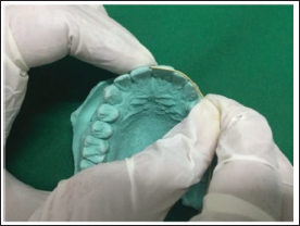
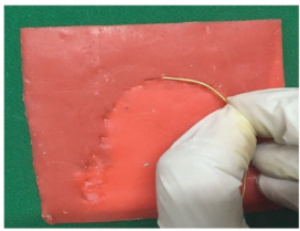
This parameter was defined as the linear distance from the mesial incisal edge of the maxillary central incisor to the cusp tip of the ipsilateral canine. A 0.7-mm brass wire was passively adapted along the curvature of the maxillary dental arch from the incisal edge of the central incisor to the canine cusp tip on each side. Care was taken to avoid stretching or compressing the wire during adaptation. The adapted wire was then carefully removed, fully straightened without distortion, and the linear length was measured using a digital vernier caliper (Mitutoyo, Japan; accuracy ± 0.01 mm). Measurements were recorded separately for the right and left sides on both dental casts and bite-mark impressions (Figures 1 and 2).
Adaptation of 0.7-mm Brass Wire Along the Maxillary Dental Arch From the Incisal Edge of the Central Incisor to the Canine Cusp Tip on a Dental Cast. The Adapted Wire Was Subsequently Removed, Straightened, and Measured Using a Digital Vernier Caliper.
Adaptation of 0.7-mm Brass Wire on a Maxillary Bitemark Impression for Recording Intra-incisor–Canine Distance. The Wire Was Then Straightened and Measured Using a Digital Vernier Caliper.
Observer Calibration and Reliability
All measurements were done by three experienced observers (two forensic odontologists and one oral pathologist), and they were carried out independently. Observers were introduced to calibration to give consistency in measurement with the use of practice casts before commencing data collection. To measure inter-observer reliability, Intraclass correlation coefficients (ICC) were used.
Statistical Analysis
SPSS version 28.0 (IBM Corp., Armonk, NY) was used to perform the data analysis. The analysis methodology included: Descriptive statistics (mean, standard deviation, and range of all measurements); testing of normality (Shapiro Wilk test); comparative analysis (independent samples t -test to compare cast and sex of the bite marks); correlation analysis (Pearson correlation values between cast and bite-mark measurements); reliability test (ICC to test inter -observer agreement); prediction model (binary logistic regression in classification of sex); and the effect size (Cohens d to estimate practical significance) were employed. p value less than .05 was considered statistically significant.
Results
Characteristics and Demographics of the Sample
There were 60 participants (30 men, 30 women), and their average age was 21.5 ± 2.1 years. An insignificant age difference (21.3 ± 2.0 years vs. 21.7 ± 2.2 years; p = .42) was found between sexes.
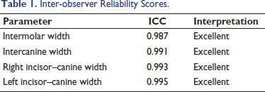
Inter-observer Reliability
All measurements displayed excellent inter-observer reliability with ICC on a scale of 0.987–0.995 (Table 1). Those values show almost absolute consensus among the observers and confirm the measurement procedures.
Inter-observer Reliability Scores.
Conventional Odontometric Parameters
Intermolar Width
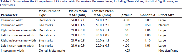
The intermolar width observed in males and females in dental casts was (mean ± SD) 54.0 ± 2.1mm and (mean ± SD) 52.0 ± 2.5 mm, respectively. The intermolar distance showed a statistically significant difference in males and females (p < .001). In bite-mark analysis, the intermolar width observed in males and females was 51.0 ± 1.6 mm and 50.0 ± 2.3 mm, respectively. A statistically significant difference in intermolar width between males and females was also observed in bite marks (p = .009).
Intercanine Width
The intercanine width observed in males and females in dental casts was (mean ± SD) Males 35.2 ± 2.3 mm and females 34.8 ± 2.1 mm. The bite marks showed: males 34.1 ± 2.0 mm, females 33.9 ± 1.9 mm. There were no significant sex differences in the intercanine width (p > .05).
Novel Incisor-to-canine Width Measurements
The width of the incisor-to-canine on dental casts varied significantly between both sexes. Males had a value of 21 ± 0.8 mm on the right maxillary arch than the 20 ± 1.1 mm in females (p = .001). In the maxillary arch on the left, the measurements were 21 ± 0.9 mm in males and 20 ± 0.9 mm in females (p = .001) (Table 2).
Summarizes the Comparison of Odontometric Parameters Between Sexes, Including Mean Values, Statistical Significance, and Effect Sizes.
Correlation Analysis
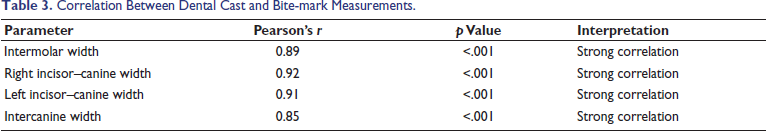
Bite-mark data turned out to be reliable, as strong positive correlations were found between similar dental cast and bite-mark measurements (Table 3).
Correlation Between Dental Cast and Bite-mark Measurements.
Predictive Modeling
Logistic Regression Analysis
Binary logistic regression models were built using significant odontometric parameters.
The combined model (Model 3) demonstrated the best classification performance with an AUC = 0.94 (95% CI = 0.87–1.00) and cross-validation accuracy of 88.3% (Table 4 and Figure 3).
Logistic Regression Models for Sex Prediction.
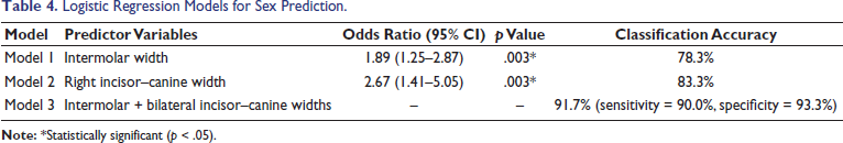
Logistic Regression Models for Sex Prediction.
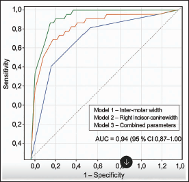
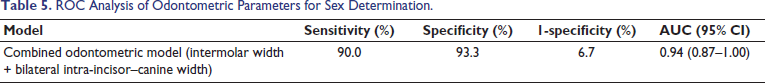
Table 5 summarizes the ROC curve analysis of the combined odontometric model, including sensitivity, specificity, and AUC values for sex determination.
ROC Analysis of Odontometric Parameters for Sex Determination.
Discussion
Our study indicates that maxillary incisor-to-canine width is an adjunct to sex determination in forensics. The major findings are high discriminatory power, and the indication of this is large effect sizes (Cohen’s d > 1.0) in sex differences. It also showed bilateral consistency, reflected by the similarities in the performance by right and left measurements; cross-method validity, represented by high correlations between cast measurements and bite-mark measurements; and improved accuracy, shown by the fact that combined parameters have over 90% classification accuracy.
Comparison with Existing Literature
Odontometric examination is viewed as the tool of identification; it is not very complicated, not very costly, and not too difficult to perform.8, 9 Dimorphism refers to the differences in size, appearance, and stature, and sexual dimorphism refers to the differences in the forms and sizes of teeth between the sexes.10, 11 Forensic dentists and anthropologists have long been fascinated by the assessment of sexual dimorphism of tooth size.
Research indicates that teeth are a good supplementary predictor of sex differences, but they cannot be utilized as the key predictor of sex. 12 It has been mentioned that the highest number of anatomical considerations must be applied in determining the sex of a skeleton. 13 The size of teeth and the parameters of the dental arch are different in individuals. Although there has already been much research on intercanine and intermolar widths, there is a lack of study on the quadrant-based odontometric analysis of sexual dimorphism. Canines are the strongest teeth in the body, especially mandibular ones, as they can hardly be pulled out, rarely become diseased, and many stay longer than any other teeth. 14
Navin Kumar and Nutan Tyagi reported that mandibular intercanine width shows clear sexual dimorphism and can be a reliable indicator of sex. 15 But our study shows that maxillary intercanine width is not sexually dimorphic. This also explains that sexual dimorphism is arch-dependent.
From a biological standpoint, combined odontometric measurements that include the size of the anterior teeth and the curvature of the maxillary arch may more accurately represent sexual differences than single linear measurements. The intra-incisor–canine width reflects the combined impact of incisor size, canine prominence, and arch width, all of which are affected by sex-related patterns of craniofacial growth. This combined effect could explain why the suggested measurement performed better in distinguishing sexes compared to using only the upper intercanine width, which did not show significant differences between sexes in this study.10, 12, 13
The Y chromosome is one of the chromosomes involved in the difference in expression of the amelogenin gene, and hence, this partially leads to the differences in the thickness of the enamel and the morphology of the crowns as observed between the two sexes. Androgens also have an effect on craniofacial development since they increase transverse arch expansion and tooth size in males. Furthermore, males and females follow different ontogenetic patterns in that they also differ in the timing and the duration of their dental and maxillofacial development. These developmental differences can enhance cumulative measures, which involve many dental elements, as in the case of intra-incisor-canine width. Another factor might be epigenetic regulation, which includes the interactions between genes and their environment that influence craniofacial development and which can be a contributory factor in population-specific expression of sexual dimorphism.16, 17
Males typically exhibit greater maxillary arch breadth and tooth crown dimensions, potentially amplifying cumulative linear measurements such as incisor–canine width.5, 12, 18 This may explain the stronger discriminatory performance of the proposed parameter compared to isolated tooth measurements.
In North India, a study conducted on 100 dental students aged 20–30 years used right and left maxillary canines and showed 64% (female) and 58% accuracy (male). Based on the presence of fragmented remains in mass disaster scenarios, the authors concluded that maxillary canines can be highly sexually dimorphic and thereby act as a secondary source of anthropometric sex identification. 11
Odontometric studies have emphasized that reliance on a single dental parameter may be limited in forensic sex determination, particularly when key teeth such as canines are absent due to trauma, disease, or post-mortem loss. In such situations, alternative measurements, including intermolar width, can serve as useful adjunctive parameters for sex assessment.12, 18 The current research supports these observations and further confirms the usefulness of intermolar width as a reliable indicator of sex.
Studies published in the Journal of Indian Academy of Forensic Medicine have demonstrated significant variation in intercanine and intermolar widths across different Indian regions, underscoring the need for region-specific standards. The findings of the present study align with previously reported Indian data, while extending the existing literature by proposing a novel maxillary intra-incisor–canine parameter that demonstrated superior discriminatory power in this cohort.5–7
Our study showed that the average maxillary intermolar width is higher in males compared to females. The observations agree with previous findings that have shown that males have broader teeth, arch widths, and greater spacing between upper and lower molars in comparison to their female counterparts.5, 18 The study applies only to the specific population and age group.
Clinical and Forensic Implications
Forensic Workflow Integration
The protocol of the proposed measurements can be incorporated into the current forensic odontology procedures. Primary measurements can be done by conventional intercanine and intermolar widths; the secondary can be done by bilateral incisor-to-canine widths in cases where the primary parameters are not available. Multiple parameter analysis can also be combined with several parameters, thus improving accuracy.
Study Limitations and Considerations
The sample demographics are limited to people between the ages of 18 and 25, which may not be very generalizable to the other age groups. A single population study might be insufficient to illustrate global differences. Since the sample size is less than 100, large samples would strengthen the research, and external validation needs to be done in separate populations. In craniofacial growth, genetic and epigenetic influences also contribute to odontometric variations. The accuracy of the classification used in this article is therefore only relevant when applied to a particular population and age group that is being studied, and its application outside this group cannot be done without external corroboration.
Methodological Considerations
In the case of incisor-to-canine, the method of using brass wire to measure is new, but there is a need to standardize the method across the laboratories in the field of forensics. Quality assurance and training protocols must be formulated to be implemented extensively.
Future Research Directions
Multi-population validation must be sought on a variety of ethnic and geographic backgrounds. The analysis of parameter stability in relation to age group and the evaluation of performance in impaired forensic samples should be expanded. The current investigation can also be extended to mandibular incisor-to-canine widths and combined with digital forensic odontology systems to enhance visibility.
Conclusions
This study demonstrates that the maxillary intra-incisor–canine width in dental casts and bite-mark analysis shows significant sexual dimorphism in young adults. This method may serve as an adjunctive tool when molars and canines are lost and where conventional measurements are not feasible. This study may serve as pilot data, and further studies on larger populations are needed to validate the findings.
Footnotes
Data Availability Statement
The datasets generated and analyzed during the current study are available from the corresponding author upon reasonable request, subject to Institutional Ethics Committee approval and participant privacy protection requirements.
Declaration of Conflicting Interests
The authors declared no potential conflicts of interest with respect to the research, authorship, and/or publication of this article.
Ethical Approval and Informed Consent
The cross-sectional study was conducted at Vinayaka Mission’s Sankarachariyar Dental College, after obtaining ethical clearance from the Institutional Ethical Committee of the institution (VMSDC/IEC/Approval No.234).
Funding
The authors disclosed receipt of the following financial support for the research, authorship, and/or publication of this article: The project received grant from ICMR STS 2022 (Short term studentship) program (Reference ID: 2022-02745).