
Abstract
Sex determination by morphological analyses of the mandible is especially important in forensic anthropology and bioarchaeology cases where the bones are still present in fragments. The present research utilises recent developments in geometric morphometrics (GMM) to examine the degree to which the mandible could be used as a tool for sex determination. Two-dimensional landmarks and semi-landmarks were plotted on 149 digital orthopantomograms of both genders between the age group of 18–50 years. Through a multivariate analytical and exploratory framework, incorporating coordinates-based method and supervised classificatory techniques, morphological changes were identified to be consistent with sex. Overall sex determination accuracy was found to be 89.3%, while the first 17 principal components (99% shape variance) yielded 93.0% accuracy. The accuracy for males was 89.3% Procrustes coordinates and 94% principal components, whereas for females was 89.2% Procrustes coordinates and 91.9% principal components. The findings of this research were highly optimistic, supporting previous studies, and suggest that this approach may be utilised to assess sexual dimorphism. The approach of geometric morphometric method proves to be a very useful tool to determine sexual dimorphism.
Keywords
Introduction
Sex determination is an integral aspect of forensic and bioarchaeological sciences since the corresponding methods for estimating age and stature are often sex specific. 1 In general, traditional morphometric methods, incorporating the analysis of linear measurements, in combination with other forms of osteological data, are used to determine sex. Sex estimation is more accurate when a complete skeleton is present, but such complete specimens are difficult to procure, particularly in cases of mass disasters and crimes. Among all the bones used to determine sexual dimorphism, the pelvis, femur, humerus and skull are usually considered.2–4 The human mandible can also exhibit the most anatomical variation in size during development, especially in the mandibular condyle and ramus. 5 Mandibular ramus measurements tend to demonstrate a greater degree of sexual dimorphism, with variations becoming more pronounced as compared to the body of mandible. 6
A broad range of work has recently utilised Geometric morphometric (GMM) approaches to address problems of biological recognition in the area of forensic and bioarchaeological sciences.7–9 GMM is the statistical analysis of shape and form as prescribed by two- and three-dimensional Cartesian landmark coordinates. 10 Central to GMM are the placement of homologous landmarks on specific locations on specimens that have structural, functional, evolutionary or developmental significance, which facilitates the examination between specimens. Through developments in computer technology, and the integration of a multivariate statistical framework, the analysis of shape geometry in biological studies is now possible.11–14
In the present work, we have developed a responsive and accurate tool for investigating shape variation in mandibular corpus anatomy using a GMM framework. The study is the first of its kind to use data from India’s population and digital orthopantomograph (OPG) data to investigate sexual dimorphism.
Material and Methods
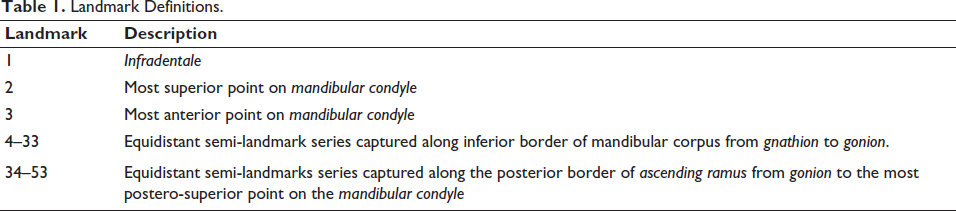
The present study is a retrospective quantitative analytical study wherein 149 digital orthopantomogram images (75 males and 74 females) between the age group of 18 and 50 years were collected. Due ethical approval (Ref.: NFSU/IEC/SFS/12/2020) was obtained from the institution. The radiographs were obtained from the diagnostic imaging centre, in the city of Ahmedabad, Gujarat, India. The parameters were as follows: Tube Potential: 66–85 kV, Tube Current: 6–10 mA, Total Filtration: 2.8 mm, time: 12 sec. Only radiographs with complete natural dentition were included in the study, whereas distorted OPGs, OPG with pathology, jaw discrepancies, or malocclusion (Class II/Class III) and prosthesis were excluded from the study. Three Type I and Type III two-dimensional landmarks (Table 1) were digitised along the symphysis and condylar region using the Thin Plate Spline (TPS) suite followed by Morpho J. The TPS software allows statistical analysis using partial wrap scores as shape variables, thereby expressing morphometric analysis as a shape deformation. The TPS suite is useful for digitising landmarks and outlines for GMM analyses. Fifty resampled equidistant sliding semilandmarks were positioned around the mandible’s inferior border and the ascending ramus’s posterior border. Semilandmarks were anchored anteriorly and posteriorly to fixed homologous points. Landmarks were mirrored to the opposite side, resulting in a total of 299 configurations, all of 53 landmarks.
Landmark Definitions.
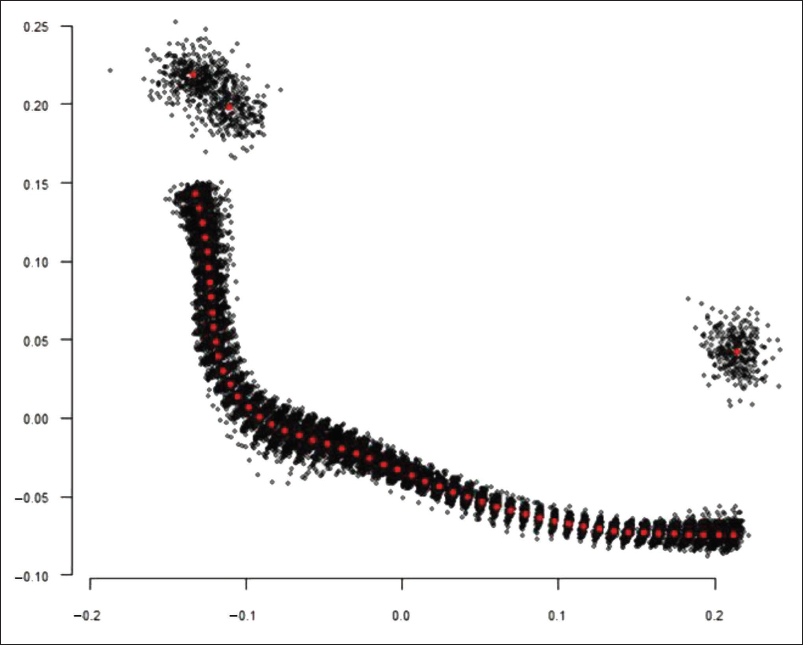
In order to extract shape variables from the digitised images, a generalised Procrustes analysis was performed.12, 14–17 Through this procedure, all specimens were converted to a common origin, scaled to unit-centroid size, and through a least-squares criterion were optimally rotated until all coordinates were aligned as closely as possible (Figure 1). A total of 36 iterations were necessary until maximum convergence was obtained (see R Script). Through this three-fold procedure, the subsequent oriented Procrustes coordinates reflect the form of each mandible.
The primary causes of shape difference between male and female mandibles were then investigated using principal component analysis (PCA). The percentage variation, along each axis was noted through a screen plot, with relative positions in the morphospace representing the range of variation within the dataset; clustering was mapped through confidence ellipses (set here to 66%). Mean shapes for each sex were also visualised. In order to explore if the mandibular shape could be successfully discriminated, a linear discriminant analysis (LDA) was performed. LDAs were performed with both the Procrustes coordinates and principal component scores examination, with leave-one-out cross-validation (jackknifing).
As an add-on to the exploratory procedure, the Procrustes coordinates were examined through Null Hypothesis Significance Testing (NHST). This was conducted through a Procrustes ANOVA, 18 with the sum-of-squares calculated through 1,000 permutations of the Procrustes process. With the sample size and statistical power, an alpha level of 0.01 (significance level of 1%) was deemed appropriate, with a null hypothesis (H0) of no difference between sex assumed.
All exploratory and analytical procedures were produced in the R Environment,
19
using both the geomorphv.3.0.7
20
and Momocs v.1.2.9
21
packages. The tidyverse
22
and extrafont packages
23
were also used for data visualisation purposes. All data, coding and metadata can be found attached with this article. Additionally, a copy of all files can be located on GitHub (
Result
The GMM is a technique to study scale and shape relationships of structures using Cartesian geometric coordinates rather than linear, areal (of area), or volumetric variables. GMM has been of great value in many biological studies, but does not appear to have been used to examine skeletal morphological variations in dentistry.
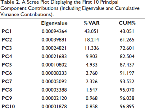
In this study, a total of 102 principal components were obtained (2k – 4 = 102 shape variables) which allowed to view for the overall shape variation in the mandibular morphology. The initial 10 principal components accounted for over 96% of shape variation in the sample, with the first eight components accounting for 95% cumulative shape variance (Table 2).
A Scree Plot Displaying the First 10 Principal Component Contributions (Including Eigenvalue and Cumulative Variance Contributions).
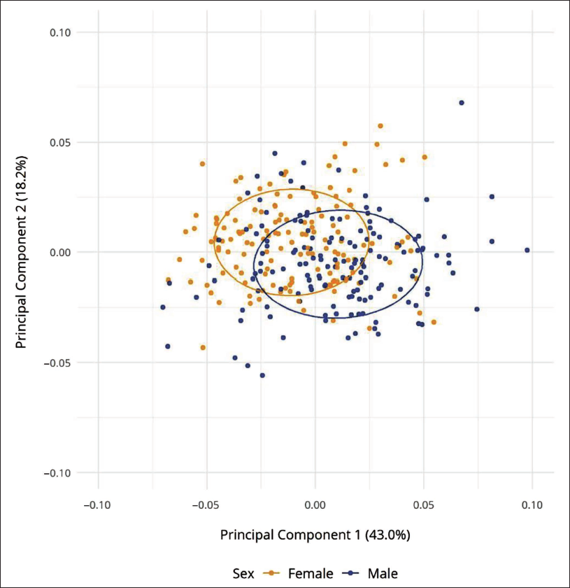
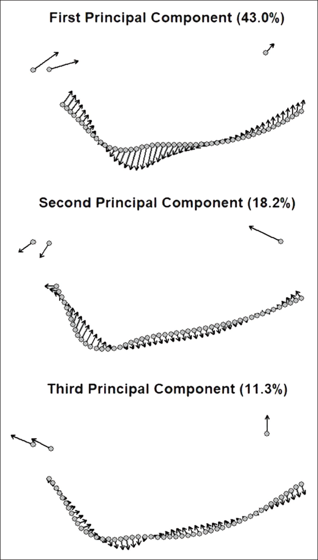
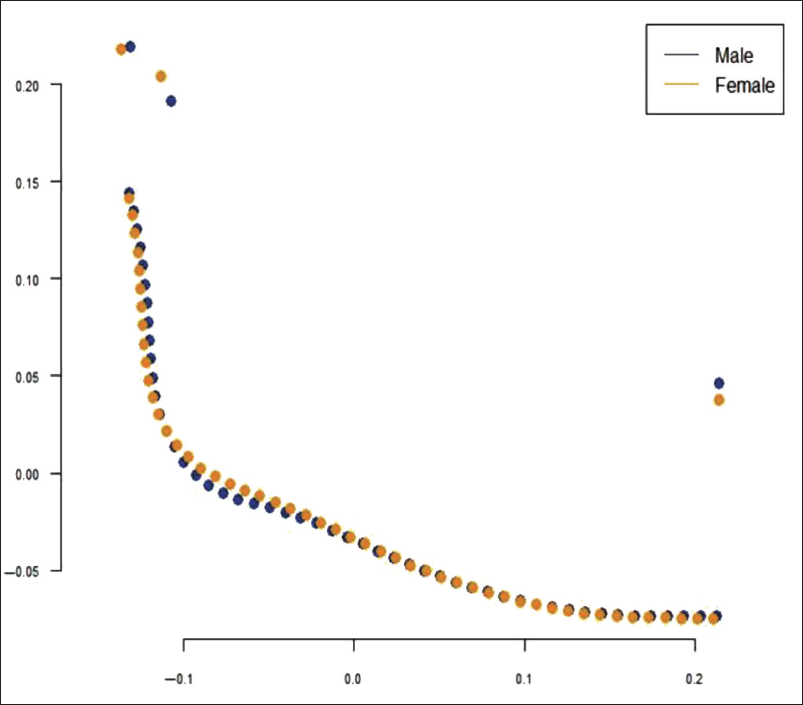
The global shape variation of principal components 1 and 2 by the plot and shape variance across the ramus and corpus is seen in Figures 2 and 3, respectively. Through the plots it is clearly visible that the variation is primarily manifested through the following aspects:
PC1: The height of ascending ramus and placement of the inferior border of the symphysis shows a considerable size difference. PC2: Gonial angle depicts high differential shape pattern compared to symphysis and ascending ramus. PC3: Evidently shows flexure of the inferior corpus along with symphysis projection.
A Principal Component Analysis (PCA) Bivariate Plot on the Covariance Matrix of k = 53 Landmarks (Cumulative Variance Explained here: 61.2%).
The Mandibular Configurations for the First Three Principal Components (Cumulative Variance Explained here: 72.5%).
Mean shape configurations (Figure 4) highlight differences in male and female mandibular profiles with varying mandibular angles, condyle points and infradentale.
Mean Shape Mandible Configurations for Indian Male and Female Populations.
A leave-one-out cross-validation of the Procrustes coordinates for k = 53 landmarks yielded a correction classification percentage of 89.3% (267/299), while a comparative analysis of the first 17 principal components (99% shape variance) yielded a correction classification percentage of 93.0% (278/299)—see the R script for classification metrics. In both instances, a higher-class accuracy for males (Procrustes coordinates: 89.3%; principal components: 94%) and lower-class accuracy for females (Procrustes coordinates: 89.2%; principal components: 91.9%) were recorded. A Procrustes ANOVA of shape residuals indicated that a null hypothesis of same populations—between male and female mandible shapes could be rejected (F: 24.85, P : .001). Predetermined fixed landmarks marked followed by equidistant semi landmarks gives maximum accuracy of the specified coordinates, as it is a technique to study scale and shape relationships of structures using Cartesian geometric coordinates rather than linear or volumetric variables.
Discussion
The human mandible is considered as the strongest bone in the facial area, and it is highly resistant to physical, chemical, and mechanical stresses, as well as being well preserved over time. This study is the first of its kind to use an Indian population and a novel approach in analysing human variation using a GMM approach with orthopantomogram images. Panoramic X-rays are most widely used to study the mandible and maxilla, which are acquired simultaneously in a single image. The current study aimed to introduce a more comprehensive method of determining sex during the human identification process, especially in the field of forensic dental radiology. Using GMM techniques, orthopantomogram enables accurate and reproducible selection of 2D images for the study of human variability.
In our study, k = 53 landmarks provided classificatory rates of 89%–93% with similar percentages for individual sexes. These results were highly comparable to the study performed by Oettlé et al. 24 from adult mandible based on ramus flexure with conclusive classification of Male: 67.8% Female: 69.9% Overall: 68.9%. A study conducted by Franklin et al. 25 on sub-adult mandible identified 55% male and 65% female and also resulted in the overall 59% with K = 21 landmarks. A similar study by Franklin et al. 25 on linear discriminant functions attained overall accuracy to be 84%, whereas for determining male and female, the accuracy was 83.3% and 84.8%, respectively. Oettlé et al. 8 in another study, especially on gonial eversion concluded overall accuracy to be 72.7% with sex determination to be 73.9% for male and 71.4% for female. Emilio et al. 14 in their study, analysed k = 53 on an OPG to obtain assessment of adult mandible based on GMM analysis of outline of inferior corpus and posterior ascending ramus with classification success of 91.0% for male and 94.0% for female; overall accuracy was 92.5%. Thus, the results were comparable to the results of the present study that was done in the Indian population.
These studies demonstrated that the adult mandible provides promising results in sexual dimorphism.26, 27 It is convenient to obtain a cluster of data across age and sex than what is available from dry bone specimens, thus can definitely be used in future studies as it is a source of biological data of great utility for research purposes. A high degree of anatomical modularity between the corpus and ramus suggests that functional ties exist between the units, and this might vary, influencing sex based morphology. The higher classification success of this proof-of-concept study has encouraged us to expand the arena of the research project, to various population-level shape differences, within group and pooled studies. In future, as a prospective study, the applicability and reliability shall be further verified on dry bone specimens too and also by incorporating other radiological parameters. Moreover, validation on three-dimensional models would be preferable to examine the model accuracy on novel data. 28 Also, accuracy rate can be increased by incorporating machine learning with such studies.
Conclusion
The results of this study may provide insight on the usefulness of OPG for sexual dimorphism in the dentate mandible, the study being first of its kind in the Indian population to analyse sexual dimorphism using GMM in OPG images, which can further be used in multivariate analysis. Furthermore, to increase the predictive accuracy and the validity of the concept established, dry bone specimens can be used in future studies.
Footnotes
Declaration of Conflicting Interests
The authors declared no potential conflicts of interest with respect to the research, authorship and/or publication of this article.
Ethical Approval
All procedures followed were in accordance with the ethical standards of the responsible committee on human experimentation (institutional and national) and with the Helsinki Declaration of 1964 and later versions.
Funding
The authors received no financial support for the research, authorship and/or publication of this article.
Informed Consent
The need for consent was waived by the institutional ethical board, since the study was a retrospective type of study.