
Abstract
Sex identification is a crucial factor in both forensic investigations and medico-legal cases, with age determination following closely. When soft tissues are missing, skeletal features are analysed. If the skull is incomplete, the mandible helps determine sex. Numerous studies have demonstrated the accuracy of panoramic radiographs in assessing anatomical measurements for sex determination. This current study investigates the use of digital orthopantomographs to analyse mandibular parameters for sex prediction specifically within the Maharashtra population. The objective is to assess the importance of the mandible in sex determination which will be achieved by analysing different morphometric parameters observed on panoramic radiographs, with the aim of identifying the most reliable predictor for sex determination. In this retrospective study, a total of 200 digital orthopantomographs (100 male and 100 female) were analysed. The collected data were then analysed using the SPSS version 20.0 statistical package. With the exception of the gonial angle, all measurements demonstrated higher values in males compared to females. Out of the eight parameters assessed, four parameters, namely condylar height, coronoid height, projective height of the ramus, and gonial angle exhibited significant differences. The overall accuracy of sex prediction based on these parameters was determined to be 72.5%. Within the sample of this study, a notable level of sexual dimorphism was observed in the gonial angle and mandibular ramus. As a result, the mandible can be considered a valuable tool for determining the sex of an individual. The utilization of digital orthopantomographs proved to be a reliable method for conducting morphometric analysis.
Introduction
In a world, where crime scenes are at an increasing rate, investigative techniques should be potent enough to solve the various criminal cases. The determination of sex is a vital aspect in forensic investigations and medico-legal cases, with age identification being subsequently prioritized. In cases of explosions, mass disasters, and air hurricanes where usually only the fragmented parts of the skeleton are found, complete accuracy of sex determination is not possible. In such situations, the pelvis and the skull bones are mainly used for the sex determination of an individual. 1 The skull, second only to the pelvis, is the most dimorphic and easily identifiable part of the skeleton in terms of sex, offering a level of accuracy of up to 92%.1, 2
In situations, where an intact skull is not found the mandibular bone is proven to play an important role in determining the sex of an individual. 3 Based on studies conducted on the mandible, it has been found that the mandibular condyle and ramus exhibit the highest degree of sexual dimorphism. These specific sites undergo substantial morphological changes in terms of size and remodeling during growth.2, 4
In dental practices, panoramic radiographs are commonly employed to evaluate the structures of both the mandible and maxilla. 5 The utilization of orthopantomography for identification purposes, as it allows for the comprehensive visualization of the jaws and associated areas in a single radiograph. 6 Multiple researchers have asserted that utilizing radiographic examination of the skull for sex prediction is a dependable method that can achieve accuracy rates ranging from 80% to 100%. 7
The present study was conducted to evaluate the accuracy of mandibular parameters in the sex determination of an individual using digital panoramic radiographs among the Maharashtra population.
Material and Methods
A retrospective study was conducted using 200 digital orthopantomographs (100 male & 100 female) taken by using SIRONA ORTHOPHOS XG Digital Panoramic Machine (69 kVp, 15 mA, 14.1 sec) in the Maharashtra population in the age range of 18–60 years.
Inclusion Criteria
Age group between 18 and 60 years
Permanent dentition
High-quality OPGs
Exclusion Criteria
Primary and mixed dentition stages
Edentulous arches
Pathological, fractured, deformed, and developmental disturbances of the mandible
OPGs were collected in JPEG format and were stored digitally and then it was exported to Adobe Photoshop 7.0 software. For the analysis part, OPGs were first life-sized in the software with the help of scale of reference, and then all the eight mandibular measurements were carried out in the software using the “Measure tool” (Figure 1).
Orthopantomograph Showing all Eight Mandibular Measurements
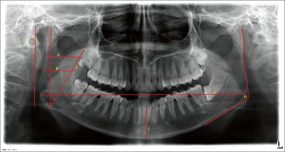
Point A-Maximum ramus breadth: It is the distance between most anterior and posterior points on the mandibular ramus. 7
Point B-Minimum ramus breadth: It is the smallest anteroposterior diameter of the mandibular ramus. 7
Point C-Condylar height: It is the distance between the most superior points on the condyle to the most protruded point on the inferior border of the ramus. 7
Point D-Projective Height of the Ramus: It is the distance between the most superior points on the condyle to the lower margin of the alveolar bone on the inferior border of the ramus. 7
Point E-Coronoid Height: It is the distance between the most superior points on the coronoid to the most protruded point on the inferior border of the ramus. 7
Point F-Height of the mandible at symphysis region: It is the distance between the midline of the central incisors to the lower border of the mandible. The mandibular symphysis region shows a significant difference between both males and females. 8
Point G-Bigonial Width: It is the distance between both gonion (Go). Go is the most inferior, posterior, and lateral point on the external angle of the mandible. It was measured horizontally from the right to the left Go. 9
Point H-Gonial Angle: A line was digitally traced on the OPGs tangential to the posterior border of the ramus and the condyle and another line was drawn tangential to the lower border of the mandible up to the distal root of the first molar. The intersection of these two lines forms the gonial angle which was measured by the software. 9
Statistical Analysis
IBM SPSS (Statistical Package for Social Sciences) version 20.0 statistical software was utilized for conducting the statistical analysis. Discriminant function analysis was employed to identify variables that differentiate between males and females, as well as to identify the most accurate predictors for determining an individual’s sex.
Results
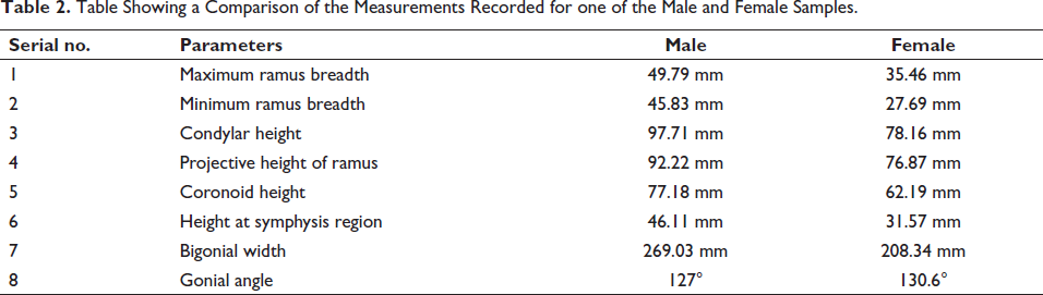
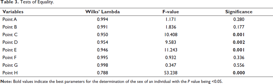
The descriptive statistics for both sexes are represented in Table 1. It shows that all the measurements recorded are higher in males as compared to females except for the gonial angle (Table 2). The f-statistic value indicates that the highest sexual dimorphism is seen with the gonial angle and the least with the bigonial width (Table 3). In this study, out of eight parameters, four parameters which are gonial angle, coronoid height, condylar height, and projective height of ramus are found to be the best parameters for the determination of the sex of an individual with the P value being <.05 (Table 3).
Descriptive Statistics.
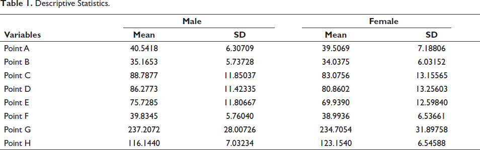
Table Showing a Comparison of the Measurements Recorded for one of the Male and Female Samples.
Tests of Equality.
From the values obtained by linear discriminant function analysis, the equation has been derived with the help of which we can determine the sex in an unknown sample (Table 4).
Linear Discriminant Function.
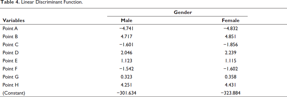
For Female = − 323.884 − 4.832 (MxRB) + 4.851 (MnRB) − 1.856 (Condylar Height) + 2.239 (PHR) + 1.115 (Coronoid Height) − 1.602 (Symphysis Region Height) + 0.358 (BW) + 4.431 (GA)
For Male = − 301.634 − 4.741 (MxRB) + 4.717 (MnRB) − 1.601 (Condylar Height) + 2.046 (PHR) + 1.123 (Coronoid Height) − 1.542 (Symphysis Region Height) + 0.323 (BW) + 4.251 (GA)
In order to classify a given sample as either male or female, a maximum of two equations were utilized. This study determined the threshold to be 0, where values exceeding this threshold are classified as male, while values below it are classified as female.
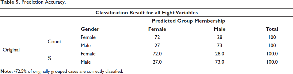
The prediction accuracy was calculated for all the variables. Out of 100 male samples 73 samples were correctly classified and out of 100 female samples 72 samples were correctly classified. The overall prediction accuracy was 72.5% (Table 5).
Prediction Accuracy.
The intra and inter-observer variability of all the variables was examined by using the chi-square test with a Pearson Correlation Coefficient (R-Value) of 0.98 and 1, respectively. The asymptomatic error was found to be very low. There were no significant differences found between intra and inter-observer analysis.
Discussion
Forensic odontology is a major branch of forensic science that deals with crime and law. Worldwide crime scenes are at an increasing rate; hence we need some strong investigative techniques to deal with criminality. The investigation starts with identification. Age estimation and sex determination are the key phases in identification. In the case of fresh bodies, it is possible to give a positive identification of gender by visual comparison. But in cases of badly decomposed or decayed bodies, it becomes difficult to identify the sex of a person.
When soft tissue parts are not available then the only source of identification is the skeleton. Skull and pelvis bones are sexually dimorphic bones of a body. The skull, second only to the pelvis, is the most dimorphic and easily identifiable part of the skeleton in terms of sex, offering a level of accuracy of up to 92%.1 In the absence of an entire skull, the mandible can serve as a source of identification.
Based on studies conducted on the mandible, it has been found that the mandibular condyle and ramus exhibit the highest degree of sexual dimorphism. These specific sites undergo substantial morphological changes in terms of size and remodeling during growth.2, 4 However, individuals of the same or similar height do not necessarily share identical mandibular morphology. Biological differences exist in the mandibular dimensions of males and females, leading to variations in morphology, which are significant in morphometric studies.10, 11
Radiography is a non-invasive technique. 1 Panoramic radiographs are routinely used for the examination of patients. Hence, it is easily available and does not involve a patient in any extra exposure dose and cost. These are good sources for retrospective studies. Panoramic radiographs examine the maxillofacial complex effectively.
In this study total of eight parameters were evaluated. Five mandibular rami parameters (maximum ramus breadth, minimum ramus breadth, condylar height, projective height of ramus and coronoid height), the height of mandible at symphysis region, bigonial width, and the gonial angle. The parameters represent the vertical and horizontal dimensions of the mandible. In this study, the mean values of all the variables were higher in males as compared to their female counterparts except in the gonial angle.
Among all eight variables, there were four variables that were statistically highly significant. Gonial angle, condylar height, the projective height of ramus, and coronoid height were highly significant in the present study with a P value <.05. In the present study, the prediction accuracy for a total of eight variables was 72% for females and 73% for males and overall it was 72.5%.
When comparing with other studies it was found that the results were almost similar to some of the studies. 12 conducted a study on the Bangalore population using mandibular rami parameters. They found all variables were significant predictors. Overall 76% of cases were correctly identified. 13 conducted a study on the Egyptian population using mandibular rami measurements and gonial angle as a parameter. They have found mandibular rami and gonial angle statistically highly significant and proved to be beneficial in gender estimation.
Nayyar et al. (2017) conducted a study on the South Indian population. They have found almost similar results to our study. Among five mandibular rami parameters, they found only three parameters, that is, condylar height, coronoid height, and projective height of ramus statistically highly significant. 7
Few studies, however, have been undertaken on the Maharashtra community14, 15; thus, the current study attempted to evaluate the efficacy of mandibular factors in predicting an individual’s gender in the Maharashtra population, with the goal of using the same in forensic analysis and anthropology.
Conclusion
From the present study, it can be concluded that mandibular ramus parameters (condylar height, coronoid height, projective height of ramus) and gonial angle can be used as aids for the gender determination of an individual. The mandibular ramus and gonial angle a valuable aids for the gender determination of a person as it is readily available, accessible, and resistant to any disintegration process.
Digital orthopantomographs were found to be reliable when all mandibular measurements were carried out for the determination of sex using image analysis software.
We recommend the use of this method in adjunct to other sex estimation techniques. However, there is a need for some research with a larger sample size and population-specific studies which will provide a uniform formula for the various populations. These will improve the usefulness of this study and help in a better understanding of the topic.
Footnotes
Declaration of Conflicting Interests
The authors declared no potential conflicts of interest with respect to the research, authorship and/or publication of this article.
Ethical Approval
The data were collected from the archives following the approval of the Institutional Ethical Committee.
Funding
The authors received no financial support for the research, authorship and/or publication of this article.