
Abstract
The cytology of peritoneal washing fluids for gastric cancer is the most basic method for judging peritoneal micrometastasis. However, the clinical value of this method is not clear at present. A retrospective analysis was performed on 277 patients with pathologically proven and surgically treated gastric cancer. The peritoneal washing fluids were collected after opening the abdomen and before the operation, and were sent to the cytology laboratory for screening of occult cancer cells in the collected washing fluids. The number of cases diagnosed as cancer cells, reactive mesothelial cells, serosal balls, and traumatic mesothelial cells were 42, 18, 27, and 190, respectively. Typical adenocarcinoma cell nests were found in eight of 10 T4b samples, whereas 34 cases of cancer cells in T3 and T4a showed that these cell nests usually contained mesothelial cells, and the three-dimensional stereoscopic sense of the nests was not obvious. In the specific subcellular morphological changes of both reactive mesothelial cells and serosal balls, the changes of both the contour of nuclear membrane and the polarity of cell alignment were present only in stage T3 and T4a. The presence or absence of mesothelial cells in the nests of cancer cells and the changes of the contour of nuclear membrane and of the polarity of cell alignment in reactive mesothelial cells or serosal balls may help us to predict the depth of invasion of cancer cells.
Keywords
Introduction
The cytology of peritoneal washing fluids for gastric cancer is used to screen the occult cancer cells in the collected washing fluids, which are obtained through washing the abdominal cavity with normal saline after opening the abdomen and before the operation. This detection method is the most basic method for judging peritoneal micrometastasis 1,2 , and has important clinical value for the clinical staging, treatment, and the prognostic judgment of gastric cancer 3,4 .
Conventional cytological methods often cause cancer cells to degenerate to a certain extent due to the lack of timely fixation of washing fluid specimens, and there are no uniform cytological diagnostic criteria, so the sensitivity is low 5 . In recent years, the detection of tumor markers in peritoneal lavage fluid has been used to indirectly speculate the existence of cancer cells. Although the sensitivity is high, the specificity is not ideal, and the method cannot be used as a basis for the treatment of peritoneal micrometastasis 6 –8 .
The introduction of liquid-based cytology at the end of the twentieth century, especially the appearance of the ThinPrep cytologic test, had brought a new dawn to the cytological examination of peritoneal washing fluids 9 –11 . In this method cells in the washing liquid are immediately put into a liquid-based bottle with a stationary liquid, and a slide is automatically prepared in an automatic preparation instrument. The effective cellular components are fully purified through membrane filtration 12 . Human interaction cannot affect the arrangement and the morphological characteristics of the cells, enabling cytologists to interpret benign and malignant cells and the severity of the lesions.
The main purpose of this study aims to interpret the accuracy of diagnosis by the morphological characteristics and arrangement of cancer cells, and to infer the invasion depth of cancer cells by the subcellular morphological characteristics of both reactive mesothelial cells and serosal balls, so as to provide a scientific basis for the clinical treatment of gastric cancer.
Materials and Methods
The study was a retrospective analysis of medical records of patients with histologically confirmed gastric cancer. All procedures in this study were conducted in accordance with the First Hospital of China Medical University’s (APPROVAL NUMBER/2016-125) approved protocols. Written informed consent was obtained from the patients for their anonymized information to be published in this article. A total of 277 peritoneal washing samples from patients who attended the department of pathology at the First Hospital of China Medical University during the period January 2017–January 2018 were included in the study. The study group comprised 202 men and 75 women, ranging in age from 29 to 82 years.
Immediately after opening the abdomen in all patients, pelvic washing was performed. A total amount of 100 ml of saline was used altogether in the three spaces (both the subphrenic spaces and pelvis). After sufficient stirring for 3 min, all the washing fluid was collected and sent to the cytopathology lab. A 30 ml representative sample was taken and centrifuged at 2000 g for 10 min; the resulting pellet was transferred to a small vial containing ThinPrep PreservCyt Solution (ThinPrep 2000; Hologic, Inc., Beijing, China). The preparation of cell slides was described elsewhere 13 .
Based on the histological diagnosis of resection specimens and the cytological diagnosis of washing cells, the diagnosis results were recorded into histological and cytological groups, respectively. Cytological and histological diagnoses were performed by experienced cytologists or histologists, and were compared with each other. According to the arrangement and morphology of the cells in the washing fluids, cytological diagnoses were divided into four groups: adenocarcinoma cells, reactive mesothelial cells, serosal balls, and traumatic mesothelial cells. Detailed diagnostic criteria are shown in Table 1. At the same time, we observed the subcellular morphology of both reactive mesothelial cells and serosal balls in detail. Histological diagnoses were subclassified according to the AJCC TNM classification (7th edition) 3 and the Japanese Classification of Gastric Carcinoma 4 . Statistical analysis was performed using SPSS 16.0 software package (SPSS, Inc. Chicago, IL, USA).
Cell Morphological Characteristics of Four Different Types in Peritoneal Washing Fluids of Patients with Gastric Cancer.

Results
The results of cytological diagnosis in the peritoneal washing fluid of patients with gastric cancer are presented in Table 1.
According to the morphological characteristics of the cells, adenocarcinoma cells were found in 42 of 277 gastric cancer douche samples; reactive mesothelial cells were found in 18 of them; serosal balls were found in 27 of them; and traumatic mesothelial cells were identified in 190 of them. Detailed morphological observations are shown in Table 1.
The positive results of cytological diagnosis of cancer cells in peritoneal washing fluid at different stages of gastric cancer are presented in Table 2.
Comparison of Histological Diagnosis with Cytological Diagnosis for Gastric Cancer.
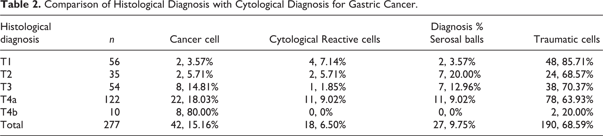
Typical adenocarcinoma cell nests were found in eight of 10 T4b samples; the aggregates of cells were arranged in a ball-like pattern and had stereoscopic morphology (Fig. 1A). While most of the cancer cells were found in the T3 and T4a samples, and these cell nests usually contained mesothelial cells, the three-dimensional sense of cell nests is not obvious (Fig. 1B). Reactive mesothelial cells and serosal balls were found in the samples with T1 to T4 stages, but mainly in the T4a stage. Traumatic mesothelial cells were found in all stages, but mainly in the first four phases (Fig. 1C).

The cell morphological characteristics of adenocarcinoma cells, Reactive mesothelial cells, and serosal balls in peritoneal washing fluids of patients with gastric cancer (Papanicolaou stain, × 400). (A) Adenocarcinoma cells. The small aggregates of cancer cells were arranged in a ball-like pattern and had stereoscopic morphology; (B) Adenocarcinoma cells. The nests of cancer cell contained a few mesothelial cells, and their stereoscopic morphology was not obvious. (C) Traumatic mesothelial cells. The cells are arranged in a flat, honeycomb structure with obvious cell size and uniform morphology. (D) Reactive mesothelial cells. The cells are arranged in a flat pattern, but they lose their honeycomb structure. The cells are of different sizes, and some nuclei are elongated. (E) Serosal balls. Mesothelial cells were arranged in a syncytial fashion, the cell boundaries were not clear, the cytoplasm was dyed green. (F) Serosal balls. Mesothelial cells were arranged in a syncytial fashion, and their size and disorder were varied.
The characteristics of subcellular features of reactive mesothelial cells and serosal balls are shown in Table 3 and Table 4.
Cell Morphological Changes of Reactive Mesothelial Cells in Different Stages of Patients with Gastric Cancer.
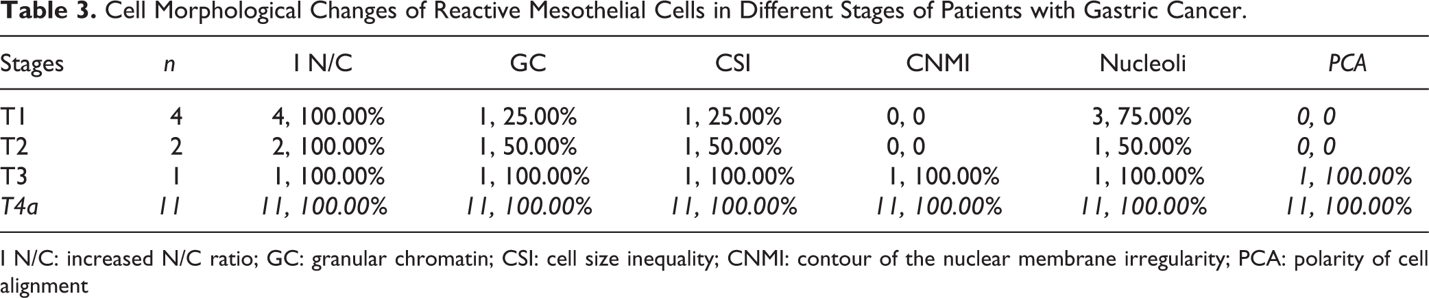
I N/C: increased N/C ratio; GC: granular chromatin; CSI: cell size inequality; CNMI: contour of the nuclear membrane irregularity; PCA: polarity of cell alignment
Morphological Changes of Serosal Balls in Different Stages of Patients with Gastric Cancer.

I N/C: increased N/C ratio; GC: granular chromatin; CSI: cell size inequality; CNMI, contour of the nuclear membrane irregularity; PCA: polarity of cell alignment
In both groups of specific subcellular morphological changes, we found that the contour of nuclear membrane and the polarity of cell alignment were present only in T3 and T4 cancers, but not in T1 and T2 cancers.
In the specific subcellular morphological changes of both reactive mesothelial cells (Fig. 1D) and serosal balls (Figs. 1E and F), we found that both N/C ratio and granular chromatin were present in all stages, whereas the changes of both contour of the nuclear membrane and the polarity of cell alignment were present only in stage T3 and T4a cancers, but not in T1 and T2 cancers.
Discussion
A large number of studies have reported that positive cytology of peritoneal washing fluids is an important indicator of poor prognosis, and also an important basis for the postoperative chemotherapy of advanced gastric cancers 14 –17 . However, it is not clear how to accurately interpret the morphological characteristics and the arrangements of cancer cells in peritoneal washing fluids. In this study, we found that there were two patterns of both the morphological characteristics and the arrangements of cancer cells in peritoneal washing fluids: one was that the cancer cells showed nesting arrangements, the three-dimensional perception was obvious, and mesothelial cells were not seen within cancer cell nests. The eight cases of T4b cancer cells in this study all showed this pattern, which suggested that the cancer cells could have broken through the serosa and formed tiny nodules on the surface of the serosa. The other pattern was that the cancer cells showed flat sheet arrangements, the three-dimensional perception was not obvious, and mesothelial cells were often found in cancer cell nests. The cancer cells of patients, both eight cases of T3 and 22 cases of T4a in this study, were all in this pattern, which suggested that the cancer cells could have just soaked the serosa and often fall off together with a few mesothelial cells. The morphological characteristics and arrangement of the above two kinds of cancer cells may be helpful for us to distinguish whether cancer cells were located in the serosa or had completely penetrated the serosa. In addition, we found two cases of cancer cells in stage T1 and T2. Although the infiltration depth of these four cases of lesions was insufficient, they all had lymph node metastasis. This result suggests that the cancer cells in washing fluids may originate from lymphatic metastasis, which is consistent with reports by Yoshida et al. and Le 18,19 .
In this study, we reported 18 cases of reactive mesothelial cells from T1 to T4a. There was no regularity in the distribution of these cases in different stages. In order to identify regular changes of subcellular morphology, we carefully observed the N/C ratio, chromatin granularity, cell size, contour of the nuclear membrane, nucleoli, and polarity of cell alignment of reactive mesothelial cells. The results showed that both N/C ratio and granular chromatin appeared in all stages from T1 to T4a; both of them showed no regular changes. However, the contour of the nuclear membrane and the polarity of cell alignment existed only in T3 and T4a, and neither appeared in the T1 and T2 cancers. These results suggest that both the changes of the nuclear membrane contour and the polarity of cell alignment may be caused by the stimulation of cancer cells, and the reactive mesothelial cells may be located around cancer cells, which is helpful for clinicians to treat and judge the prognosis. It also suggested that if mesothelial cells have only N/C ratio and chromatin granularity changes, these cells should not be classified as reactive mesothelial cells in the future.
In this study, 27 cases of the serosal balls were found in the fluid from T1 to T4a cancer. The composition and morphology of serosal balls had been found in the peritoneal washing fluid of ovarian cancer, but whether their subcellular morphology is related to the depth of invasion of the tumor has not been reported 20 . In order to identify the regular changes of subcellular morphology, we also carefully observed the N/C ratio, chromatin granularity, cell size, contour of the nuclear membrane, nucleoli, and polarity of cell alignment of serosal balls. The results showed that both N/C ratio and nucleoli appeared all stages from T1 to T4a, and neither were regular changes. However, the contour of the nuclear membrane and the polarity of cell alignment existed only in T3 and T4a, and neither appeared in T1 and T2. These results suggest that these two atypical changes may also be caused by the stimulation of cancer cells. These mesothelial cells might also be located around cancer cells, and this result is helpful for clinicians to treat and judge the prognosis. It also suggests that if the cells of serosal balls exhibit only subcellular changes of N/C ratio and nucleoli, these cells might be normal mesothelial cells and need not be reported in the diagnoses.
In addition, the results of this study revealed that the most of washing fluids in T4a and T4b showed negative results, indicating that this method could only elucidate a few lesions; identification of most lesions will rely on development of new techniques to make up for the shortcomings of this method. This might include following the sampling methods of cervical and bronchial lesions 21,22 , slight brush of the high-incidence areas of omentum lesions before washing, and fully improving the sensitivity of cytological diagnosis on the premise of ensuring that the technique is minimally invasive or noninvasive. This method will be further studied in the future.
Conclusion
The presence or absence of mesothelial cells in the nests of cancer cells and changes of the contour of nuclear membrane and of the polarity of cell alignment in reactive mesothelial cells or serosal balls may help us to judge the depth of invasion of cancer cells.
Footnotes
Ethical Approval
This study was approved by our institutional review board.
Statement of Human and Animal Rights
All procedures in this study were conducted in accordance with the First Hospital of China Medical University's (APPROVAL NUMBER/2016-125) approved protocols.
Statement of Informed Consent
Written informed consent was obtained from the patients for their anonymized information to be published in this article.
Declaration of Conflicting Interests
The author(s) declared no potential conflicts of interest with respect to the research, authorship, and/or publication of this article.
Funding
The author(s) disclosed receipt of the following financial support for the research, authorship, and/or publication of this article: This work was supported by grants from the National Natural Science Foundation of China to Guang-Ping Wu, Grant No.81171650 and 81672082.