
Abstract
In this experiment, CO was used as a gas mixture in a reversible relationship with O2. CO was added in a gas form mixed with O2. An isolated donor rat heart was obtained, exposed to a gas mixture such as PO2 = 1,800 hPa and PCO = 200 hPa, and PO2 = 1,000 hPa and PCO = 1,000 hPa in a 2 ATA high-pressure chamber and preserved in a refrigerator at 4°C. This report demonstrates that significant reproducibility has been verified. The heart was removed from the refrigerator 24 h later and heterotopic heart transplantation was performed in the right neck of a recipient rat and the pulsating of the transplanted heart was detected by an electrocardiogram.
Keywords
Introduction
Clinical transplantation therapy for the human lung, heart, liver, and kidneys is a common practice in clinical use (6). The shortage of organs for patients who are awaiting transplantation is increasing yearly, and the situation continues to get worse. The supply of transplanted organs is insufficient, mainly because organs cannot be preserved for a long period as in a blood bank. Currently, the preservation of human organs for transplantation mainly consists of cryopreservation, with a limit of 4–24 h of preservation. Organs cannot be preserved for a long period because the cell membranes are damaged due to the low temperature (4°C) and ischemia (5,9,10). This system would improve considerably if transplanted organs could be preserved for a long period as with red blood cells and other microorganisms. As a result, it is necessary to establish new methods for long-term organ preservation (1).
In previous experiments, 4–18 h has been the limit, even with cryopreservation and resuscitation of isolated rat, rabbit, baboon, and human hearts using University of Wisconsin solution (UW) as a simple method of immersion into a preservative solution (8,16). Body tissue slightly decomposes even at low temperatures, and from the perspective of eliminating waste products caused thereby, a method of perfusing the tissue has been attempted. Consequently, preservation times longer than that of the simple immersion method have been reported, but this has not been commonly used, because the equipment is massive and expensive. Similarly, to improve this situation, while focusing on tissue decomposition at low temperatures, others have attempted to supply O2 to the preservation solution and perfusion solution, and extended preservation times have been reported (14).
In these attempts, Kuroda et al. filled the heart chamber with UW to provide immersion preservation while supplying mixed gas (PCO2 = 50 hPa and PO2 = 950 hPa) to perfluorocarbon (PFC) solution, an inert fluid in which a large amount of O2 can be dissolved and maintained viability for 24 (100%) to 48 h (four of five subjects) (7). Seki et al. focused on cryptobiosis, which reduces decomposition by decreasing the amount of water in a living body in order to adapt to an extreme environment such as dryness and low temperature. Seki et al. showed that, under such conditions, tardigrades can regenerate even under ultrahigh pressure (12).
Since 1998, Seki et al. have conducted experiments aimed at reducing the amount of water using a PFC solution for the preservation of isolated rat organs before resuscitation and obtained good results at times, but the reproducibility was insufficient. Subsequently, focusing attention on CO2 gas, which has anesthetic and decomposition-inhibiting affects on living organisms, they attempted to preserve isolated rat hearts in an environment with a reduced amount of water and PCO2 = 200 hPa and they thus obtained results with verified reproducibility (13). When the isolated rat heart was aeration dried at 100 hPa of partially compressed CO2 after being immersed in PFC, the preservation time could be extended to 72 h (17). In this experiment, CO2 was replaced in a gas mixture with CO in a reversible relationship with O2. CO is highly toxic, so symptoms of poisoning develop in humans even at a concentration of 0.01% (100 ppm) CO in the air. If it were 0.08–0.12%, then a coma would be induced, thus potentially leading to respiratory failure and heart failure. When the concentration is 0.08%, 50% of the blood hemoglobin becomes COHb. If the concentration is 0.15%, it is likely to cause death. Moreover, if it is 0.19% (1,900 ppm), it will cause death within a short period (2).
This report found that an isolated rat heart was exposed to CO gas at high partial compression exceeding 200 hPa (200,000 ppm when converted to atmospheric pressure, equivalent to 100 times the amount lethal to humans) to 1,000 ppm and preserved for 24 h, followed by resuscitation before being heterotopically transplanted into a recipient rat.
Materials and Methods
Inbred rats LEW/SsN Slc (male, 6 weeks old) that had been raised for organ transplantation by Japan SLC Inc. were used as donors and recipients. Diethyl ether inhalational anesthesia was administered to the rat and 1.0 U of normal saline plus 100 U of heparin sodium was administered intravenously. The surgical site was shaved and sterilized with 75% ethanol. The chest was opened with a median incision, the left and right costal parts were incised dorsally, and the chest wall was inverted cranially to expose the cardiac region. The heart was isolated and moved to a petri dish filled with heparin-containing normal saline (room temperature). The ascending aorta and pulmonary artery were ligated, while keeping them as long as possible, and the blood was removed and the heart was perfused from the broken end of the ascending aorta using heparin-containing normal saline (room temperature). Afterwards, the superior vena cava and inferior vena cava were ligated and cut off.
After the pulmonary vein and bronchial tube were ligated as one piece, the donor isolated heart with the lungs removed was weighed and then a Krebs-Henseleit (KH) solution was injected into the isolated rat heart chamber, followed by preservation in a 2 ATA high-pressure chamber pressurized with a gas mixture of PO2 = 1,800 hPa and PCO = 200 hPa or PO2 = 1,000 hPa and PCO = 1,000 hPa and then placed in a refrigerator (4°C). KH solution containing antibiotics and warfarin was used. A beaker filled with distilled water was placed in a high-pressure chamber and the humidity in the chamber was maintained at over 90% (Fig. 1). Twenty-four hours later, after diethyl ether inhalational anesthesia had been provided to the recipient rat, the right neck was shaved and sterilized with 75% ethanol and a 3-cm longitudinal incision was made in that area. The connective tissue under the skin was burned off with an electrical scalpel to expose an extended length of the external jugular vein and common carotid artery. After the inner side of the external jugular vein had been blocked with a microclip, the peripheral side of the external jugular vein was ligated and cut off. Subsequently, after the inner side of the common carotid artery had been blocked with a microclip, the inner side of the common carotid artery was legated and cut off. After cuffs fitting the size of the external jugular vein and common carotid artery were each fitted, the intravascular lumen was flushed with heparin-containing normal saline.

A hyperbaric chamber was used for the preservation of the isolated heart. A beaker containing distilled water was placed in the chamber to maintain the humidity inside the chamber at over 90%. The heart was filled with KH solution containing antibiotics and warfarin sodium and it was hung as shown in the illustration. The mixture of CO and O2 was pressurized to 2 ATA and the chamber was put in a refrigerator maintained at 4°C for 24 h.
After preparing the recipient rat, the preserved heart was transferred from the chamber to a refrigerator and temporarily immersed in normal saline and then end-to-end anastomosed at the recipient rat's common carotid artery to the aorta and the external jugular vein to the pulmonary artery, respectively. The microclips of the external jugular vein and common carotid artery were removed to allow blood to pass through and flow to the transplanted heart to be pumped and the donor heart was observed. Thereafter, the incised skin on the neck was sutured. After the surgery, 1.0 ml of normal saline containing 100 U of heparin sodium was administered intravenously to the recipient to prevent blood from clotting in the transplanted heart. Thereafter, an electrocardiogram of the transplanted heart was recorded each week. Drinking water containing antibiotics was given to the recipient rat and postobservation was conducted in the breeding room. About 8 weeks later, the pulsation of the hearts of the recipient rat and the donor rat was recorded by an electrocardiogram.
Results
The isolated rat heart, which was resuscitated after being preserved for 24 h in a 2 ATA high-pressure chamber pressurized with a mixture of O2 and CO, was end-to-end anastomosed at the recipient rat's common carotid artery and aorta and the external jugular vein and pulmonary artery, respectively. Thirty-three heterotopic heart transplants were attempted and 14 pulsating donor hearts could be recorded with an electrocardiogram. Among these, 11 cases showed resuscitation of the ventricle and the atrium and 3 cases showed resuscitation of the atrium only (Fig. 2, Table 1). The pulsation was checked 8 weeks later and an electrocardiogram could be recorded in 11 cases (Fig. 3). As a control experiment, the preservation and resuscitation experiment was conducted under the same conditions using gas mixtures of pure O2, pure He, standard air, PHe = 1,400 hPa + PCO = 600 hPa and PCO2 = 1,400 hPa + PCO = 600 hPa and heterotopic heart transplantation was subsequently performed. Only those preserved in the gas mixture of PHe = 1,400 hPa + PCO = 600 hPa showed slight resuscitation (pulsating of the atrium: four of five subjects), but none of the donor hearts in the five remaining cases in the control experiments demonstrated any pulsation.
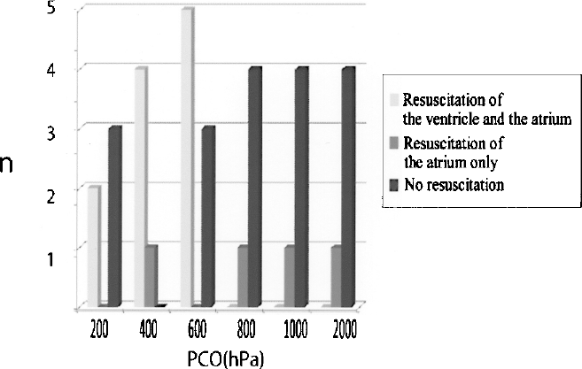
Graph of the resulting number of individuals resuscitated in this experimental method, grouped by partially compressed CO gas pressure aerated for preservation.
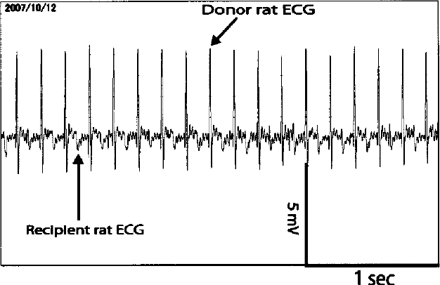
Isolated rat heart to be preserved for 24 h at 400 hPa of partially compressed CO, followed by heterotopic heart transplantation in the neck and resuscitation and 8 weeks later, electrocardiograms of the donor heart and recipient hearts were recorded on October 12, 2007.
Preservation Method and Revival Rate

The table shows the resulting proportion of resuscitation in this experimental method, grouped by the type of gas aerated for preservation. n shows the number of hearts, classified into those with resuscitation of both the ventricle and the atrium, with the resuscitation of the atrium only and without resuscitation, which is represented as the revival rate. In the gas mixture of O2 and CO, two fifths (40%) of those preserved in CO (200 hPa) were resuscitated. Four fifths (80%) of those preserved in CO (400 hPa) were resuscitated. Five eighths (62.5%) of those preserved in CO (600 hPa) were resuscitated. None (0/5; 0%) of those preserved in CO (800 hPa) were resuscitated. None (0/5; 0%) of those preserved in CO (1,000 hPa) were resuscitated. None (0/5; 0%) of those preserved in CO (2,000 hPa) were resuscitated. None (0/5; 0%) of those preserved in O2, He, and standard air at 2000 hPa were resuscitated. As for those preserved in the gas mixture of CO (600 hPa) and O2 (1,400 hPa) with O2 replaced by He and CO2, only the atrium showed pulsation when replaced with He. All (10/10; 100%) of the controls with no preservation time were resuscitated.
Discussion
Seki et al. demonstrated that tardigrades could be resuscitated and survive even after exposure to an extremely high hydrostatic environment under 600 MPa of pressure and indicated that this can be applied to the preservation of multicellular organisms. This physiological mechanism of preserving and resuscitating tardigrades was applied to mammalian organ preservation (12). A tardigrade is a multicellular organism that is composed of about 40,000 cells and also has nerve cells. Among the cells comprising mammals, techniques for the long-term preservation and resuscitation only of single cells such as blood, sperm, and ovum have been developed and are in practical use. The technique of preserving tissue, which is a collection of cells and organs (body organs) comprising several tissues up to 24 h has been in practical use and transplantation needs to be performed within that period (6). The major problem in mammalian organ preservation is that it is necessary to verify which tissue survives within each organ after preservation. Methods for verifying that include the histologically anatomical method, a transplantation method in which transplantation is actually performed for verification, and an electrophysiological method. But regardless of which method is employed, it is necessary to verify whether respective tissue cells survive. In this laboratory, heterotopic heart transplantation in the neck of a rat was employed as a method for verifying the resuscitation of an isolated rat heart.
Heterotopic heart transplantation does not function in pumping blood out of the heart, but as a major merit, unlike other organs, it is possible to objectively judge whether the heart survives via external and electrocardiographic records.
Focusing attention on CO2 gas, which has anesthetic and decomposition-inhibiting properties, heart preservation was performed in an environment with a reduced amount of water and PCO2 = 400 hPa in order to obtain results with verified reproducibility (13). This organ preservation and resuscitation technique produced a new field called semibiology, wherein the vital activity is stopped and resuscitated. An automobile can be repaired to be usable again when it breaks down, because there are design drawings, parts, and repair techniques. In the case of humans, design drawings (anatomical drawings) and repair techniques (surgery) have been already completed. However, there are still no spare parts (organs). The parts depend on the supply obtainable from human brain-dead donors, but the maximum preservation time of organs is 24 h. The organ preservation and resuscitation technique using CO2 gas succeeded in extending the preservation time of parts (organs) from 24 h to 72 h. If the preservation time of organs were at least 1 year, human life would be semipermanent, as in the case of a semiconductor. It is believed that human life could eventually become semipermanent as with automobiles if semibiology techniques could someday be developed. The natural phenomenon in which plants and animals in the natural world put themselves into drought dormancy almost every year and awake in the next spring has already been applied to the cells and tissues of plants and animals, seeking practical use. Studies are going to determine if this natural phenomenon, repeated in the natural world almost every year, can be applied to isolated mammal organs.
In this experiment, CO gas was used to preserve an isolated rat heart. The replacement of CO2 in a gas mixture containing O2 and CO in a dynamic equilibrium relationship was incorporated in this study. A rat isolated heart that had been exposed to 600 hPa of partially compressed CO for 24 h was preserved before being heterotopically transplanted into the right neck of a recipient rat and resuscitated, after which the transplanted heart survived for more than about 3 months. CO gas binds with the Fe2+ in cytochrome oxidase, an enzyme that living organisms require and acts to suppress the activity of this enzyme in order to generate energy from glucose (15). In addition, CO gas may have suppressed intracellular decomposition and prevented necrosis. According to the law of entropy increase, the dynamic equilibrium theory states that decomposed components are degraded purposely in advance and reconstruction can be constantly achieved before such clutter accumulates, with all molecules being replaced at a fast rate. It is believed that this mechanism establishes a dynamic equilibrium between the O2 and CO in cells and tissues within a heart preserved under high pressure and intracellular decomposition is suppressed and necrosis is prevented by binding to the Fe2+ of cytochrome oxidase, thereby suppessing the activity of this enzyme (3,4,11). The resuscitation rate was highest when was heart is preserved under 400 hPa of partially compressed CO in a gas mixture of CO and O2 in the hyperbaric environment of 0.2 MPa of pressure. When the isolated heart was dry preserved in a 100% concentration of CO, there was pulsating of the heart, but none survived. In addition, He, which is an inert gas, CO2 and CO gas (600 hPa) were mixed to preserve the isolated heart, and although pulsating was observed immediately after transplantation and resuscitation, unfortunately none survived.
Dry preservation of single cells and tissues of plants and animals is already in practical use, but resuscitation after the dry preservation of isolated mammalian organs has not been implemented until now. When the isolated heart is exposed to 0.2 MPa of pressure in a hyperbaric environment, the amount of water in an isolated heart is gradually lost, thus leading to a dry condition. It has been found that the resuscitation rate varies depending on the gases used as a desiccant in this case. It is known that CO2 is suitable for dry preservation and resuscitation (2,13). The results of this experiment indicated that CO gas can be applied to the preservation and resuscitation of the organs of humans and other mammals. In addition, this preservation method is being applied to further extend the preservation time in order to allow for effective preservation and resuscitation.