
Abstract
Introduction
Doxorubicin (DOX), a member of the anthracycline family, has been one of the most widely used anticancer drugs since it was first approved by the U.S Food and Drug Administration (FDA) for clinical use. 1 DOX is a highly effective chemotherapeutic used to treat many adult and pediatric cancers, such as solid tumors, leukemia, lymphomas and breast cancer.2–4 On the other hand, DOX can cause injuries to multiple organs, including the heart, liver, and brain or kidney.5,6 Cardiotoxicity is the most prominent side effect of DOX. It induces a dose-related cardiotoxicity, which is among the most common etiologies of cancer chemotherapy-associated heart failure.1,7,8 Pathological manifestations of DOX-induced cardiotoxicity include vast cytoplasmic vacuolization, sarcoplasmic reticulum swelling, and myofibrillar disarray. 6 DOX-induced cardiotoxicity has been extensively studied in the last several decades and multiple underlying mechanisms have been proposed. These mechanisms include oxidative stress, topoisomerase inhibition, ferroptosis, cardiogenetics, mitochondrial bio-energetics, and autophagy modulation, etc.1,9–16
Autophagy is Greek for “eating of self”, a term first coined in 1967 by Christian de Duve when observing rat liver treated with glucagon, a pancreatic hormone, and the following degradation of mitochondria and other intracellular structures within lysosomes. 17 Autophagy is a unique membrane trafficking process whereby newly formed membranes, termed phagophores, engulf parts of the cytoplasm leading to the production of double-membraned autophagosomes that get delivered to lysosomes for degradation. 18 Autophagy is a highly conserved process in response to extra- or intra- cellular stress and signals such as starvation, growth factor deprivation, and pathogen infection. 19 This self-digestion not only provides nutrients to maintain vital cellular functions during fasting, but also can rid the cell of superfluous or damaged organelles, misfolded proteins, and invading microorganisms. 18 Therefore, autophagy is an important biological process for maintaining cellular homeostasis and normal functions of cells. Persistent decreases or increases in autophagy can lead to diseases such as cancer, liver diseases, diabetes, kidney diseases, inflammatory disease, neurodegenerative diseases, and heart disease.20–28
Studies have demonstrated DOX influences autophagy in cardiomyocytes.12–16,29 However, little is known about the effects of this chemotherapeutic in cardiac fibroblasts. Fibroblasts are the largest cell population in the heart and the main cell type responsible for the synthesis, deposition, and degradation of cardiac extracellular matrix (ECM). 30 In this study, we examined and compared the effects of DOX on autophagy regulation in primary cardiac fibroblasts and an immortalized NIH3T3 embryonic fibroblast cell line.
Materials and methods
Cell culture
Primary cardiac fibroblasts (CFs) from BALB/c mice were obtained from Cell Biologics (Chicago, Illinois, USA) and cultured in fibroblast medium provided by the vendor, which contained fibroblasts growth factor, hydrocortisone, antibiotics-antimycotics, 2 mM
Assessment of cell proliferation following DOX treatment
The effect of DOX on the proliferation of CFs and NIH3T3 cells was assessed using alamarBlue assay (Thermo Fisher Scientific, Waltham, MA, USA). Briefly, cells were seeded at a density of 1 × 10⁴ cells per well in a 96-well flat bottom plate and were treated with DOX (0-5 µM) for 24 h. The alamarBlue reagent (10% v/v) was added to cells for the last 3 h of treatment. Resazurin, the active ingredient of alamarBlue reagent, is reduced to fluorescent resorufin upon entering living cells. 31 At the end of incubation, the fluorescence intensity of each well was measured using a microplate reader with 560 nm excitation and 590 nm emission.
Quantification of autophagy protein markers by western blot
NIH3T3s and CFs were seeded at a density of 2 × 10⁵ cells per well in 6-well plates. Cells were treated with DOX at a concentration of 1 μM or a vehicle control for 24 h. Bafilomycin a1 (BAF) was added to half of the wells at a concentration of 100 nM and a vehicle control was added to the other half during the last 2 hours of treatment to monitor autophagy flux. BAF is an antibiotic that inhibits V-ATPase-dependent acidification and Ca-P60 A/SERCA-dependent autophagosome-lysosome fusion, which in turn causes the accumulation of LC3II protein. 32 BAF has been used to monitor autophagy flux due to this effect. At the end of treatment, the culture media was removed; the cells were washed with cold PBS three times, lysed on the plates in RIPA buffer containing protease and phosphatase inhibitors on ice for 15 min, and centrifuged at 4°C. The supernatants were transferred to clean 1.5 mL low protein binding microcentrifuge vials and stored at −80°C until analysis. Protein concentrations in the cell lysates were determined using a Pierce™ BCA protein assay kit (Thermo Fisher Scientific, Waltham, MA, USA) prior to Western blot procedures described below.
Information of the primary and secondary antibodies used in this study.

Assessment of autophagy-related gene expression by RT-qPCR
Primary cardiac fibroblasts and NIH3T3 cells were seeded at a density of 2 × 10⁵ cells per well in 6-well plates. After overnight incubation, the cells were treated with 1 μM DOX or a vehicle control for 24 h. Total RNA was extracted from cells using an RNeasy Mini Kit (Qiagen, Germantown, MD, USA). RNA concentrations were determined with a NanoDrop spectrophotometer (Thermo Fisher Scientific, Waltham, MA, USA). A mouse autophagy signaling pathway RT2 Profiler™ PCR Array (Qiagen, Germantown, MD, USA) was used to quantify the relative expression levels of autophagy-related genes following the manufacturer’s instruction. Briefly, 100 ng RNA from each sample were reverse transcribed to cDNA using an RT2 first strand kit (Qiagen, Germantown, MD, USA). One hundred nanograms of cDNA was then mixed with SYBR Green mastermix. Real-time PCR was performed on a LightCycler® 96 (Roche Diagnostics Corporation, Indianapolis, IN, USA). A web-based tool from Qiagen, RT2 Profiler PCR Data Analysis, was used for differential gene expression analysis.
Statistical analysis
The values were presented as mean ± SE. Differences between the two groups were evaluated using the Student t test. The level of significance was selected to be p < 0.05. R statistical analysis software was used to perform the statistical analysis.
Results
Effect of DOX on cell proliferation
The alarmaBlue assay results showed that DOX induced a similar effect on the proliferation of in CFs and NIH3T3 cells (Figure 1). A significant decrease in cell numbers was observed after 24 h treatment with DOX at 2.5 μM concentration compared to control groups (Figure 1). DOX induced an approximate 30% inhibition of cell proliferation in CFs and NIH3T3 cells were viable at 1 μM of. This concentration was used in the subsequent gene expression and western blotting experiments. Cell proliferation assessment of primary cardiac fibroblasts isolated from BALB/c mice and NIH3T3 mouse embryonic fibroblasts. Cells were treated with a series of concentrations of DOX (0-5 µM) for 24 h. Values represent mean ± standard error of six replicates (n = 6).
DOX induced a significant increase in LC3II levels in NIH3T3 cells, but not primary cardiac fibroblasts
LC3II protein is commonly used as a marker to monitor autophagy activity in cells. To determine whether DOX affects autophagy in fibroblasts, we quantified the level of LC3II in both CFs and NIH3T3 cells after 24 h of treatment with DOX at 1 μM. The results showed that DOX significantly increased LC3II levels in NIH3T3 cells with the presence of BAF during the last 2 hours of treatment. Similar effects were not observed in CFs (Figure 2). These results indicate DOX could increase autophagy activity in fibroblasts in a cell-dependent manner. DOX significantly increased LC3II level in NIH3T3 cells, but not in CFs. Western blot analysis of the effect of DOX on LC3II level in NIH3T3 cells (a) and CFs (b). Values represent mean ± standard error four replicates (n = 4). * p-value <.05 (Student’s t test).
DOX significantly decreased P62 levels in NIH3T3 cells, but not primary cardiac fibroblasts
Also known as sequestosome-1, p62 is a scaffold protein that accumulates when autophagy is inhibited.33–35 Its level decreases when autophagy is induced.
33
Thus, it has been used as a protein marker for monitoring autophagy activity. In this study, we quantified the level of p62 in both CFs and NIH3T3 cells after 24 h of treatment with DOX at 1 μM. We observed that p62 levels were decreased in NIH3T3 cells following DOX treatment (Figure 3). In contrast, p62 level remained unchanged in primary cardiac fibroblasts (Figure 3). These observations were consistent with LC3II results. DOX significantly decreased P62 levels in NIH3T3 cells, but not in CFs. Western blot analysis of the effect of DOX on P62 levels in NIH3T3 cells (a) and CFs (b). Values represent mean ± standard error of four replicates (n = 4). * p-value <.05 (Student’s t test).
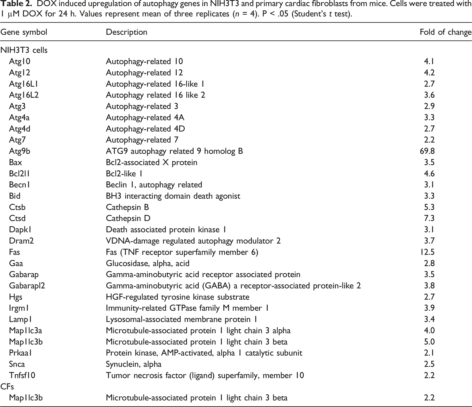
DOX induced different patterns in autophagy gene expression in NIH3T3 cells and primary cardiac fibroblasts
We examined the effects of DOX on the expression of 84 selected genes involved in autophagy using RT-qPCR. DOX induced significant upregulations in autophagy gene expression in NIH3T3 cells (Figure 4, Table 2). In contrast, the effects of DOX on autophagy genes were minimal in primary cardiac fibroblasts (Figure 4, Table 2). Consistent with changes in protein markers observed in the cells, these findings indicate that DOX upregulates autophagy signaling in a cell type-specific manner. Effects of DOX on the expression of selected genes in autophagy signaling pathways in (a) NIH3T3 and (b) primary cardiac fibroblasts from mice. Cells were treated with 1 µM DOX for 24 h. Experiments were performed in triplicate (n = 3). Student’s t test with Bonferroni correction was used for statistical analysis. DOX induced upregulation of autophagy genes in NIH3T3 and primary cardiac fibroblasts from mice. Cells were treated with 1 µM DOX for 24 h. Values represent mean of three replicates (n = 4). P < .05 (Student’s t test).
Discussion
Autophagy plays an important role in maintaining cardiac function as it regulates proper ATP production in the myocardium.23,36 Impairment of proper mitochondrial autophagy can lead to cardiac dysfunction. 23 An autophagy-inducing peptide known as Tat-Beclin-1 was reported to reduce mitochondrial dysfunction and the progression of heart failure by increasing autophagy and mitophagy in a pressure-loaded mouse model. 37 These suggest that autophagy and mitophagy contribute to the proper maintenance of cardiac function and the modulation of autophagy and mitophagy may affect cardiac function after the onset of heart disease.
Toxicity of DOX in cardiomyocytes has been extensively studied in the last several decades, and multiple mechanisms have been proposed, including oxidative stress, topoisomerase inhibition, ferroptosis, cardiogenetics, mitochondrial bio-energetics, autophagy modulation, etc.1,9–11 Studies have demonstrated that DOX influences autophagy in cardiomyocytes.12–16,29 In this study, we investigated the potential effects of DOX on autophagy activity in primary cardiac fibroblasts isolated from Balb/c mice and compared with a NIH3T3 embryonic fibroblast cell line.
Fibroblasts are diverse mesenchymal cells that participate in tissue homeostasis and disease by producing complex extracellular matrix and creating signaling niches through biophysical and biochemical cues. 32 In this study, we examined LC3II and p62 – two commonly used protein markers for monitoring autophagy activity – in NIH3T3 and primary cardiac fibroblasts. LC3 is a soluble protein that is distributed ubiquitously in mammalian tissues. 38 During autophagy, a cytosolic form of LC3 (LC3I) is conjugated to phosphatidylethanolamine to form LC3-phosphatidylethanolamine conjugate (LC3II). 38 p62 is a classical receptor of autophagy that is involved in the proteasomal degradation of ubiquitinated proteins during the autophagy process.33–35 To facilitate the investigation of autophagy flux, BAF was added to the cell culture during the last 2 hours of treatment in this study. We observed that LC3II levels significantly increased following the DOX treatment in NIH3T3 cells with presence of BAF during the last 2 hours of treatment. In the absence of BAF, although a similar trend of LC3II levels was observed in NIH3T3 cells treated with DOX, the changes were not statistically significant. Interestingly, BAF did not induced significantly accumulation of LCII in either cell type. This may be due to the timing and dose of the treatment. There is evidence indicating the manner that BAF impairs LC3II degradation differs at different times and concentrations in specific experimental systems. 39 In addition to the observed increase in LC3II level, p62 levels significantly decreased following the DOX treatment in NIH3T3 cells. These results indicate that DOX stimulated autophagy activity in these fibroblasts. In contrast, similar effects were not observed in primary cardiac fibroblasts. No significant increase of LC3II and decrease of p62 were observed, which indicate that DOX treatment did not influence autophagy activity in these cells. Taken together, these results reveal that DOX affects autophagy in a cell-specific manner. DOX is known to cause toxicity in multiple organs. 32 Autophagy modulation in cardiomyocytes by DOX is among the many mechanisms of its cardiotoxicity that have been investigated.12–16,29 However, conflicting results on the effects of DOX on autophagy and its role in cardiotoxicity has been reported. 29 While some of the studies observed that DOX stimulated autophagy in primary cardiomyocytes isolated from neonatal or adult rats,16,40–42 others showed that DOX inhibited autophagy in primary cardiomyocyte cultures of neonatal mice.43,44 The discrepancies between studies appear to be due to a cell-type specific effect. 29
In addition, this study examined the effects of DOX on expression of key genes that encode components of the molecular machinery and key regulators modulating autophagy in response to both extracellular and intracellular signals. Similar to findings in the autophagy protein marker analysis, DOX treatment induced the upregulation of a significant number of genes involved in autophagy in NIH3T3 cells. In contrast, only one gene was upregulated in primary cardiac fibroblasts.
In summary, the results from this study revealed that DOX modulated autophagy activity in a cell-specific manner at the dose (1 µM) and treatment duration (24 h) tested in this study. DOX did not induce significant changes in primary cardiac fibroblasts. Thus, autophagy regulation may not play a role in cardiotoxicity mediated by fibroblasts. However, DOX is known to lead to injuries to other organs, such as liver, kidney, and brain. Its effect on autophagy in other cells and organs need to be clarified.
Footnotes
Acknowledgements
We acknowledge support from the Institutional Development Awards (IDeA) from the National Institute of General Medical Sciences of the National Institutes of Health under Grants #P20GM103408, and P20GM109095. We also acknowledge support from The Biomolecular Research Center at Boise State, BSU-Biomolecular Research Center, RRID: SCR_019174, with funding from the National Science Foundation, Grants #0619793 and #0923535; the M. J. Murdock Charitable Trust; Lori and Duane Stueckle, and the Idaho State Board of Education.
Declaration of conflicting interests
The author(s) declared no potential conflicts of interest with respect to the research, authorship, and/or publication of this article.
Funding
The author(s) disclosed receipt of the following financial support for the research, authorship, and/or publication of this article: We acknowledge support from the Institutional Development Awards (IDeA) from the National Institute of General Medical Sciences of the National Institutes of Health under Grants #P20GM103408 and P20GM109095. We also acknowledge support from The Biomolecular Research Center at Boise State, BSU-Biomolecular Research Center, RRID: SCR_019174, with funding from the National Science Foundation, Grants #0619793 and #0923535; the M. J. Murdock Charitable Trust; Lori and Duane Stueckle, and the Idaho State Board of Education.